Label-efficient Multi-organ Segmentation Method with Diffusion Model
Publication
Metrics
AI Quick Summary
This paper proposes a label-efficient multi-organ segmentation method using a pre-trained diffusion model, which generates additional 2D CT images from unlabeled data. The approach achieves competitive segmentation performance with minimal labeled data, demonstrating significant improvements in scenarios with limited labeled datasets.
Paper Preview
Abstract
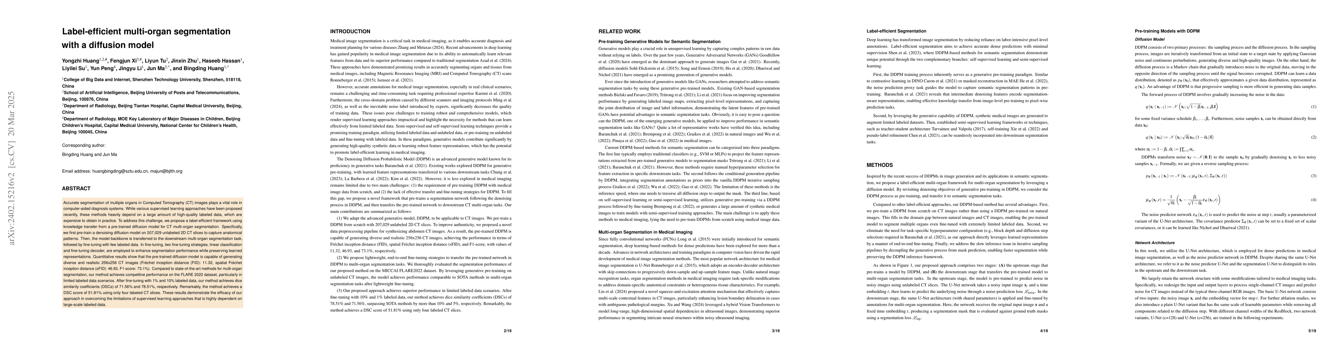
Accurate segmentation of multiple organs in Computed Tomography (CT) images plays a vital role in computer-aided diagnosis systems. Various supervised-learning approaches have been proposed recently. However, these methods heavily depend on a large amount of high-quality labeled data, which is expensive to obtain in practice. In this study, we present a label-efficient learning approach using a pre-trained diffusion model for multi-organ segmentation tasks in CT images. First, a denoising diffusion model was trained using unlabeled CT data, generating additional two-dimensional (2D) CT images. Then the pre-trained denoising diffusion network was transferred to the downstream multi-organ segmentation task, effectively creating a semi-supervised learning model that requires only a small amount of labeled data. Furthermore, linear classification and fine-tuning decoder strategies were employed to enhance the network's segmentation performance. Our generative model at 256x256 resolution achieves impressive performance in terms of Fr\'echet inception distance, spatial Fr\'echet inception distance, and F1-score, with values of 11.32, 46.93, and 73.1\%, respectively. These results affirm the diffusion model's ability to generate diverse and realistic 2D CT images. Additionally, our method achieves competitive multi-organ segmentation performance compared to state-of-the-art methods on the FLARE 2022 dataset, particularly in limited labeled data scenarios. Remarkably, even with only 1\% and 10\% labeled data, our method achieves Dice similarity coefficients (DSCs) of 71.56\% and 78.51\% after fine-tuning, respectively. The method achieves a DSC score of 51.81\% using just four labeled CT scans. These results demonstrate the efficacy of our approach in overcoming the limitations of supervised learning heavily reliant on large-scale labeled data.
AI Key Findings
Get AI-generated insights about this paper's methodology, results, significance, and more — seven facets brought into focus.
Impact
Paper Details
Authors
PDF Preview
Key Terms
Citation Network
Current paper (gray), citations (green), references (blue)
Display is limited for performance on very large graphs.
Discussion 0