Publication

Published: Feb 25, 2026
Categories: cs.LG, cs.AI, math.NA

Metrics

Source: ArXiv

Quick Actions

View PDF

Paper Preview

Abstract

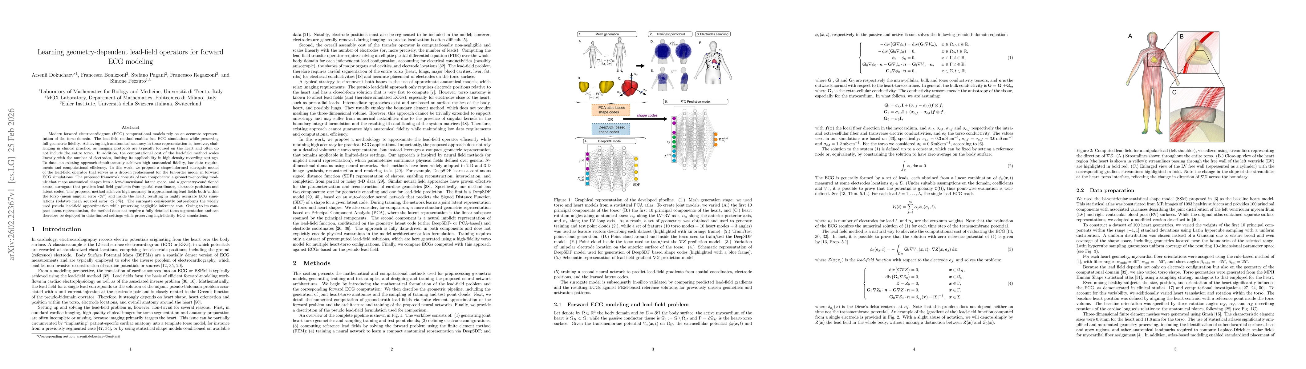
Modern forward electrocardiogram (ECG) computational models rely on an accurate representation of the torso domain. The lead-field method enables fast ECG simulations while preserving full geometric fidelity. Achieving high anatomical accuracy in torso representation is, however, challenging in clinical practice, as imaging protocols are typically focused on the heart and often do not include the entire torso. In addition, the computational cost of the lead-field method scales linearly with the number of electrodes, limiting its applicability in high-density recording settings. To date, no existing approach simultaneously achieves high anatomical fidelity, low data requirements and computational efficiency. In this work, we propose a shape-informed surrogate model of the lead-field operator that serves as a drop-in replacement for the full-order model in forward ECG simulations. The proposed framework consists of two components: a geometry-encoding module that maps anatomical shapes into a low-dimensional latent space, and a geometry-conditioned neural surrogate that predicts lead-field gradients from spatial coordinates, electrode positions and latent codes. The proposed method achieves high accuracy in approximating lead fields both within the torso (mean angular error 5°) and inside the heart, resulting in highly accurate ECG simulations (relative mean squared error <2.5%. The surrogate consistently outperforms the widely used pseudo lead-field approximation while preserving negligible inference cost. Owing to its compact latent representation, the method does not require a fully detailed torso segmentation and can therefore be deployed in data-limited settings while preserving high-fidelity ECG simulations.
AI Key Findings

Generated Feb 27, 2026

Methodology

The study builds a shape‑informed surrogate for the lead‑field operator by first encoding joint heart‑torso geometry with a low‑dimensional latent space (DeepSDF or PCA) and then training a neural network to predict lead‑field gradients from spatial coordinates, electrode positions, and the latent code.

Key Results

  • Lead‑field gradients predicted with mean angular error <4° and ECG relative L2 error <2% on a 10‑patient test set.
  • Surrogate inference is ~24× faster than full FEM lead‑field computation, enabling near‑real‑time simulations.
  • DeepSDF‑based encoding outperforms PCA, yielding lower angular and ECG errors, especially for precordial leads.
  • The surrogate accurately captures discontinuities at the heart‑torso interface and preserves clinically relevant ECG morphology.
  • Pseudo lead‑field approximation shows significantly higher errors, confirming the surrogate’s superiority.

Significance

This work delivers a fast, accurate forward ECG model that requires only sparse anatomical data, making it suitable for inverse ECG, personalized digital twins, and high‑density electrode arrays where traditional FEM is computationally prohibitive.

Technical Contribution

A decoupled, geometry‑conditioned neural surrogate that learns the lead‑field operator as a Green’s function, enabling rapid, high‑fidelity ECG forward simulations with minimal anatomical input.

Novelty

First use of a DeepSDF latent representation for joint heart‑torso geometry in ECG lead‑field modeling, achieving both high accuracy and computational speed while requiring only sparse imaging data, unlike prior pseudo‑lead‑field or full FEM approaches.

Limitations

  • The model assumes fixed myocardial fiber orientation and tissue conductivity, limiting applicability to cases with altered electrophysiology.
  • It is trained on a limited set of torso geometries and does not account for additional organs (lungs, blood cavities) that affect conductivity.
  • The current implementation is CPU‑based; GPU acceleration could further improve speed but is not demonstrated.

Future Work

  • Incorporate variable conductivity and fiber anisotropy into the latent space to handle pathological tissue changes.
  • Extend the framework to other bioelectric inverse problems (EEG, magnetoencephalography) and to multi‑organ geometries.
  • Integrate the surrogate into real‑time inverse ECG pipelines and explore adaptive mesh refinement for further efficiency.
  • Deploy GPU‑accelerated inference to support dense electrode arrays and real‑time clinical workflows.

Paper Details

Paper ID: 2602.22367
Comments: 20 pages, 9 figures
Categories:
cs.LG cs.AI math.NA q-bio.TO

Similar Papers

Found 4 papers

Multimodal Lead-Specific Modeling of ECG for Low-Cost Pulmonary Hypertension Assessment

Mohammod N. I. Suvon, Shuo Zhou, Prasun C. Tripathi et al.

2025-03-03

Lightweight Deep Learning Architecture for Multi-Lead ECG Arrhythmia Detection.

Elsheikhy, Donia H, Hassan, Abdelwahab S, Yhiea, Nashwa M et al.

2025-09-05

Comments (0)

This website uses cookies to ensure you get the best experience. Learn more