Publication
Metrics
AI Quick Summary
This paper demonstrates the effectiveness of Res-CR-Net, a fully convolutional neural network initially designed for microscopy image segmentation, in accurately segmenting lung fields in chest X-rays, showing comparable or superior performance to U-Net-based architectures.
Paper Preview
Abstract
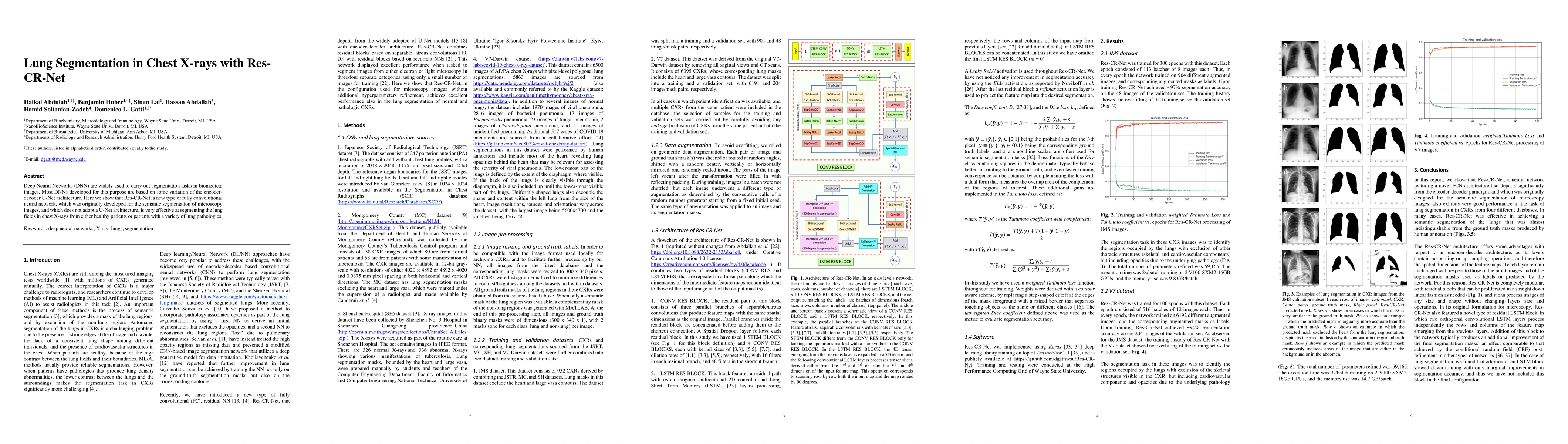
Deep Neural Networks (DNN) are widely used to carry out segmentation tasks in biomedical images. Most DNNs developed for this purpose are based on some variation of the encoder-decoder U-Net architecture. Here we show that Res-CR-Net, a new type of fully convolutional neural network, which was originally developed for the semantic segmentation of microscopy images, and which does not adopt a U-Net architecture, is very effective at segmenting the lung fields in chest X-rays from either healthy patients or patients with a variety of lung pathologies.
AI Key Findings
Get AI-generated insights about this paper's methodology, results, significance, and more — seven facets brought into focus.
Impact
Paper Details
Authors
PDF Preview
Key Terms
Citation Network
Current paper (gray), citations (green), references (blue)
Display is limited for performance on very large graphs.
Discussion 0