A hybrid mechanistic learning framework combining a mathematical tumor growth model (ODEs) with a guided denoising diffusion model (DDIM) to generate future MRI scans from prior scans.
Mechanistic Learning with Guided Diffusion Models to Predict Spatio-Temporal Brain Tumor Growth
Publication
Metrics
Quick Answers
What methodology did the authors use?
A hybrid mechanistic learning framework combining a mathematical tumor growth model (ODEs) with a guided denoising diffusion model (DDIM) to generate future MRI scans from prior scans. More in Methodology →
What are the key results?
The framework generates realistic follow-up MRIs aligned with predicted tumor growth and patient anatomy. — Tumor growth probability maps capture clinically relevant growth extent and directionality with 95th percentile Hausdorff Distance. More in Key Results →
Why is this work significant?
This approach provides accurate spatio-temporal tumor growth predictions, aiding clinical decision-making in neuro-oncology, especially for pediatric gliomas where data is limited. More in Significance →
What are the main limitations?
Requires multiple imaging timepoints for accurate modeling — Guided diffusion network relies on multiparametric MRI as input More in Limitations →
Abstract
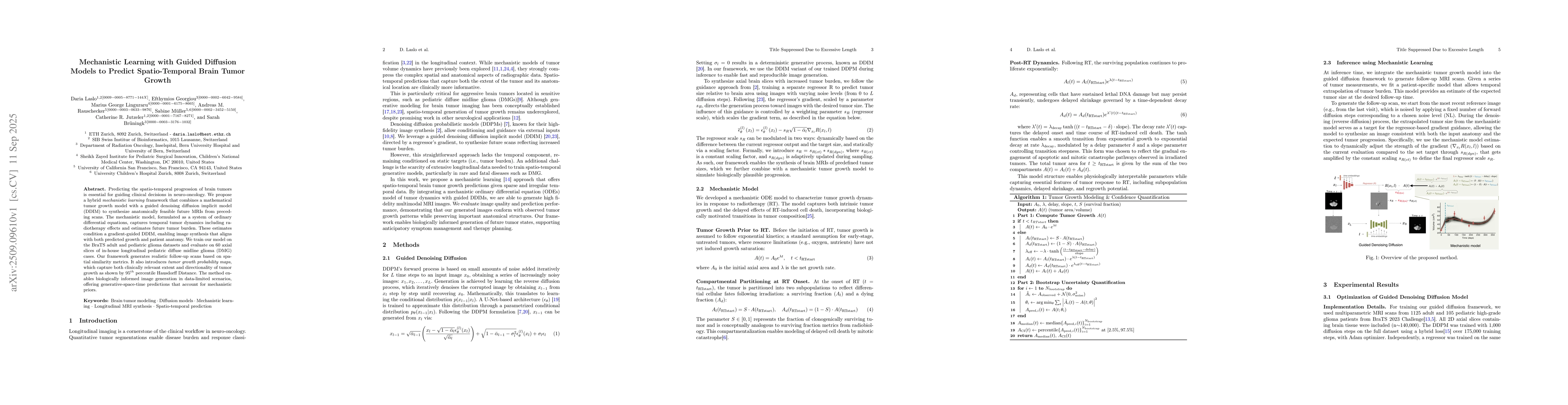
Predicting the spatio-temporal progression of brain tumors is essential for guiding clinical decisions in neuro-oncology. We propose a hybrid mechanistic learning framework that combines a mathematical tumor growth model with a guided denoising diffusion implicit model (DDIM) to synthesize anatomically feasible future MRIs from preceding scans. The mechanistic model, formulated as a system of ordinary differential equations, captures temporal tumor dynamics including radiotherapy effects and estimates future tumor burden. These estimates condition a gradient-guided DDIM, enabling image synthesis that aligns with both predicted growth and patient anatomy. We train our model on the BraTS adult and pediatric glioma datasets and evaluate on 60 axial slices of in-house longitudinal pediatric diffuse midline glioma (DMG) cases. Our framework generates realistic follow-up scans based on spatial similarity metrics. It also introduces tumor growth probability maps, which capture both clinically relevant extent and directionality of tumor growth as shown by 95th percentile Hausdorff Distance. The method enables biologically informed image generation in data-limited scenarios, offering generative-space-time predictions that account for mechanistic priors.
Key Findings, in focus
Seven facets of this paper, analysed and brought into focus by AI.
This approach provides accurate spatio-temporal tumor growth predictions, aiding clinical decision-making in neuro-oncology, especially for pediatric gliomas where data is limited.
- The framework generates realistic follow-up MRIs aligned with predicted tumor growth and patient anatomy.
- Tumor growth probability maps capture clinically relevant growth extent and directionality with 95th percentile Hausdorff Distance.
- Mechanistic models enable biologically informed image generation in data-limited scenarios.
This approach provides accurate spatio-temporal tumor growth predictions, aiding clinical decision-making in neuro-oncology, especially for pediatric gliomas where data is limited.
Integration of mechanistic ODE tumor models with generative DDIM models for biologically informed image synthesis
First hybrid framework combining mechanistic tumor growth models with guided diffusion models for spatio-temporal MRI prediction in neuro-oncology
- Requires multiple imaging timepoints for accurate modeling
- Guided diffusion network relies on multiparametric MRI as input
- Explore single-contrast MRI application via ablation study
- Investigate patient-specific fine-tuning of diffusion process
Discussion 0