01
MethodologyHow they did it
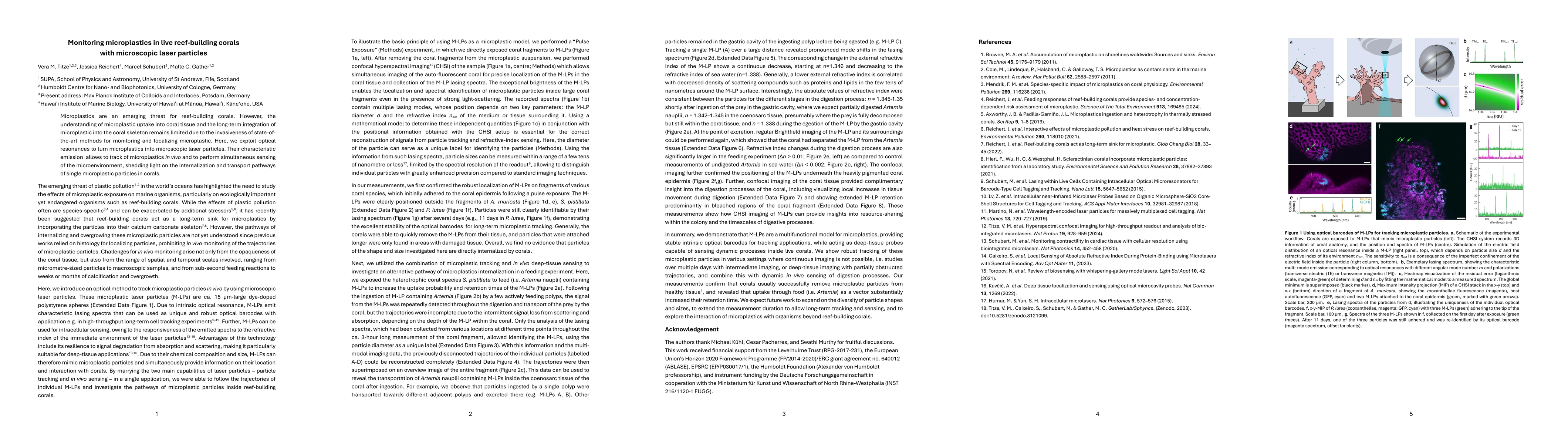
The research utilized a multi-modal confocal imaging and hyperspectral imaging (CHSI) setup to track microplastic particles (M-LPs) in live corals. Corals were exposed to M-LPs, and their lasing spectra were recorded to determine particle diameter and external refractive index. Feeding measurements were conducted under the CHSI microscope, and mathematical analysis of lasing spectra was used for combined tracking and sensing.
Discussion 0