Monte Carlo study of a 3D Compton imaging device with GEANT4
Publication
Metrics
AI Quick Summary
Researchers used a Monte Carlo simulation to study a 3D Compton imaging device for brain imaging, finding a resolution of about 2mm and contrast of 12% with a 600 keV gamma beam, and estimated an effective dose of 1 mSv for a complete brain scan.
Paper Preview
Abstract
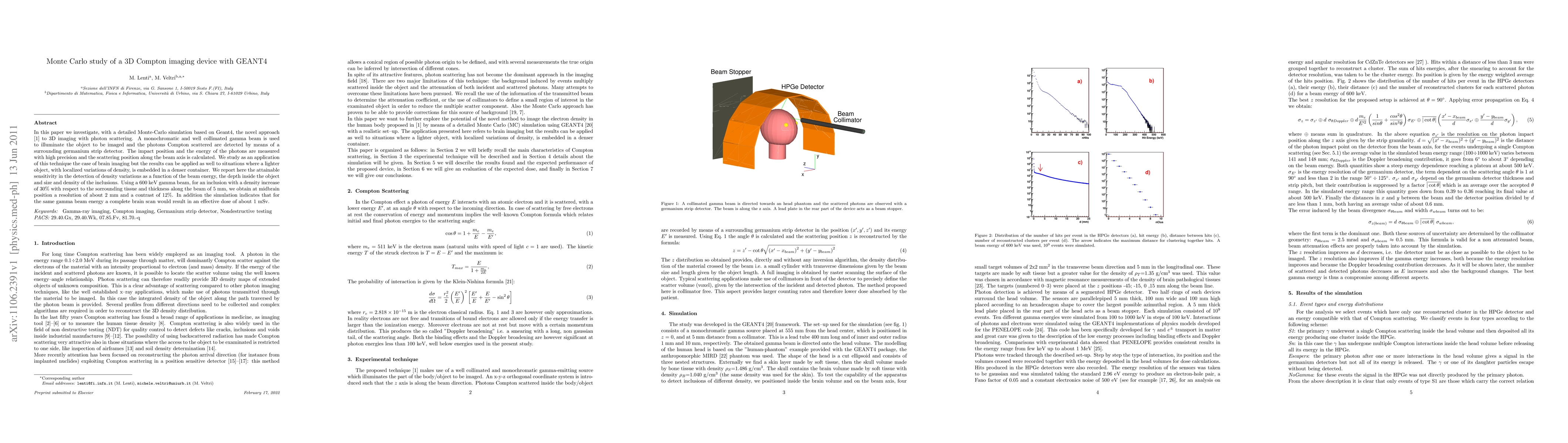
In this paper we investigate, with a detailed Monte-Carlo simulation based on Geant4, the novel approach [Nucl. Instrum. Methods A588 (2008) 457] to 3D imaging with photon scattering. A monochromatic and well collimated gamma beam is used to illuminate the object to be imaged and the photons Compton scattered are detected by means of a surrounding germanium strip detector. The impact position and the energy of the photons are measured with high precision and the scattering position along the beam axis is calculated. We study as an application of this technique the case of brain imaging but the results can be applied as well to situations where a lighter object, with localized variations of density, is embedded in a denser container. We report here the attainable sensitivity in the detection of density variations as a function of the beam energy, the depth inside the object and size and density of the inclusions. Using a 600 keV gamma beam, for an inclusion with a density increase of 30% with respect to the sorrounding tissue and thickness along the beam of 5 mm, we obtain at midbrain position a resolution of about 2 mm and a contrast of 12%. In addition the simulation indicates that for the same gamma beam energy a complete brain scan would result in an effective dose of about 1 mSv.
AI Key Findings
Get AI-generated insights about this paper's methodology, results, significance, and more — seven facets brought into focus.
Impact
Paper Details
PDF Preview
Key Terms
Citation Network
Current paper (gray), citations (green), references (blue)
Display is limited for performance on very large graphs.
Discussion 0