Morphologies of compressed active epithelial monolayers
Publication
Metrics
AI Quick Summary
This paper uses a 3D active vertex model to explore the shapes of compressed epithelial monolayers under active junctional noise, finding that specific compressive strains induce distinct fold patterns. The study reveals that strong junctional tension fluctuations can fluidize the tissue, leading to villus morphology even without compressive strain, and analyzes the effects of strain rate and tissue thickness modulation.
Paper Preview
Abstract
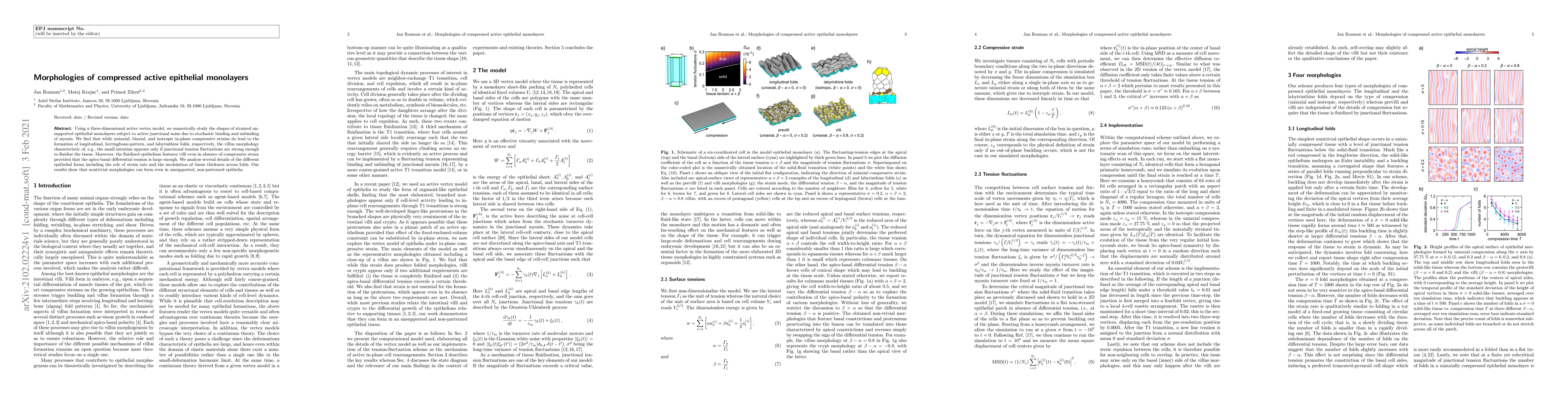
Using a three-dimensional active vertex model, we numerically study the shapes of strained unsupported epithelial monolayers subject to active junctional noise due to stochastic binding and unbinding of myosin. We find that while uniaxial, biaxial, and isotropic in-plane compressive strains do lead to the formation of longitudinal, herringbone-pattern, and labyrinthine folds, respectively, the villus morphology characteristic of, e.g., the small intestine appears only if junctional tension fluctuations are strong enough to fluidize the tissue. Moreover, the fluidized epithelium features villi even in absence of compressive strain provided that the apico-basal differential tension is large enough. We analyze several details of the different epithelial forms including the role of strain rate and the modulation of tissue thickness across folds. Our results show that nontrivial morphologies can form even in unsupported, non-patterned epithelia.
AI Key Findings
Get AI-generated insights about this paper's methodology, results, significance, and more — seven facets brought into focus.
Impact
Paper Details
Authors
PDF Preview
Key Terms
Citation Network
Current paper (gray), citations (green), references (blue)
Display is limited for performance on very large graphs.
Discussion 0