Multi-planar 2D-U-Net Segmentation of 3D-CT Abdominal Organs augmented by Spatial Occurrence Maps
Publication
Metrics
Paper Preview
Abstract
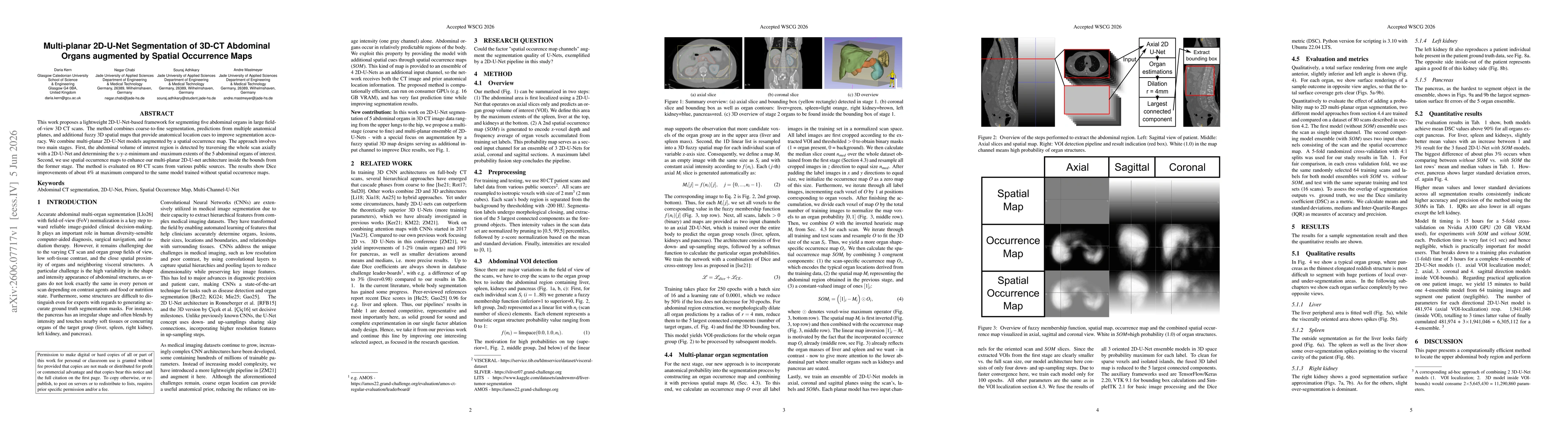
This work proposes a lightweight 2D-U-Net-based framework for segmenting five abdominal organs in large field-of-view 3D CT scans. The method combines coarse-to-fine segmentation, predictions from multiple anatomical planes, and additional fuzzy 3D spatial maps that provide anatomical location cues to improve segmentation accuracy. We combine multi-planar 2D-U-Net models augmented by a spatial occurrence map. The approach involves two main stages. First, the abdominal volume of interest region is detected by traversing the whole scan axially with a 2D-U-Net and determining the x-y-z-minimum and -maximum extents of the 5 abdominal organs of interest. Second, we use spatial occurrence maps to enhance our multi-planar 2D-U-net architecture inside the bounds from the former stage. The method is evaluated on 80 CT scans from various public sources. The results show Dice improvements of about 4% at maximum compared to the same model trained without spatial occurrence maps.
AI Key Findings
Get AI-generated insights about this paper's methodology, results, significance, and more — seven facets brought into focus.
Discussion 0