Publication
Metrics
AI Quick Summary
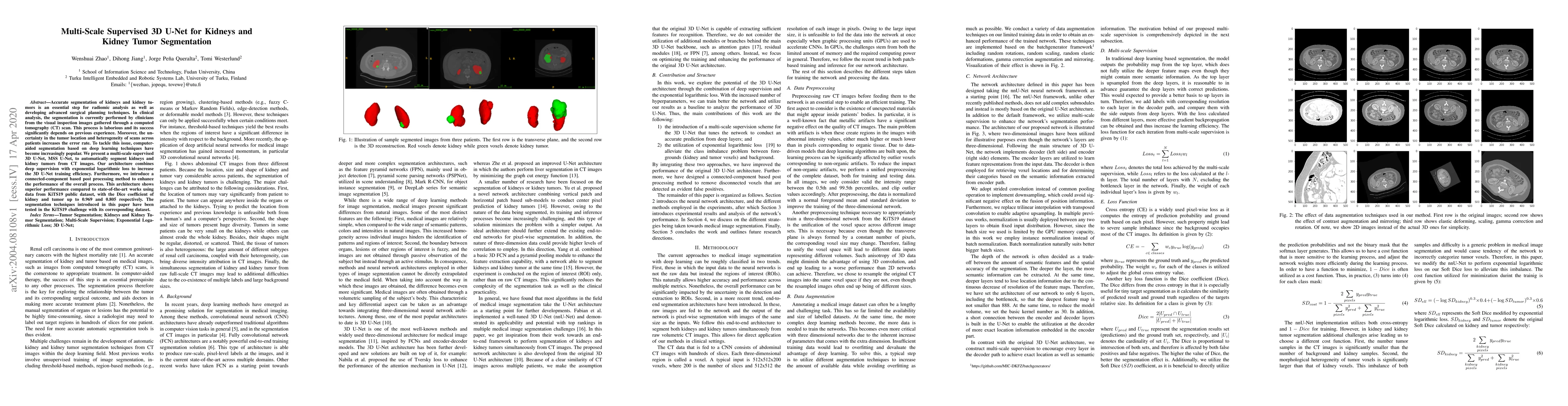
This paper proposes a multi-scale supervised 3D U-Net, MSS U-Net, for the automatic segmentation of kidneys and kidney tumors from CT scans, achieving high Dice coefficients of 0.969 and 0.805 respectively. The method enhances training efficiency and post-processing for superior accuracy compared to existing techniques.
Paper Preview
Abstract
Accurate segmentation of kidneys and kidney tumors is an essential step for radiomic analysis as well as developing advanced surgical planning techniques. In clinical analysis, the segmentation is currently performed by clinicians from the visual inspection images gathered through a computed tomography (CT) scan. This process is laborious and its success significantly depends on previous experience. Moreover, the uncertainty in the tumor location and heterogeneity of scans across patients increases the error rate. To tackle this issue, computer-aided segmentation based on deep learning techniques have become increasingly popular. We present a multi-scale supervised 3D U-Net, MSS U-Net, to automatically segment kidneys and kidney tumors from CT images. Our architecture combines deep supervision with exponential logarithmic loss to increase the 3D U-Net training efficiency. Furthermore, we introduce a connected-component based post processing method to enhance the performance of the overall process. This architecture shows superior performance compared to state-of-the-art works using data from KiTS19 public dataset, with the Dice coefficient of kidney and tumor up to 0.969 and 0.805 respectively. The segmentation techniques introduced in this paper have been tested in the KiTS19 challenge with its corresponding dataset.
AI Key Findings
Get AI-generated insights about this paper's methodology, results, significance, and more — seven facets brought into focus.
Impact
Paper Details
Authors
PDF Preview
Key Terms
Citation Network
Current paper (gray), citations (green), references (blue)
Display is limited for performance on very large graphs.
Discussion 0