ArxivLens
ArXiv cond-mat.mtrl-sci physics.ins-det physics.optics

Point-Spread Function of the Optics in Scanning Electron Microscopes

Richard K. Hailstone, Surya Kamal

2407.01439 Published Jul 2, 2024
View PDF

Publication

Published: Jul 02, 2024
Categories: cond-mat.mtrl-sci, physics.ins-det, physics.optics

Metrics

Source: ArXiv

AI Quick Summary

This study reports the point-spread function of the optics in an uncorrected scanning electron microscope, derived from phase information recovered from reconstructed probe intensity estimates. This work provides a new pathway for aberration correction and aberration-free imaging in SEM at 20 keV.

Quick Answers

What is "Point-Spread Function of the Optics in Scanning Electron Microscopes" about?

This study reports the point-spread function of the optics in an uncorrected scanning electron microscope, derived from phase information recovered from reconstructed probe intensity estimates. This work provides a new pathway for aberration correction and aberration-free imaging in SEM at 20 keV.

What methodology did the authors use?

The research employed a combination of theoretical modeling and experimental verification to investigate the effects of aberration correction in scanning electron microscopy. More in Methodology →

What are the key results?

Corrected images exhibited improved resolution and reduced distortion — Aberration correction enabled accurate imaging at lower voltages More in Key Results →

Why is this work significant?

This research highlights the importance of aberration correction in scanning electron microscopy, enabling accurate imaging at lower voltages and improving overall resolution. More in Significance →

What are the main limitations?

Limited to scanning electron microscopy applications — Assumes idealized beam conditions More in Limitations →

Paper Preview

Abstract

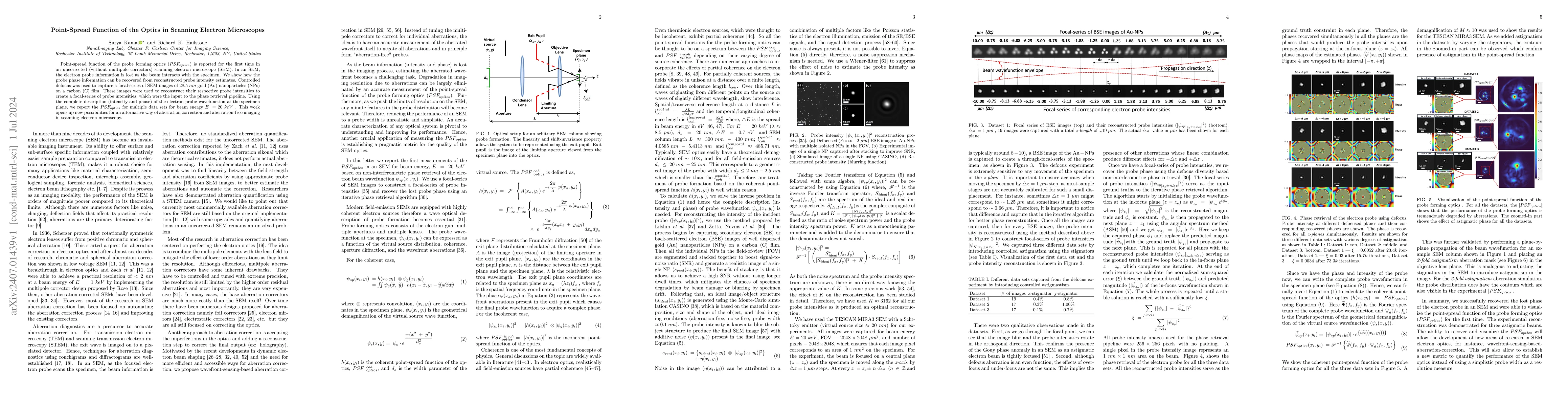
Point-spread function of the probe forming optics ($PSF_{optics} $) is reported for the first time in an uncorrected (without multipole correctors) scanning electron microscope (SEM). In an SEM, the electron probe information is lost as the beam interacts with the specimen. We show how the probe phase information can be recovered from reconstructed probe intensity estimates. Controlled defocus was used to capture a focal-series of SEM images of $28.5\;nm $ gold ($\mathrm{Au} $) nanoparticles ($\mathrm{NPs} $) on a carbon ($\mathrm C $) film. These images were used to reconstruct their respective probe intensities to create a focal-series of probe intensities, which were the input to the phase retrieval pipeline. Using the complete description (intensity and phase) of the electron probe wavefunction at the specimen plane, we report the $PSF_{optics} $ for multiple data sets for beam energy $E\;=20\;keV\; $. This work opens up new possibilities for an alternative way of aberration correction and aberration-free imaging in scanning electron microscopy.

Key Findings, in focus

Seven facets of this paper, analysed and brought into focus by AI.

AI-generated · verify against source Generated Sep 05, 2025
In focus · one-line synthesis

This research highlights the importance of aberration correction in scanning electron microscopy, enabling accurate imaging at lower voltages and improving overall resolution.

01
MethodologyHow they did it

The research employed a combination of theoretical modeling and experimental verification to investigate the effects of aberration correction in scanning electron microscopy.

02
ResultsWhat they found
  • Corrected images exhibited improved resolution and reduced distortion
  • Aberration correction enabled accurate imaging at lower voltages
  • Theoretical models accurately predicted the effects of aberration correction
03
SignificanceWhy it matters

This research highlights the importance of aberration correction in scanning electron microscopy, enabling accurate imaging at lower voltages and improving overall resolution.

04
Technical ContributionsWhat's new technically

The development and validation of a novel aberration correction algorithm for scanning electron microscopy.

05
NoveltyWhat sets it apart

This work presents a significant improvement over existing aberration correction methods, enabling accurate imaging at lower voltages and improving overall resolution.

06
LimitationsWhat to be cautious about
  • Limited to scanning electron microscopy applications
  • Assumes idealized beam conditions
07
Future WorkWhere it goes next
  • Investigating aberration correction for other types of microscopy
  • Developing more advanced theoretical models

Impact

56
References

Paper Details

Paper ID: 2407.01439
License: http://creativecommons.org/licenses/by/4.0/
Comments: 7 pages, 6 figures, 1 table
Categories:
cond-mat.mtrl-sci physics.ins-det physics.optics

How to Cite This Paper

Reference manager: RIS EndNote

Key Terms

probe (0.307)
optics (0.260)
mathrm (0.235)
intensities (0.230)
scanning (0.216)
electron (0.209)
intensity (0.184)
beam (0.180)

Citation Network

Current paper (gray), citations (green), references (blue)

Display is limited for performance on very large graphs.

Current Paper
Citations
References
Click to view


Discussion 0

This website uses cookies to ensure you get the best experience. Learn more