The research utilized Geant4 Monte-Carlo simulations to analyze scintillation light in a monolithic hemi-ellipsoidal CsI crystal, creating a look-up table (LUT) to map crystal points to expected light patterns on the surface. A Poisson-statistics-based algorithm was developed for localizing gamma-ray event locations.
Scintillation Event Localization in Hemi-Ellipsoid Detector for SPECT, a simulation study using Geant4 Monte-Carlo
Publication
Metrics
AI Quick Summary
This study investigates the localization of scintillation events in a hemi-ellipsoid detector for Cardiac SPECT using Geant4 Monte-Carlo simulations. The results show an average localization error of 0.71 mm in the detector and <1 mm when back-projected to the object midplane, indicating high resolution and potential for high sensitivity in Cardiac SPECT applications.
Quick Answers
What is "Scintillation Event Localization in Hemi-Ellipsoid Detector for SPECT, a simulation study using Geant4 Monte-Carlo" about?
This study investigates the localization of scintillation events in a hemi-ellipsoid detector for Cardiac SPECT using Geant4 Monte-Carlo simulations. The results show an average localization error of 0.71 mm in the detector and <1 mm when back-projected to the object midplane, indicating high resolution and potential for high sensitivity in Cardiac SPECT applications.
What methodology did the authors use?
The research utilized Geant4 Monte-Carlo simulations to analyze scintillation light in a monolithic hemi-ellipsoidal CsI crystal, creating a look-up table (LUT) to map crystal points to expected light patterns on the surface. A Poisson-statistics-based algorithm was developed for localizing gamma-ray event locations. More in Methodology →
What are the key results?
The average localization error within the detector was 0.71 (+/-0.44) mm, with a maximum of 1.36 (+/-0.67) mm at the apex. — Back-projected errors in object-space averaged 0.4 (+/-0.22) mm, demonstrating high resolution due to the detector's magnification. More in Key Results →
Why is this work significant?
This study is significant as it demonstrates a hemi-ellipsoid curved detector capable of achieving sub-mm localization errors for Cardiac SPECT, combining better intrinsic resolution with high magnification to reduce overall error. More in Significance →
What are the main limitations?
The study assumed perfect pinhole collimator resolution recovery, which may not be achievable in practice. — The research focused on CsI(Tl) crystals, and results may not directly translate to other scintillation materials. More in Limitations →
Abstract
A high sensitivity Cardiac SPECT system using curved crystals with pinhole collimation was proposed previously (Dey, IEEE TNS 2012, Bhusal et al, Med. Phys. 2019). Here, we hypothesize that a high curvature hemi-ellipsoid detector results in measurable differences in light distribution from events at different depths in the crystal. This was tested by analyzing the scintillation light in hemi-ellipsoid detector using Monte-Carlo (Geant4) and evaluation of both the localization error at detector and the back-projected errors in 
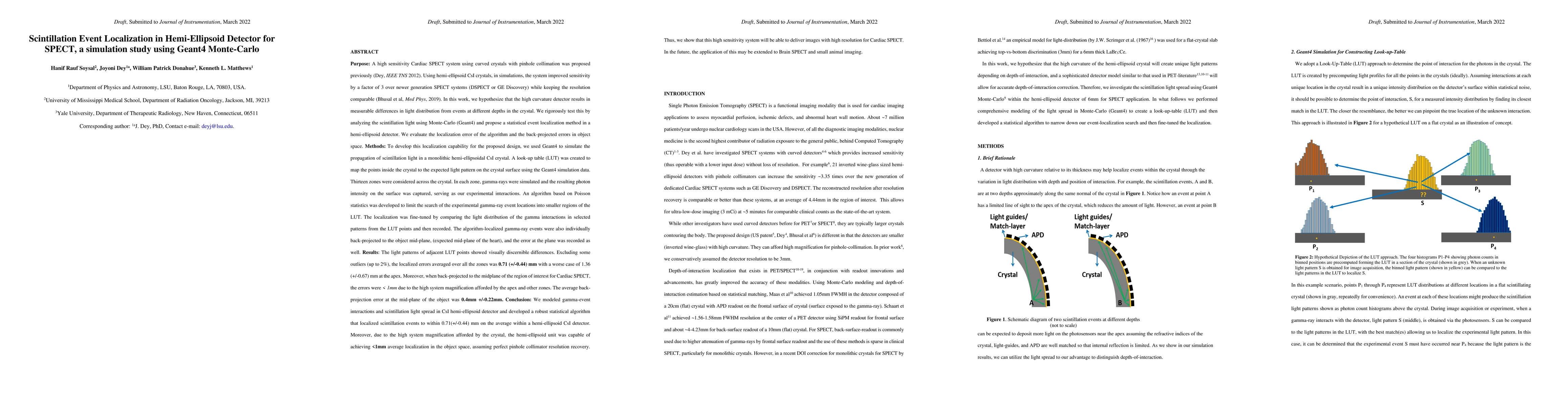
object-space. We used Geant4 to simulate the propagation of scintillation light in a monolithic hemi-ellipsoidal CsI crystal. A look-up table (LUT) was created to map the points inside the crystal to the expected light pattern on the crystal surface using Geant4. In thirteen zones across the crystal, gamma-rays were simulated and the resulting scintillator light intensity on the surface was captured, serving as our experimental interactions. A Poisson-statistics-based algorithm was developed to limit the search of the gamma-ray event locations into small regions of the LUT and fine-tuned by interpolating between selected LUT points by comparing the light distribution of the gamma interactions and LUT light patterns. The localized events were individually back-projected to the object mid-plane, and the errors recorded. Excluding some outliers (up to 2%), the localized errors averaged over all the zones was 0.71 (+/-0.44) mm with a worse case of 1.36 (+/-0.67) mm at the apex. When back-projected to the midplane of the object for Cardiac SPECT, the errors were <1mm and average error was 0.4(+/-0.22) mm, due to the high system magnification afforded by the detector. Thus, for our high sensitivity system, we were also able to achieve high resolution, assuming perfect pinhole collimator resolution recovery for Cardiac SPECT application.
Key Findings, in focus
Seven facets of this paper, analysed and brought into focus by AI.
This study is significant as it demonstrates a hemi-ellipsoid curved detector capable of achieving sub-mm localization errors for Cardiac SPECT, combining better intrinsic resolution with high magnification to reduce overall error.
- The average localization error within the detector was 0.71 (+/-0.44) mm, with a maximum of 1.36 (+/-0.67) mm at the apex.
- Back-projected errors in object-space averaged 0.4 (+/-0.22) mm, demonstrating high resolution due to the detector's magnification.
- 99% and 98% of localization errors were within 2 mm when excluding or including Compton-scattered gamma-rays, respectively.
This study is significant as it demonstrates a hemi-ellipsoid curved detector capable of achieving sub-mm localization errors for Cardiac SPECT, combining better intrinsic resolution with high magnification to reduce overall error.
The development of a robust localization algorithm using a LUT and Poisson statistics for curved crystal detectors in SPECT imaging.
The research introduces a novel approach to event localization in hemi-ellipsoid detectors, combining curved geometry with advanced simulation and algorithmic techniques to achieve ultra-high resolution in SPECT imaging.
- The study assumed perfect pinhole collimator resolution recovery, which may not be achievable in practice.
- The research focused on CsI(Tl) crystals, and results may not directly translate to other scintillation materials.
- Investigate the impact of collimator resolution loss on overall system performance.
- Explore the applicability of this design for Brain SPECT and small animal imaging.
Impact
Paper Details
Authors
PDF Preview
Key Terms
Citation Network
Current paper (gray), citations (green), references (blue)
Display is limited for performance on very large graphs.
Discussion 0