Segmentation of carotid vessel wall using U-Net and segmentation average network
Publication
Metrics
AI Quick Summary
This research introduces a convolutional neural network (CNN) utilizing three U-Nets to segment the common carotid artery from 3D carotid ultrasound images, enhancing vessel wall volume and thickness quantification. The segmentation maps are further refined using a novel segmentation average network, resulting in improved Dice similarity coefficient, sensitivity, and AUC.
Paper Preview
Abstract
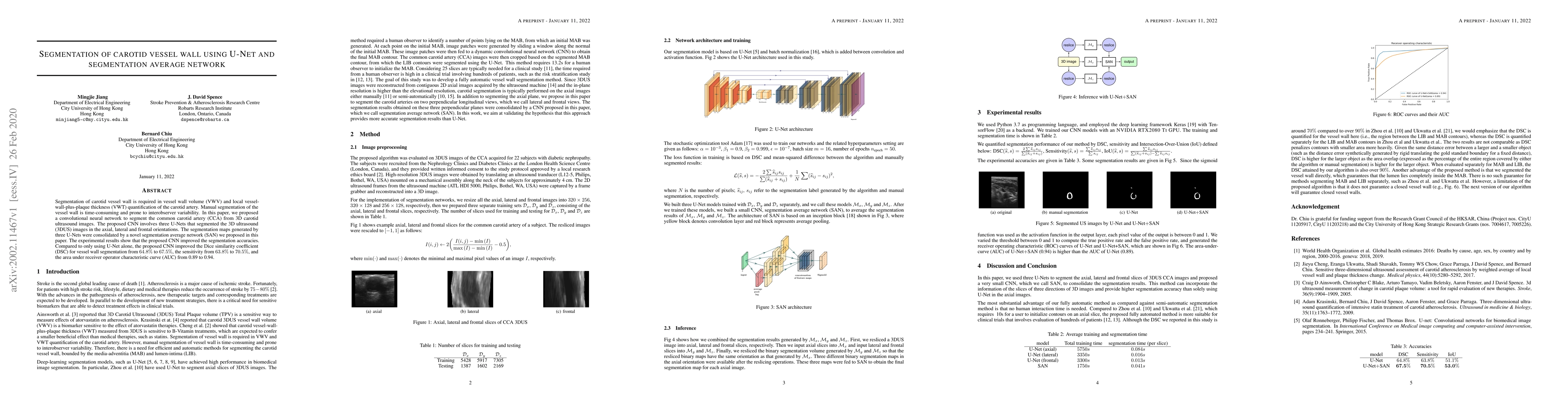
Segmentation of carotid vessel wall is required in vessel wall volume (VWV) and local vessel-wall-plus-plaque thickness (VWT) quantification of the carotid artery. Manual segmentation of the vessel wall is time-consuming and prone to interobserver variability. In this paper, we proposed a convolution neural network to segment the common carotid artery (CCA) from 3D carotid ultrasound images. The proposed CNN involves three U-Nets that segmented the 3D ultrasound (3DUS) images in the axial, lateral and frontal orientations. The segmentation maps generated by three U-Nets were consolidated by a novel segmentation average network (SAN) we proposed in this paper. The experimental results show that the proposed CNN improved the Dice similarity coefficient (DSC) for vessel wall segmentation from 64.8% to 67.5%, the sensitivity from 63.8% to 70.5%, and the area under receiver operator characteristic curve (AUC) from 0.89 to 0.94.
AI Key Findings
Get AI-generated insights about this paper's methodology, results, significance, and more — seven facets brought into focus.
Impact
Paper Details
Authors
PDF Preview
Key Terms
Citation Network
Current paper (gray), citations (green), references (blue)
Display is limited for performance on very large graphs.
Discussion 0