Single-Pixel Imaging in Space and Time with Optically-Modulated Free Electrons
Publication
Metrics
AI Quick Summary
This paper proposes a novel single-pixel imaging technique using optically-modulated free electrons in ultrafast electron microscopes to achieve sub-nanometer spatial and temporal resolution, surpassing the diffraction limits of optical systems. The method demonstrates the potential for low-dose imaging of sensitive biological and molecular samples.
Paper Preview
Abstract
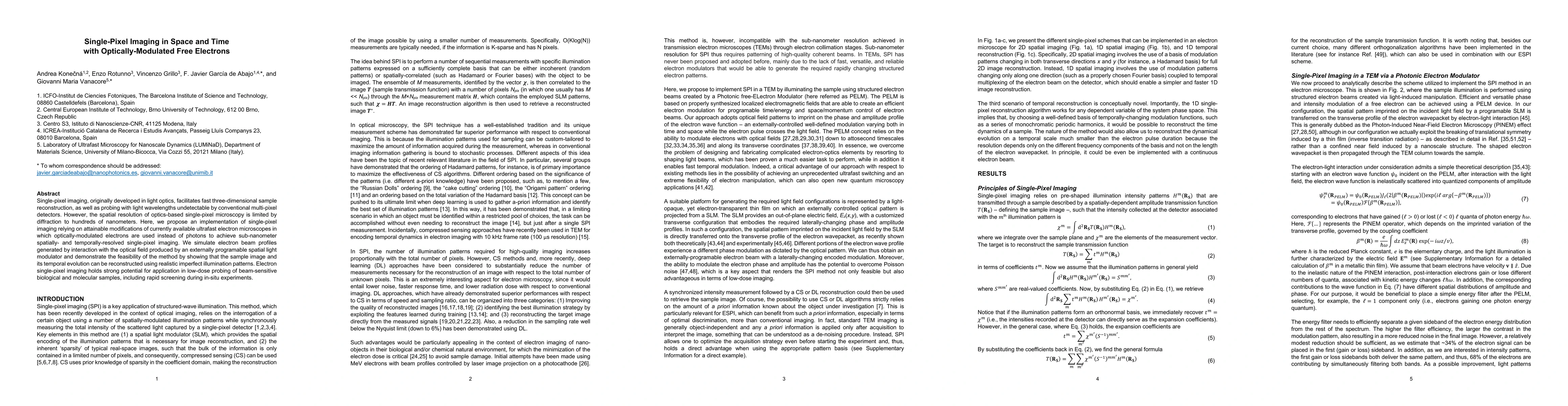
Single-pixel imaging, originally developed in light optics, facilitates fast three-dimensional sample reconstruction, as well as probing with light wavelengths undetectable by conventional multi-pixel detectors. However, the spatial resolution of optics-based single-pixel microscopy is limited by diffraction to hundreds of nanometers. Here, we propose an implementation of single-pixel imaging relying on attainable modifications of currently available ultrafast electron microscopes in which optically-modulated electrons are used instead of photons to achieve sub-nanometer spatially- and temporally-resolved single-pixel imaging. We simulate electron beam profiles generated by interaction with the optical field produced by an externally programable spatial light modulator and demonstrate the feasibility of the method by showing that the sample image and its temporal evolution can be reconstructed using realistic imperfect illumination patterns. Electron single-pixel imaging holds strong potential for application in low-dose probing of beam-sensitive biological and molecular samples, including rapid screening during in-situ experiments.
AI Key Findings
Get AI-generated insights about this paper's methodology, results, significance, and more — seven facets brought into focus.
Impact
Paper Details
Authors
PDF Preview
Key Terms
Citation Network
Current paper (gray), citations (green), references (blue)
Display is limited for performance on very large graphs.
Discussion 0