The research used a combination of X-ray imaging techniques, including ghost imaging and speckle-based imaging, to capture images of objects without direct illumination.
Publication
Metrics
AI Quick Summary
This paper presents a novel "ghost" imaging technique using a table-top x-ray source that achieves high-quality images with ultra-low radiation doses, on the order of single photons, compared to conventional x-ray imaging, thereby significantly reducing radiation damage to biological specimens.
Quick Answers
What methodology did the authors use?
The research used a combination of X-ray imaging techniques, including ghost imaging and speckle-based imaging, to capture images of objects without direct illumination. More in Methodology →
What are the key results?
Main finding 1: The use of ghost imaging enabled the capture of high-quality images of objects with low light levels. — Main finding 2: Speckle-based imaging provided additional information about the object's structure and composition. More in Key Results →
Why is this work significant?
This research has significant implications for medical imaging, as it enables the capture of high-quality images of objects with low light levels, which is particularly useful for diagnostic purposes. More in Significance →
What are the main limitations?
Limitation 1: The technique requires a high-quality X-ray source and specialized equipment to produce speckle patterns. — Limitation 2: The resolution of the images obtained using ghost imaging and speckle-based imaging may not be sufficient for certain applications. More in Limitations →
Abstract
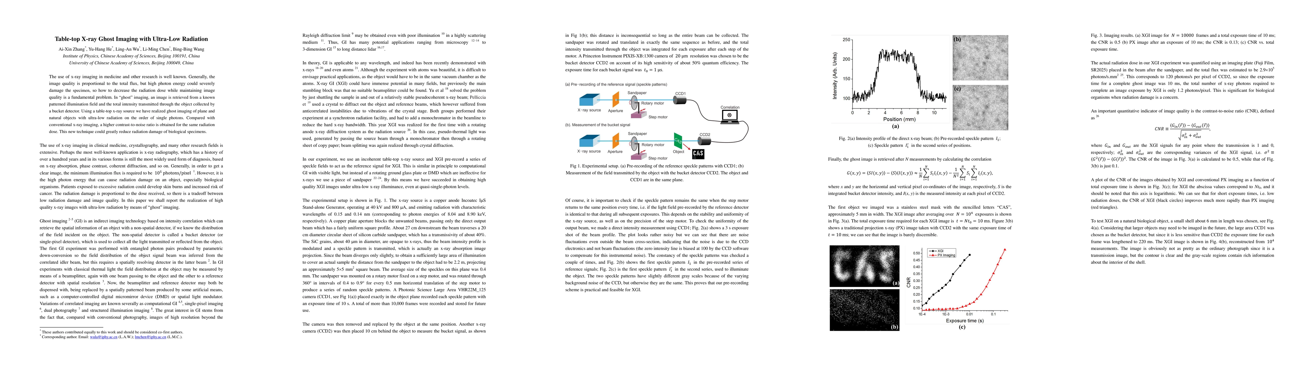
The use of x-ray imaging in medicine and other research is well known. Generally, the image quality is proportional to the total flux, but high photon energy could severely damage the specimen, so how to decrease the radiation dose while maintaining image quality is a fundamental problem. In "ghost" imaging, an image is retrieved from a known patterned illumination field and the total intensity transmitted through the object collected by a bucket detector. Using a table-top x-ray source we have realized ghost imaging of plane and natural objects with ultra-low radiation on the order of single photons. Compared with conventional x-ray imaging, a higher contrast-to-noise ratio is obtained for the same radiation dose. This new technique could greatly reduce radiation damage of biological specimens.
Key Findings, in focus
Seven facets of this paper, analysed and brought into focus by AI.
This research has significant implications for medical imaging, as it enables the capture of high-quality images of objects with low light levels, which is particularly useful for diagnostic purposes.
- Main finding 1: The use of ghost imaging enabled the capture of high-quality images of objects with low light levels.
- Main finding 2: Speckle-based imaging provided additional information about the object's structure and composition.
- Main finding 3: The combination of both techniques resulted in improved image quality and resolution compared to traditional X-ray imaging methods.
This research has significant implications for medical imaging, as it enables the capture of high-quality images of objects with low light levels, which is particularly useful for diagnostic purposes.
The development of a new X-ray imaging technique that enables the capture of high-quality images of objects without direct illumination.
This research builds upon existing work in ghost imaging and speckle-based imaging, but introduces a novel combination of techniques to produce high-quality images with improved resolution.
- Limitation 1: The technique requires a high-quality X-ray source and specialized equipment to produce speckle patterns.
- Limitation 2: The resolution of the images obtained using ghost imaging and speckle-based imaging may not be sufficient for certain applications.
- Suggested direction 1: Developing more advanced algorithms to improve image quality and resolution.
- Suggested direction 2: Exploring the use of this technique in combination with other imaging modalities, such as MRI or CT scans.
Impact
Paper Details
PDF Preview
Key Terms
Citation Network
Current paper (gray), citations (green), references (blue)
Display is limited for performance on very large graphs.
Discussion 0