ArxivLens

Publication

Published: Feb 17, 2025
Categories: physics.optics

Metrics

Source: ArXiv

AI Quick Summary

This paper proposes a low-cost 3D imaging technique using surface plasmon coupled emission (SPCE) to visualize biological cells, eliminating the need for expensive microscopes. The developed method reconstructs accurate 3D cell images from SPCE patterns with RMSE errors of $\lesssim 1.4%$ for circular cells and $\lesssim 2.8%$ for irregular shapes, showcasing its potential for simple, affordable cell imaging.

Quick Answers

What methodology did the authors use?

The research proposes a method to obtain 3D cell images using surface plasmon coupled emission (SPCE) patterns from fluorescently labeled biological cells, eliminating the need for conventional microscopes or extensive data processing. More in Methodology →

What are the key results?

3D cell structures have been successfully reconstructed from detected SPCE patterns with RMSE within a few percent. — For circular-shaped cells, RMSE is ≲1.4%, and for irregular cell shapes, RMSE is ≲2.8%. More in Key Results →

Why is this work significant?

This low-cost, simple optical arrangement technique is highly desirable for biomedical and microbiology studies, providing an alternative to expensive equipment. More in Significance →

What are the main limitations?

The technique is in its initial stages of development. — Determining the dipole position from the near-field image remains challenging. More in Limitations →

Paper Preview

Abstract

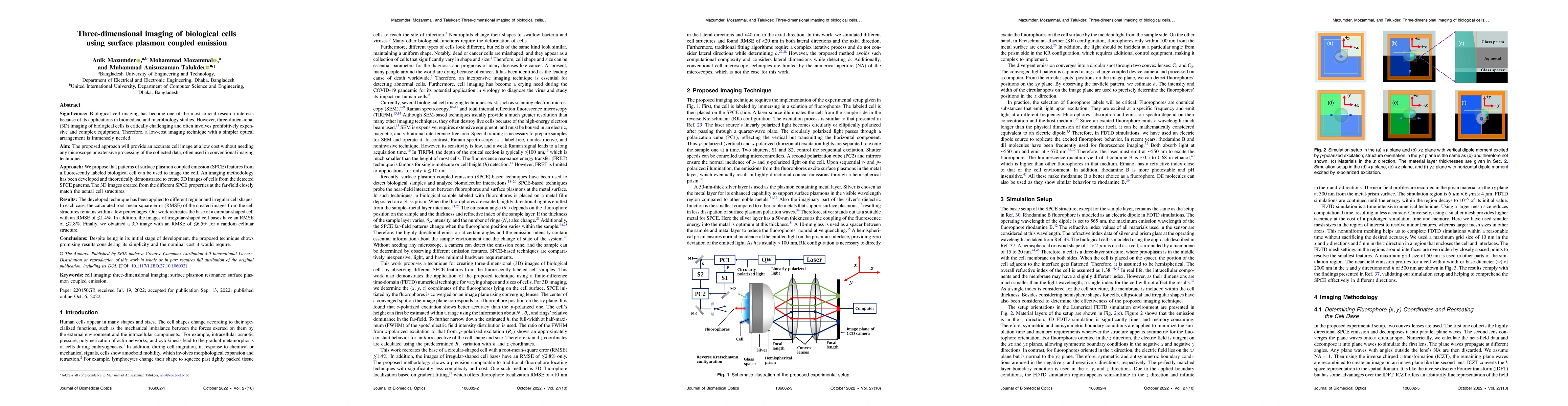
Biological cell imaging has become one of the most crucial research interests due to its wide-ranging applications in biomedical and microbiology studies. However, three-dimensional (3D) imaging of biological cells remains critically challenging and often requires prohibitively expensive and complex equipment. Therefore, a low-cost imaging technique with a simpler optical arrangement is highly desirable. We propose an approach to obtain accurate 3D cell images using surface plasmon coupled emission (SPCE) patterns from a fluorescently labeled biological cell, eliminating the need for conventional microscopes or extensive data processing. An imaging methodology has been developed and theoretically demonstrated to reconstruct 3D cell structures from detected SPCE patterns. The reconstructed 3D images closely match the actual cell geometries. The technique has been applied to both regular and irregular cell shapes. In each case, the root-mean-square error (RMSE) between the reconstructed images and the actual structures remains within a few percent. For a circular-shaped cell base, the RMSE is $\lesssim 1.4%$, while for irregular cell bases, the RMSE is $\lesssim 2.8%$. Finally, a 3D image of a random cellular structure is obtained with an RMSE of $\lesssim 6.5%$. Despite being in its initial stages of development, the proposed technique demonstrates promising results considering its simplicity and low cost.
Key Findings, in focus

Seven facets of this paper, analysed and brought into focus by AI.

AI-generated · verify against source Generated Jun 11, 2025
In focus · one-line synthesis

This low-cost, simple optical arrangement technique is highly desirable for biomedical and microbiology studies, providing an alternative to expensive equipment.

01
MethodologyHow they did it

The research proposes a method to obtain 3D cell images using surface plasmon coupled emission (SPCE) patterns from fluorescently labeled biological cells, eliminating the need for conventional microscopes or extensive data processing.

02
ResultsWhat they found
  • 3D cell structures have been successfully reconstructed from detected SPCE patterns with RMSE within a few percent.
  • For circular-shaped cells, RMSE is ≲1.4%, and for irregular cell shapes, RMSE is ≲2.8%.
  • A 3D image of a random cellular structure was obtained with an RMSE of ≲6.5%.
03
SignificanceWhy it matters

This low-cost, simple optical arrangement technique is highly desirable for biomedical and microbiology studies, providing an alternative to expensive equipment.

04
Technical ContributionsWhat's new technically

The development of an imaging methodology to reconstruct 3D cell structures from surface plasmon coupled emission patterns.

05
NoveltyWhat sets it apart

This work presents a novel approach for 3D cell imaging using SPCE, offering a simpler and more cost-effective alternative to existing techniques.

06
LimitationsWhat to be cautious about
  • The technique is in its initial stages of development.
  • Determining the dipole position from the near-field image remains challenging.
07
Future WorkWhere it goes next
  • Further optimization of the technique for practical applications.
  • Testing the method on a wider variety of cell types and sizes.

Impact

62
References

Paper Details

Paper ID: 2502.11334
Categories:
physics.optics

How to Cite This Paper

Reference manager: RIS EndNote

Citation Network

Current paper (gray), citations (green), references (blue)

Display is limited for performance on very large graphs.

Current Paper
Citations
References
Click to view


Discussion 0

This website uses cookies to ensure you get the best experience. Learn more