01
MethodologyHow they did it
Brief description of the research methodology used
The paper introduces a novel two-photon X-ray ghost microscope that uses ghost imaging to produce high-resolution, point-to-point images of an object's internal structure, potentially surpassing the limitations of conventional X-ray microscopy. This method could enable deeper imaging and nanometer resolution, with applications in physics, material science, and medical imaging.
The paper introduces a novel two-photon X-ray ghost microscope that uses ghost imaging to produce high-resolution, point-to-point images of an object's internal structure, potentially surpassing the limitations of conventional X-ray microscopy. This method could enable deeper imaging and nanometer resolution, with applications in physics, material science, and medical imaging.
Brief description of the research methodology used More in Methodology →
Main finding 1 — Main finding 2 More in Key Results →
Why this research is important and its potential impact More in Significance →
Limitation 1 — Limitation 2 More in Limitations →
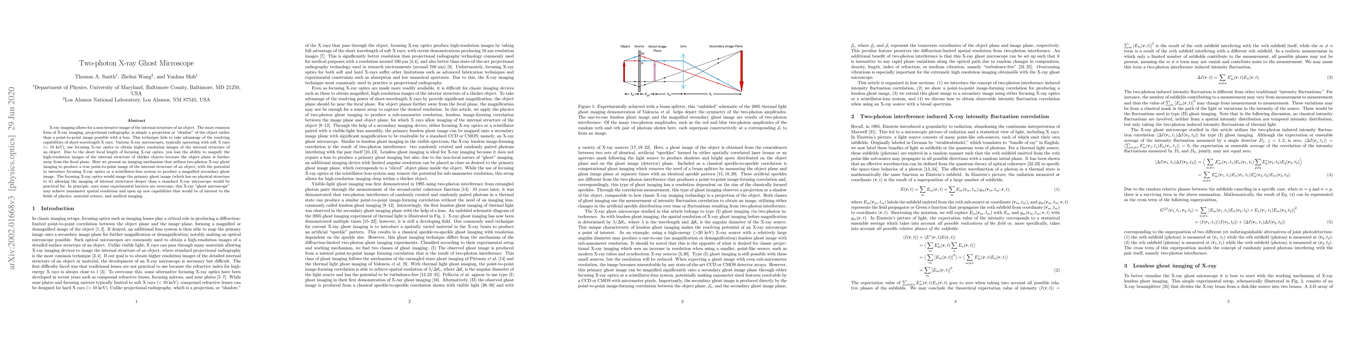
X-ray imaging allows for a non-invasive image of the internal structure of an object. The most common form of X-ray imaging, projectional radiography, is simply a projection or "shadow" of the object rather than a point-to-point image possible with a lens. This technique fails to take advantage of the resolving capabilities of short-wavelength X rays. Various X-ray microscopes, typically operating with soft X rays (< 10 keV), use focusing X-ray optics to obtain higher resolution images of the internal structure of an object. Due to the short focal length of focusing X-ray optics, it becomes difficult to focus on the internal structure of larger objects in such a way to provide significant magnification to be resolvable. Here we present an imaging mechanism that utilizes two-photon X-ray ghost imaging to produce a true point-to-point image of the internal structure of an object, with the potential to introduce focusing X-ray optics or a scintillator-lens pairing to produce a magnified secondary ghost image. The focusing X-ray optics would image the primary ghost image (which has no physical structure to it) allowing the imaging of internal structures deeper than a standard X-ray microscope would allow. In principle, once some experimental barriers are overcome, this X-ray "ghost microscope" may achieve nanometer spatial resolution and open up new capabilities that would be of interest to the fields of physics, material science, and medical imaging.
Seven facets of this paper, analysed and brought into focus by AI.
Why this research is important and its potential impact
Brief description of the research methodology used
Why this research is important and its potential impact
Main technical or theoretical contribution
What makes this work novel or different from existing research
Current paper (gray), citations (green), references (blue)
Display is limited for performance on very large graphs.
Discussion 0