Publication
Metrics
AI Quick Summary
This paper explores the optical readout of Micromegas detectors for X-ray imaging, demonstrating effective radiography and high granularity for low-energy X-rays. The study also presents applications in neutron imaging and beta imaging for non-destructive examination and oncology drug studies, respectively.
Paper Preview
Abstract
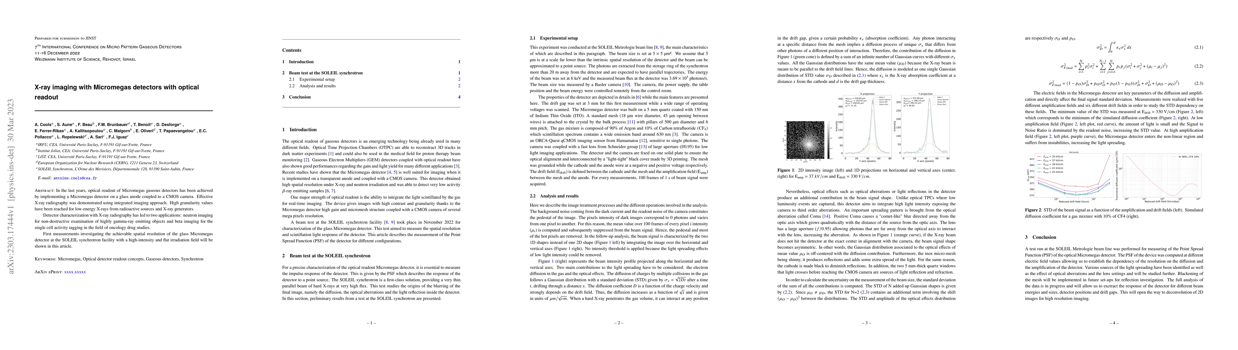
In the last years, optical readout of Micromegas gaseous detectors has been achieved by implementing a Micromegas detector on a glass anode coupled to a CMOS camera. Effective X-ray radiography was demonstrated using integrated imaging approach. High granularity values have been reached for low-energy X-rays from radioactive sources and X-ray generators. Detector characterization with X-ray radiography has led to two applications: neutron imaging for non-destructive examination of highly gamma-ray emitting objects and beta imaging for the single cell activity tagging in the field of oncology drug studies. First measurements investigating the achievable spatial resolution of the glass Micromegas detector at the SOLEIL synchrotron facility with a high-intensity and flat irradiation field will be shown in this article.
AI Key Findings
Get AI-generated insights about this paper's methodology, results, significance, and more — seven facets brought into focus.
Impact
Paper Details
Authors
PDF Preview
Key Terms
Citation Network
Current paper (gray), citations (green), references (blue)
Display is limited for performance on very large graphs.
Discussion 0