Academic Profile
Statistics
Similar Authors
Papers on arXiv
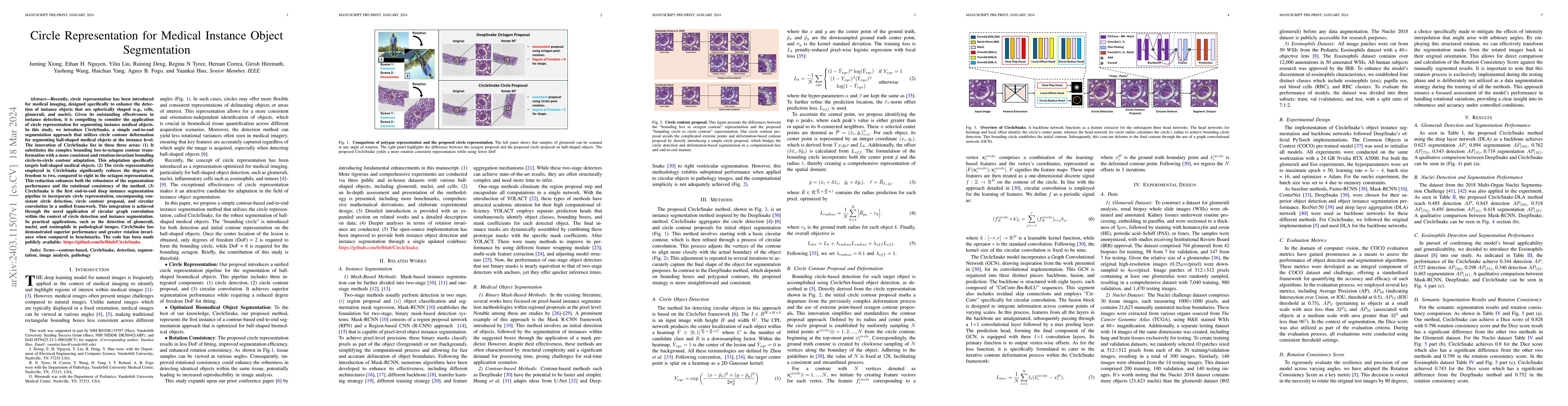
Recently, circle representation has been introduced for medical imaging, designed specifically to enhance the detection of instance objects that are spherically shaped (e.g., cells, glomeruli, and n...
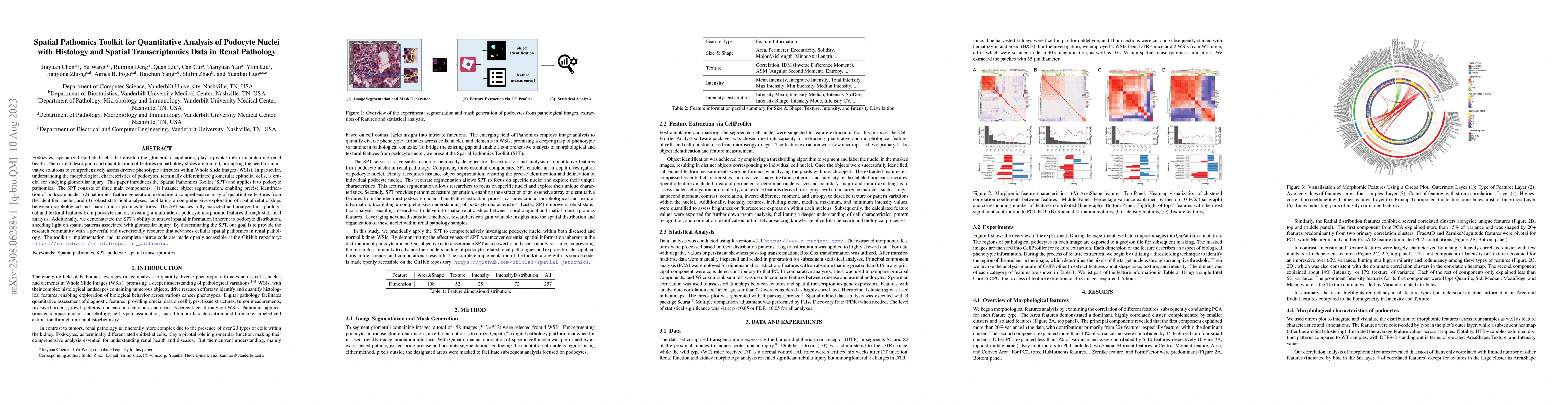
Podocytes, specialized epithelial cells that envelop the glomerular capillaries, play a pivotal role in maintaining renal health. The current description and quantification of features on pathology ...
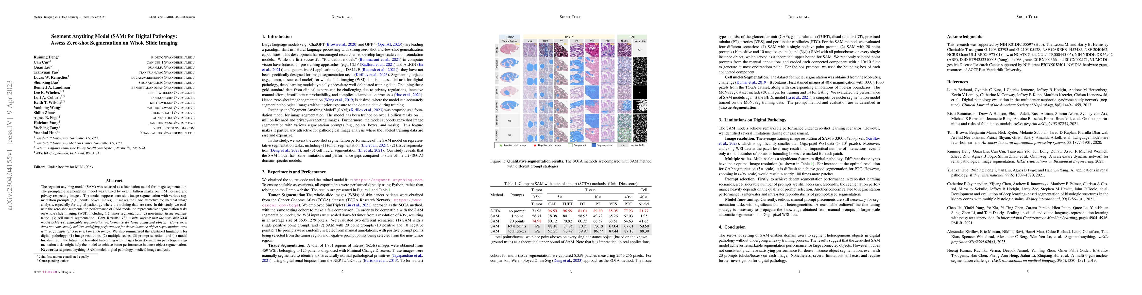
The segment anything model (SAM) was released as a foundation model for image segmentation. The promptable segmentation model was trained by over 1 billion masks on 11M licensed and privacy-respecti...
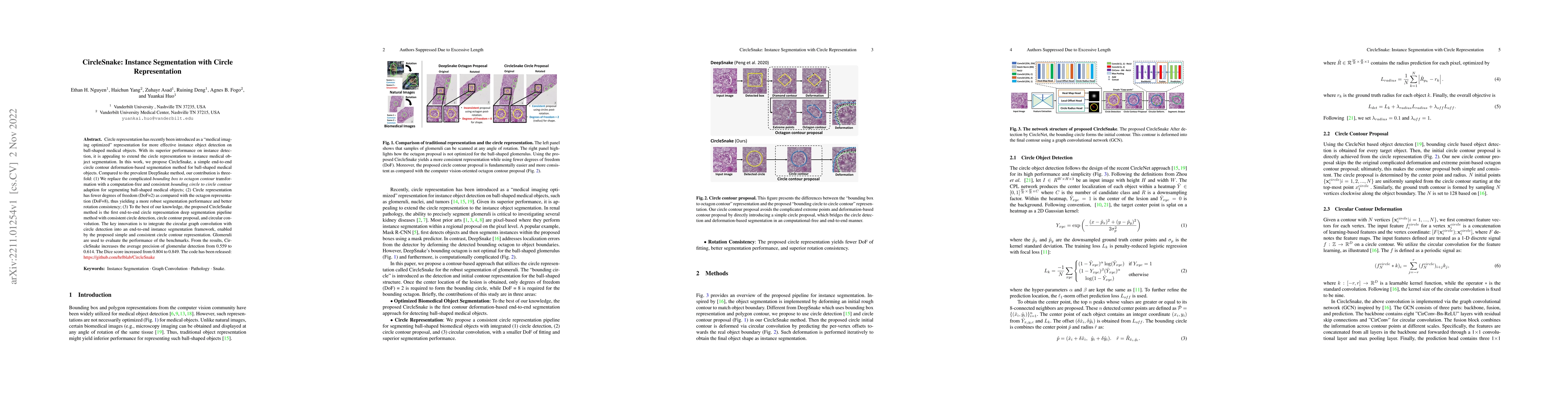
Circle representation has recently been introduced as a medical imaging optimized representation for more effective instance object detection on ball-shaped medical objects. With its superior perfor...
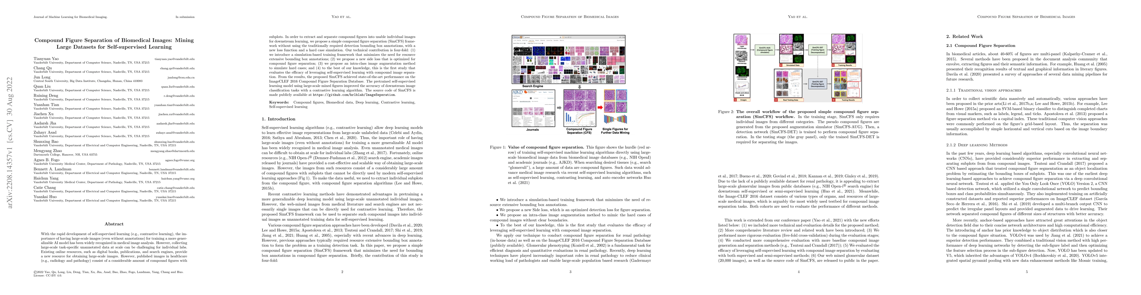
With the rapid development of self-supervised learning (e.g., contrastive learning), the importance of having large-scale images (even without annotations) for training a more generalizable AI model...
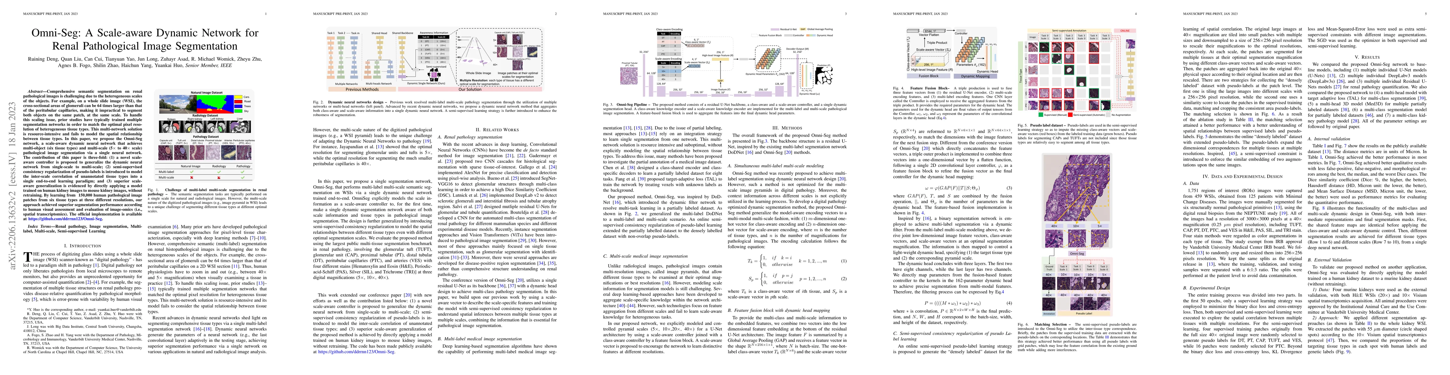
Comprehensive semantic segmentation on renal pathological images is challenging due to the heterogeneous scales of the objects. For example, on a whole slide image (WSI), the cross-sectional areas o...
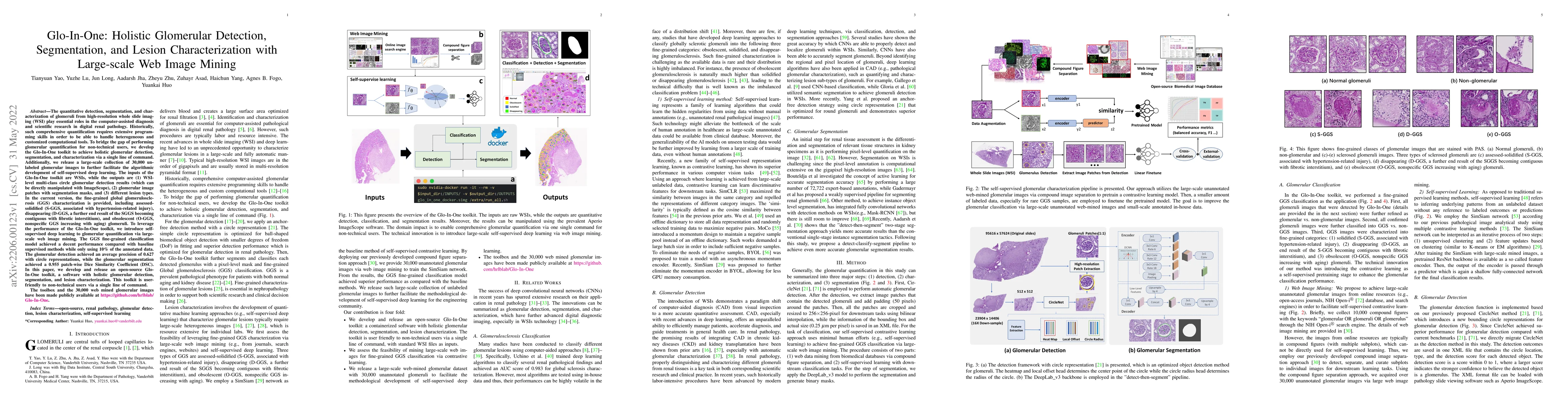
The quantitative detection, segmentation, and characterization of glomeruli from high-resolution whole slide imaging (WSI) play essential roles in the computer-assisted diagnosis and scientific rese...
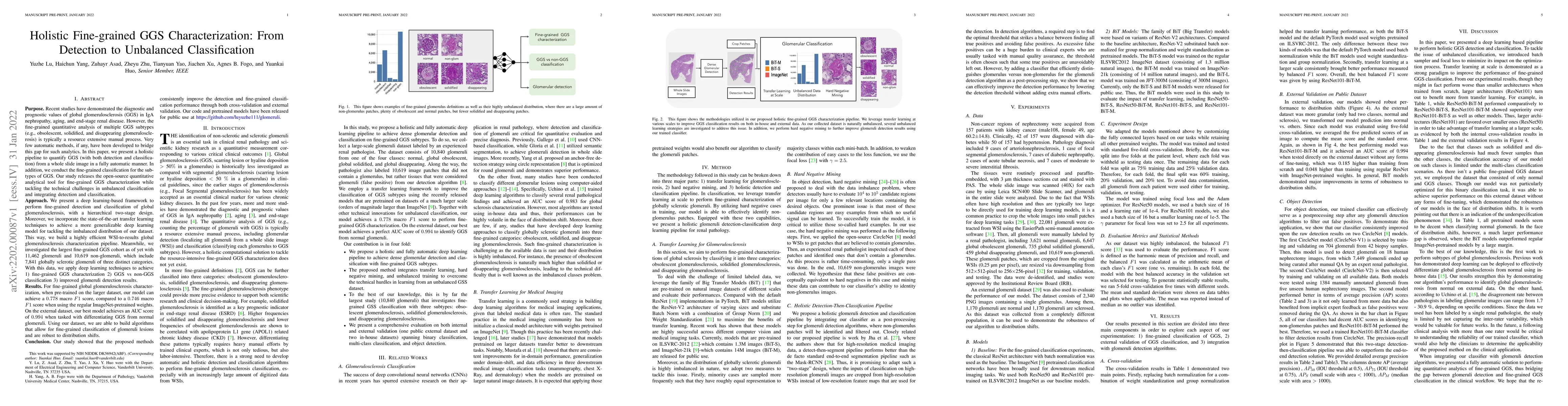
Recent studies have demonstrated the diagnostic and prognostic values of global glomerulosclerosis (GGS) in IgA nephropathy, aging, and end-stage renal disease. However, the fine-grained quantitativ...
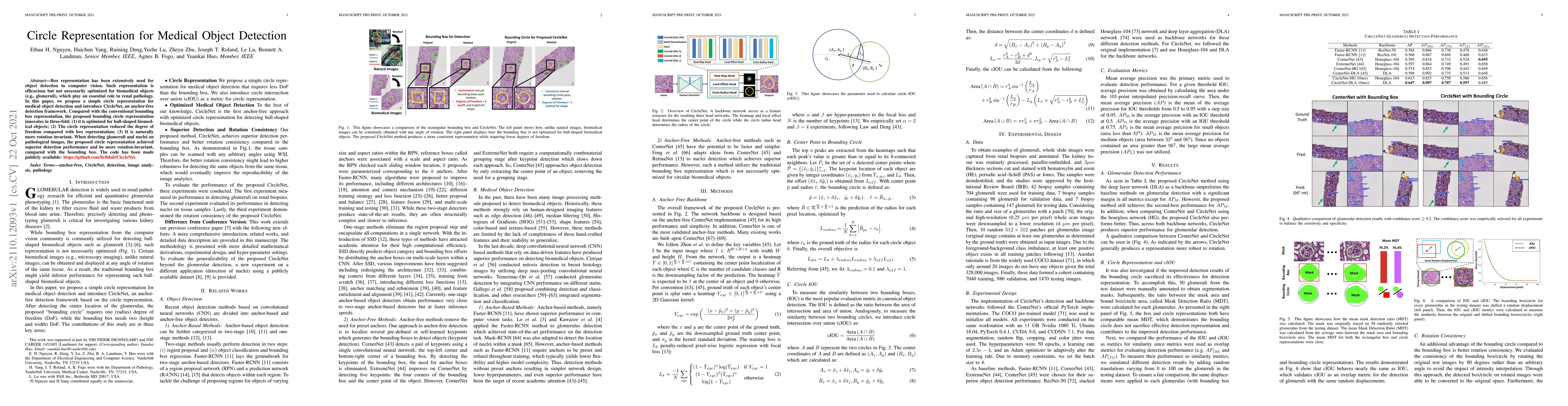
Box representation has been extensively used for object detection in computer vision. Such representation is efficacious but not necessarily optimized for biomedical objects (e.g., glomeruli), which...
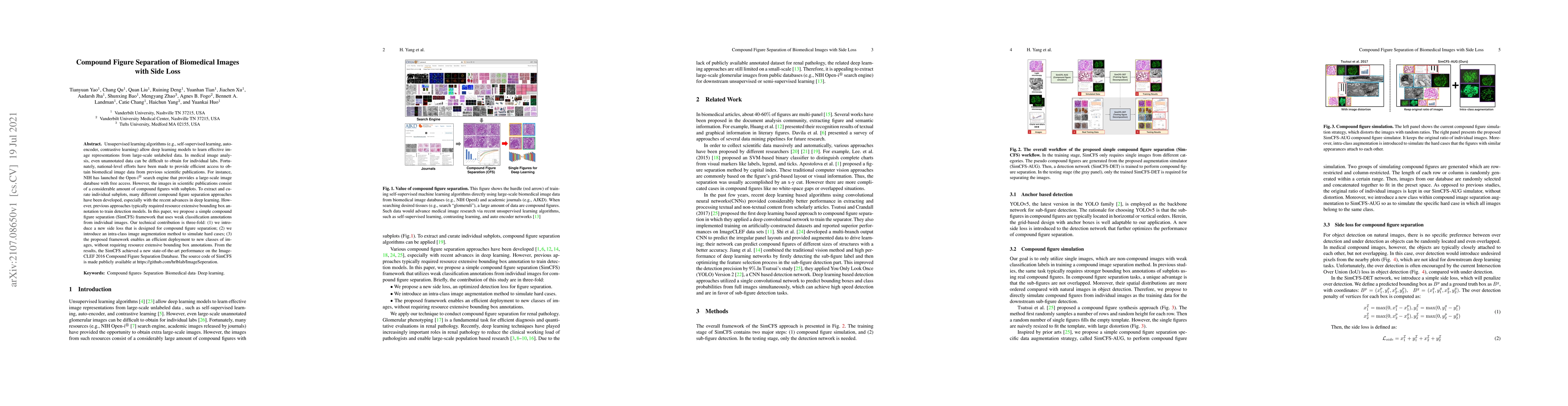
Unsupervised learning algorithms (e.g., self-supervised learning, auto-encoder, contrastive learning) allow deep learning models to learn effective image representations from large-scale unlabeled d...
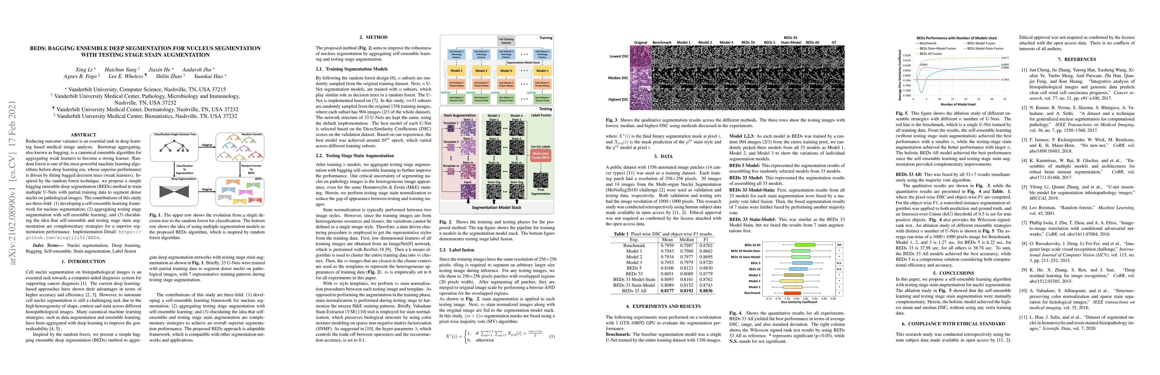
Reducing outcome variance is an essential task in deep learning based medical image analysis. Bootstrap aggregating, also known as bagging, is a canonical ensemble algorithm for aggregating weak lea...
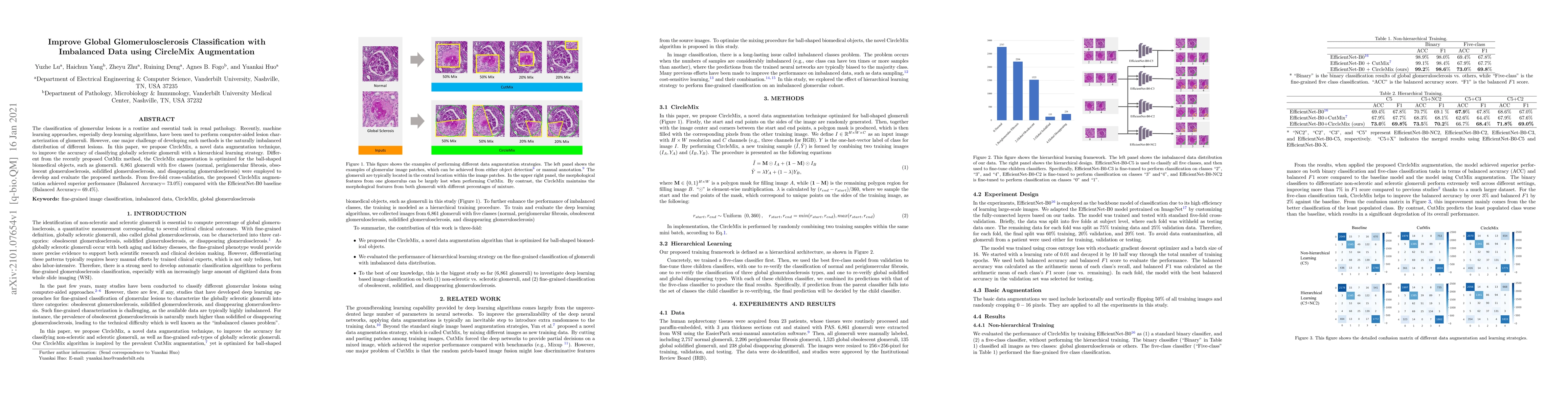
The classification of glomerular lesions is a routine and essential task in renal pathology. Recently, machine learning approaches, especially deep learning algorithms, have been used to perform com...
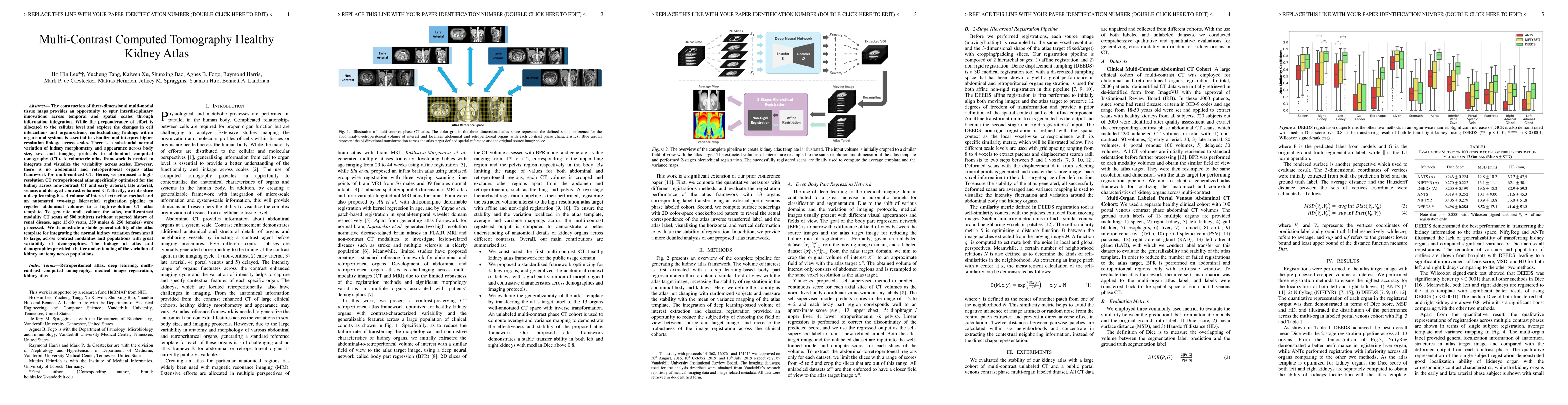
The construction of three-dimensional multi-modal tissue maps provides an opportunity to spur interdisciplinary innovations across temporal and spatial scales through information integration. While ...
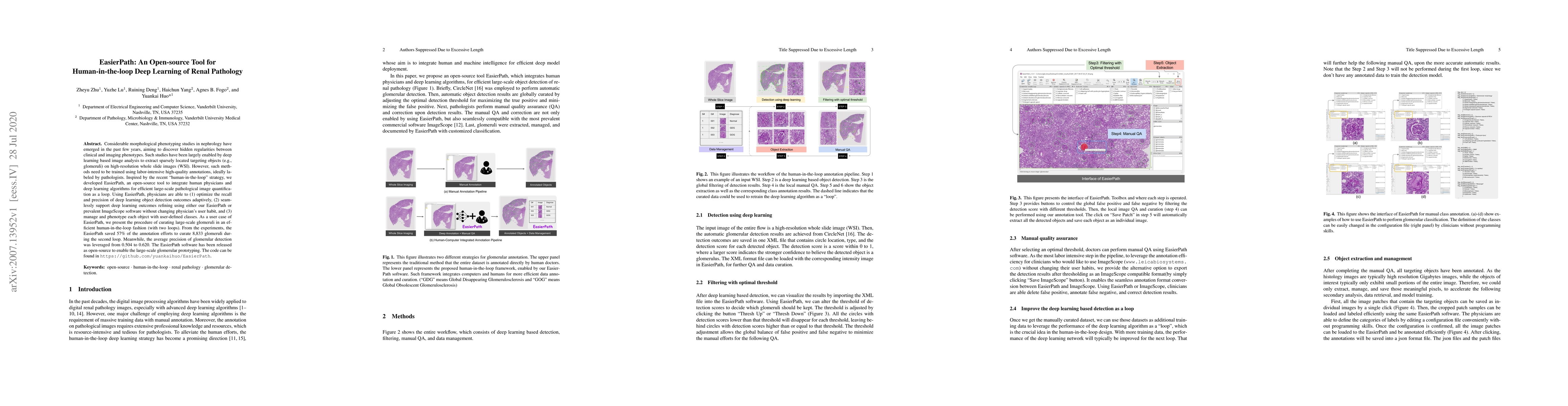
Considerable morphological phenotyping studies in nephrology have emerged in the past few years, aiming to discover hidden regularities between clinical and imaging phenotypes. Such studies have bee...
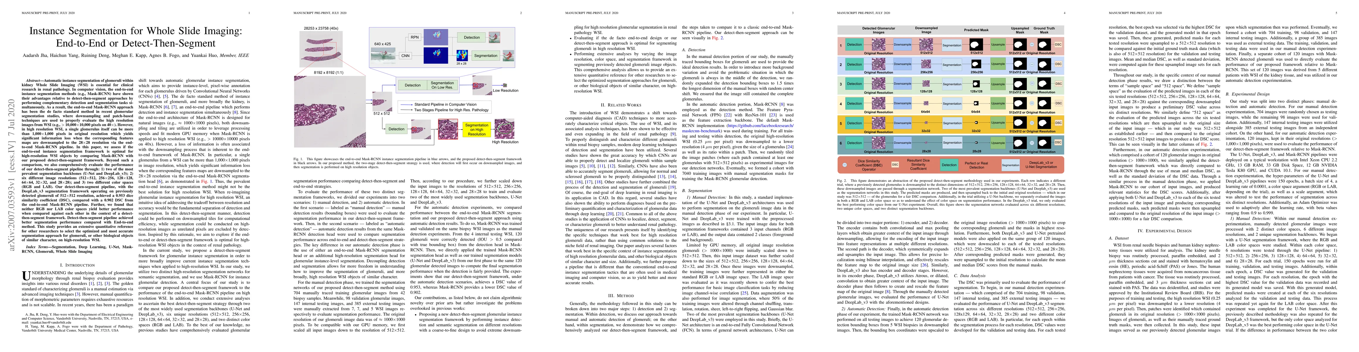
Automatic instance segmentation of glomeruli within kidney Whole Slide Imaging (WSI) is essential for clinical research in renal pathology. In computer vision, the end-to-end instance segmentation m...
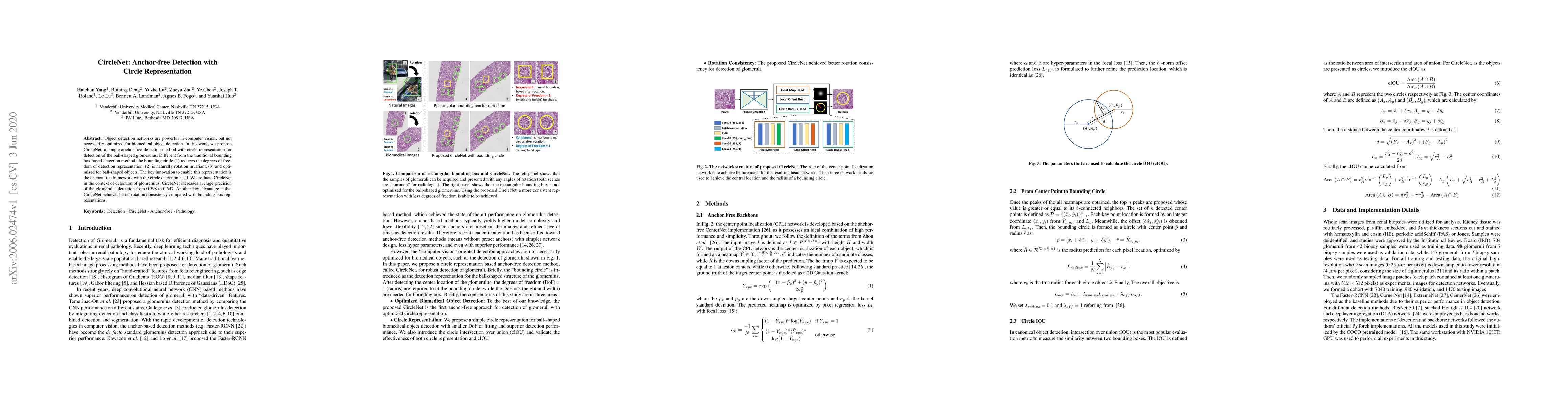
Object detection networks are powerful in computer vision, but not necessarily optimized for biomedical object detection. In this work, we propose CircleNet, a simple anchor-free detection method wi...