ArxivLens

Quick Summary:

The Glo-In-One toolkit simplifies glomerular detection, segmentation, and lesion characterization in digital renal pathology through a single command, releasing 30,000 unlabeled images for self-supervised deep learning. It achieves high-precision detection and segmentation, outperforming baseline methods with minimal annotated data.

arXiv Id: 2206.00123
Date Published: 2022-06-02
Date Updated: 2022-06-02

Categories:

Key Terms (TF-IDF):

quantification (0.245)
segmentation (0.229)
supervised (0.186)
self supervised (0.185)
circle (0.176)
detection (0.170)
achieved (0.168)
characterization (0.149)
deep learning (0.148)
large scale (0.137)
image (0.124)
deep (0.120)
skills (0.118)
patches (0.117)
self (0.116)
roles (0.116)
methods using (0.116)
patch (0.115)
mining (0.115)
performance compared (0.114)
License: http://creativecommons.org/licenses/by/4.0/

Summary:

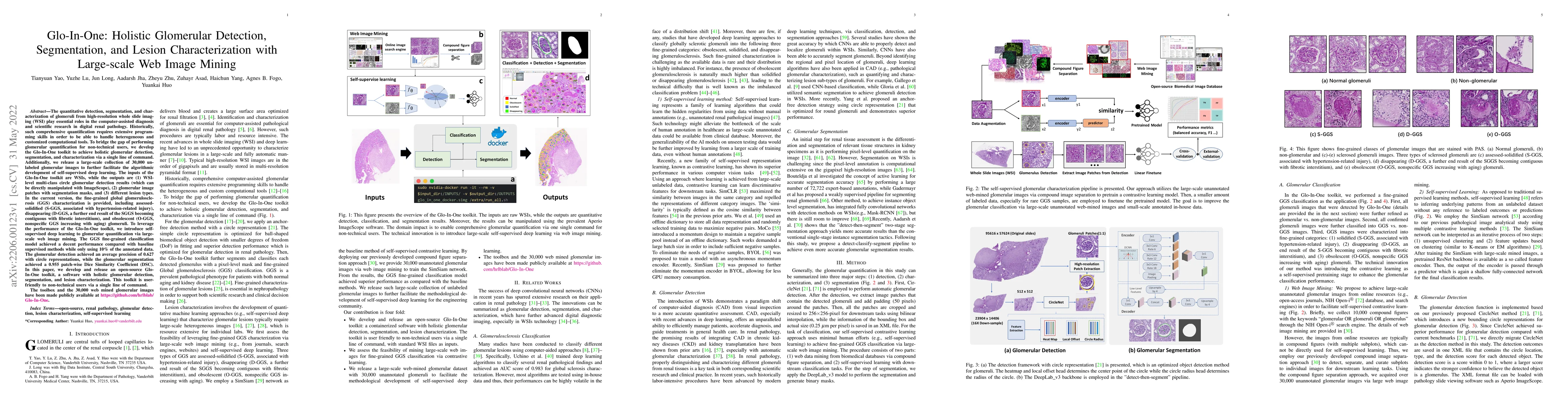
The quantitative detection, segmentation, and characterization of glomeruli from high-resolution whole slide imaging (WSI) play essential roles in the computer-assisted diagnosis and scientific research in digital renal pathology. Historically, such comprehensive quantification requires extensive programming skills in order to be able to handle heterogeneous and customized computational tools. To bridge the gap of performing glomerular quantification for non-technical users, we develop the Glo-In-One toolkit to achieve holistic glomerular detection, segmentation, and characterization via a single line of command. Additionally, we release a large-scale collection of 30,000 unlabeled glomerular images to further facilitate the algorithmic development of self-supervised deep learning. The inputs of the Glo-In-One toolkit are WSIs, while the outputs are (1) WSI-level multi-class circle glomerular detection results (which can be directly manipulated with ImageScope), (2) glomerular image patches with segmentation masks, and (3) different lesion types. To leverage the performance of the Glo-In-One toolkit, we introduce self-supervised deep learning to glomerular quantification via large-scale web image mining. The GGS fine-grained classification model achieved a decent performance compared with baseline supervised methods while only using 10% of the annotated data. The glomerular detection achieved an average precision of 0.627 with circle representations, while the glomerular segmentation achieved a 0.955 patch-wise Dice Similarity Coefficient (DSC).

Citation & Reference Network Visualization

This graph shows how this paper is connected to other research. The current paper is shown in dark gray, papers that cite it are in green, and papers it references are in blue. You can:

Current Paper
Papers citing this work (Citations)
Papers cited by this work (References)

• Hover over any node to see the paper title

• Click a node to view that paper's details

• Drag nodes to rearrange the graph

• Use the zoom controls (bottom right) to zoom in/out

Citations and References

Title Authors Year Actions
GlomSAM: Hybrid customized SAM for multi-glomerular detection and segmentation in immunofluorescence images
Shengyu Pan, Xuanli Tang, Bingxian Chen, Xiaobo Lai, Wei Jin
2025
View Paper
Glo-net: A dual task branch based neural network for multi-class glomeruli segmentation.
Xiangxue Wang, Jingkai Zhang, Yuemei Xu, Yang Huang, Wenlong Ming, Yiping Jiao, Bicheng Liu, Xiangshan Fan, Jun Xu
2025
View Paper
Glo-In-One-v2: Holistic Identification of Glomerular Cells, Tissues, and Lesions in Human and Mouse Histopathology
Lining Yu, Mengmeng Yin, Ruining Deng, Quan Liu, Tianyuan Yao, Can Cui, Junlin Guo, Yu Wang, Yaohong Wang, Shilin Zhao, Haichun Yang, Yuankai Huo
2024
View Paper
Unsupervised learning for labeling global glomerulosclerosis
Hrafn Weishaupt, J. Besusparis, Cleo-Aron Weis, S. Porubsky, A. Laurinavičius, S. Leh
2024
View Paper
An image inpainting-based data augmentation method for improved sclerosed glomerular identification performance with the segmentation model EfficientNetB3-Unet
Songping He, Yi Zou, Bin Li, Fangyu Peng, Xia Lu, Hui Guo, Xin Tan, Yanyan Chen
2024
View Paper
Time for a full digital approach in nephropathology: a systematic review of current artificial intelligence applications and future directions
G. Cazzaniga, Mattia Rossi, A. Eccher, Ilaria Girolami, Vincenzo L’Imperio, Hien Van Nguyen, Jan U. Becker, María Gloria Bueno García, M. Sbaraglia, A. D. Dei Tos, G. Gambaro, F. Pagni
2023
View Paper
From WSI-level to patch-level: Structure prior-guided binuclear cell fine-grained detection
G. Hu, Baomin Wang, Boxian Hu, Dan Chen, Lihua Hu, Cheng Li, Yu An, G. Hu, G. Jia
2023
View Paper
Standardized CycleGAN training for unsupervised stain adaptation in invasive carcinoma classification for breast histopathology
Nicolas Nerrienet, Rémy Peyret, Marie Sockeel, Stéphane Sockeel
2023
View Paper
Special Section Guest Editorial: Advances in High-Dimensional Medical Image Processing
I. Išgum, B. Landman, T. Vrtovec
2022
View Paper

Comments (0)

This website uses cookies to ensure you get the best experience. Learn more