Academic Profile
Statistics
Similar Authors
Papers on arXiv
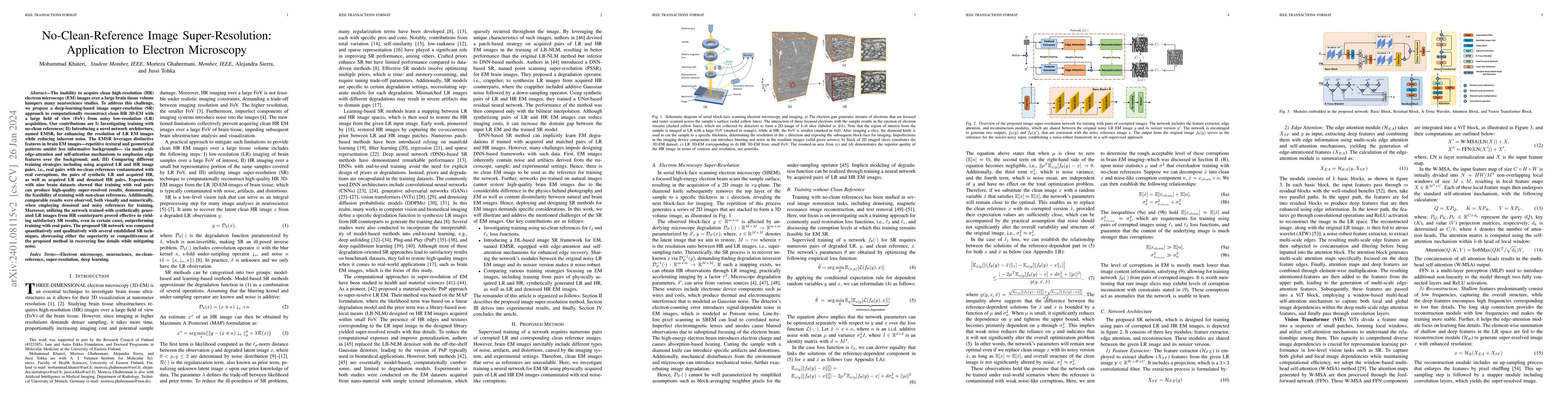
The inability to acquire clean high-resolution (HR) electron microscopy (EM) images over a large brain tissue volume hampers many neuroscience studies. To address this challenge, we propose a deep-l...
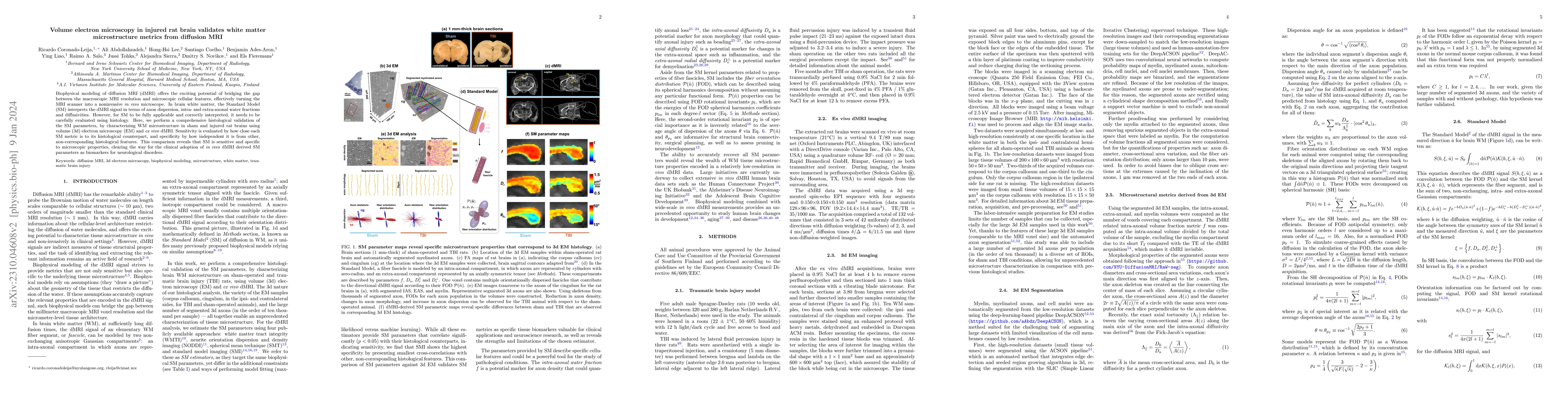
Biophysical modeling of diffusion MRI (dMRI) offers the exciting potential of bridging the gap between the macroscopic MRI resolution and microscopic cellular features, effectively turning the MRI s...
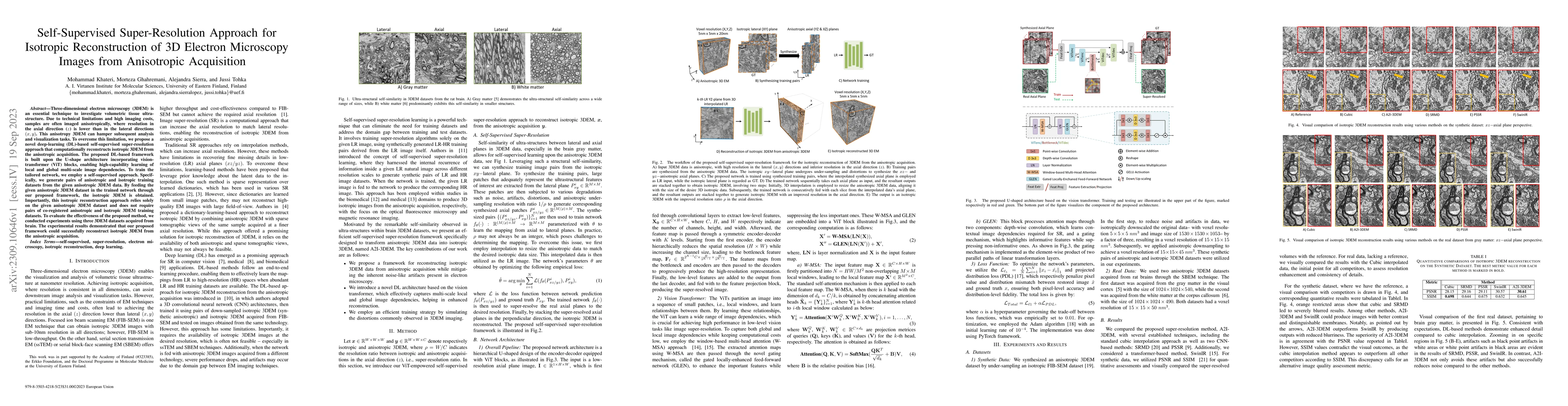
Three-dimensional electron microscopy (3DEM) is an essential technique to investigate volumetric tissue ultra-structure. Due to technical limitations and high imaging costs, samples are often imaged...
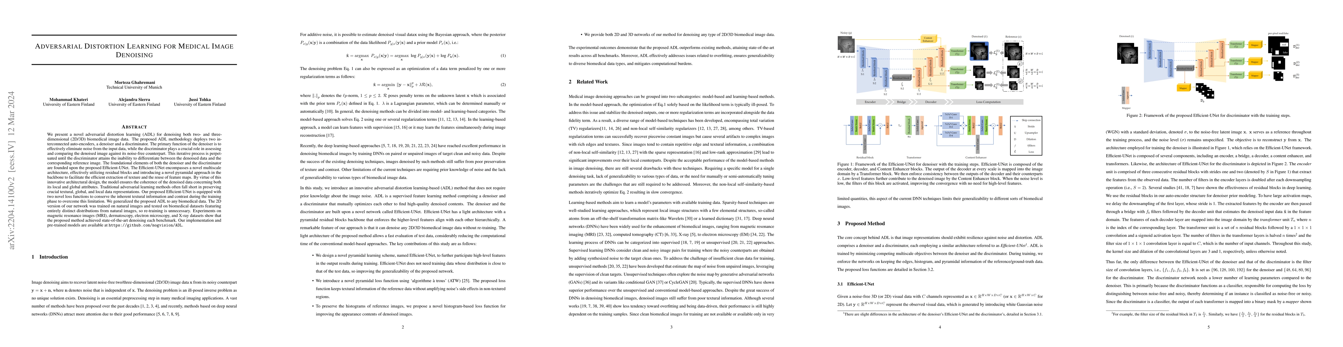
We present a novel adversarial distortion learning (ADL) for denoising two- and three-dimensional (2D/3D) biomedical image data. The proposed ADL consists of two auto-encoders: a denoiser and a disc...
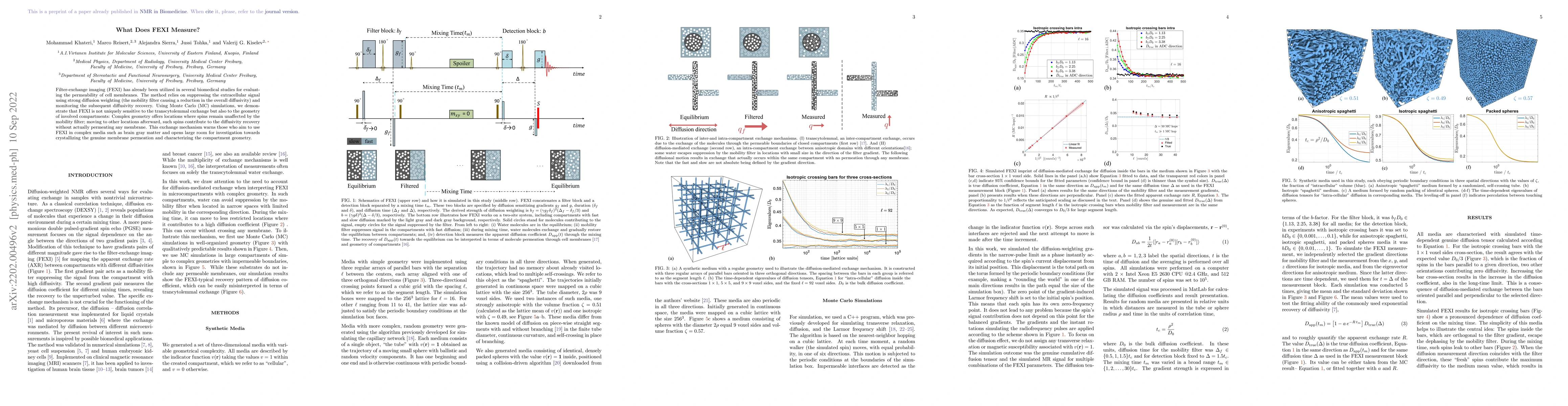
Filter-exchange imaging (FEXI) has already been utilized in several biomedical studies for evaluating the permeability of cell membranes. The method relies on suppressing the extracellular signal us...
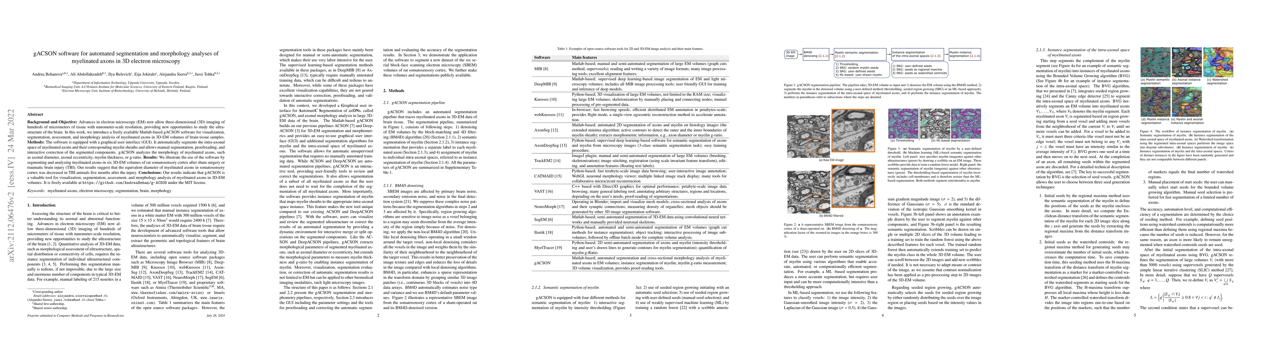
Background and Objective: Advances in electron microscopy (EM) now allow three-dimensional (3D) imaging of hundreds of micrometers of tissue with nanometer-scale resolution, providing new opportunit...
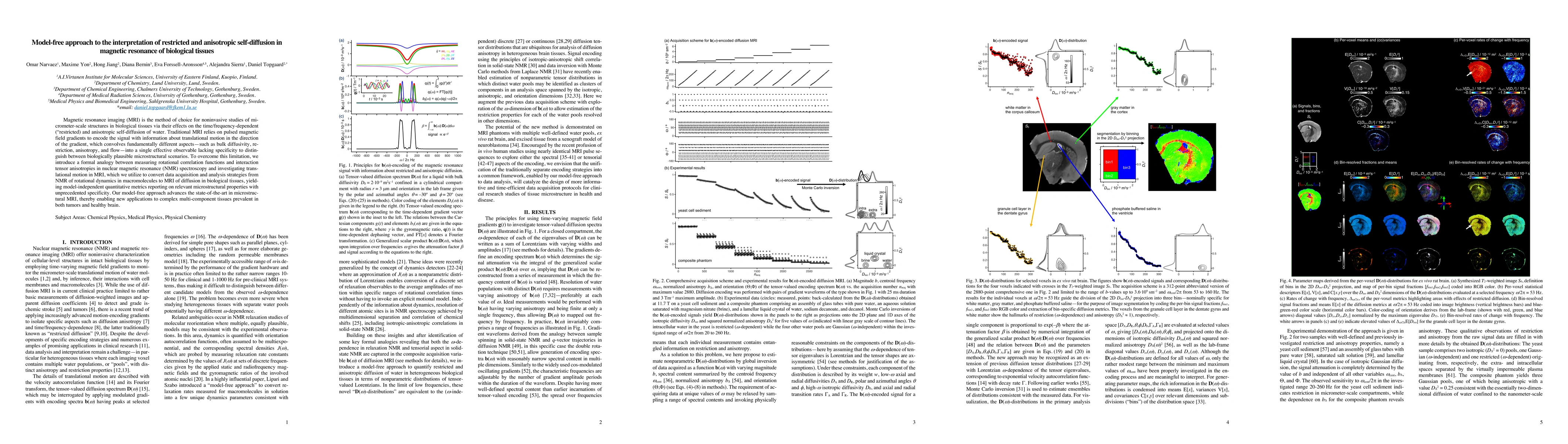
Magnetic resonance imaging (MRI) is the method of choice for noninvasive studies of micrometer-scale structures in biological tissues via their effects on the time/frequency-dependent ("restricted")...
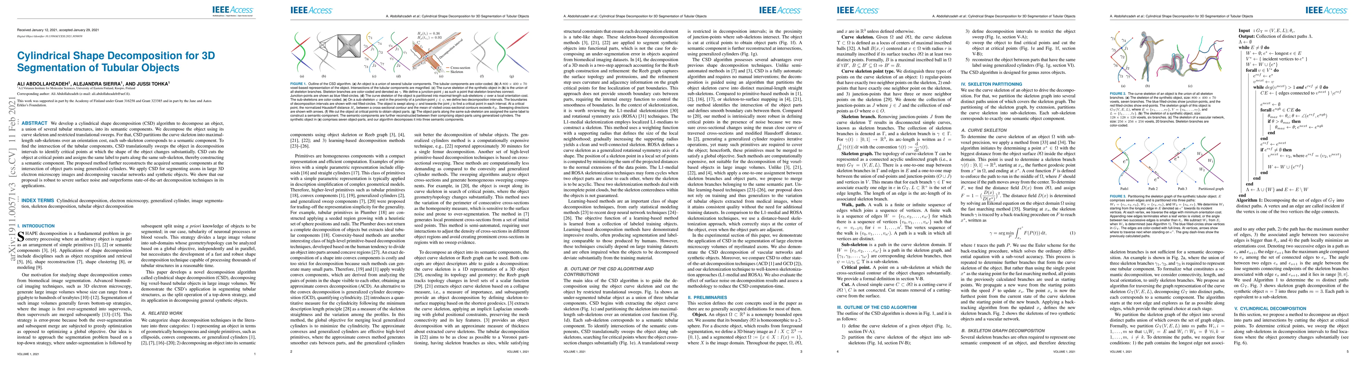
We develop a cylindrical shape decomposition (CSD) algorithm to decompose an object, a union of several tubular structures, into its semantic components. We decompose the object using its curve skel...
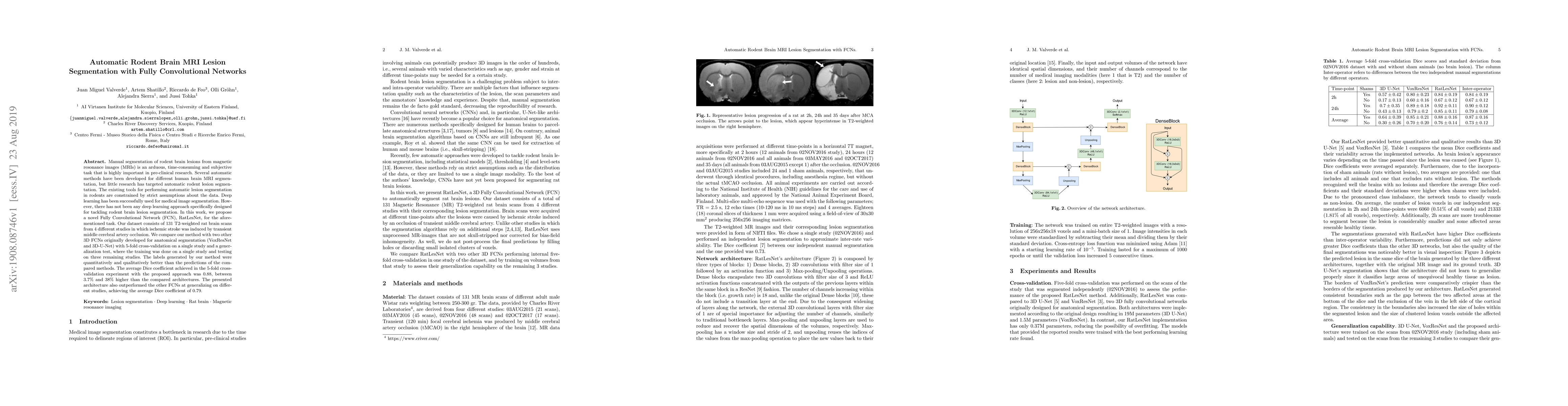
Manual segmentation of rodent brain lesions from magnetic resonance images (MRIs) is an arduous, time-consuming and subjective task that is highly important in pre-clinical research. Several automat...
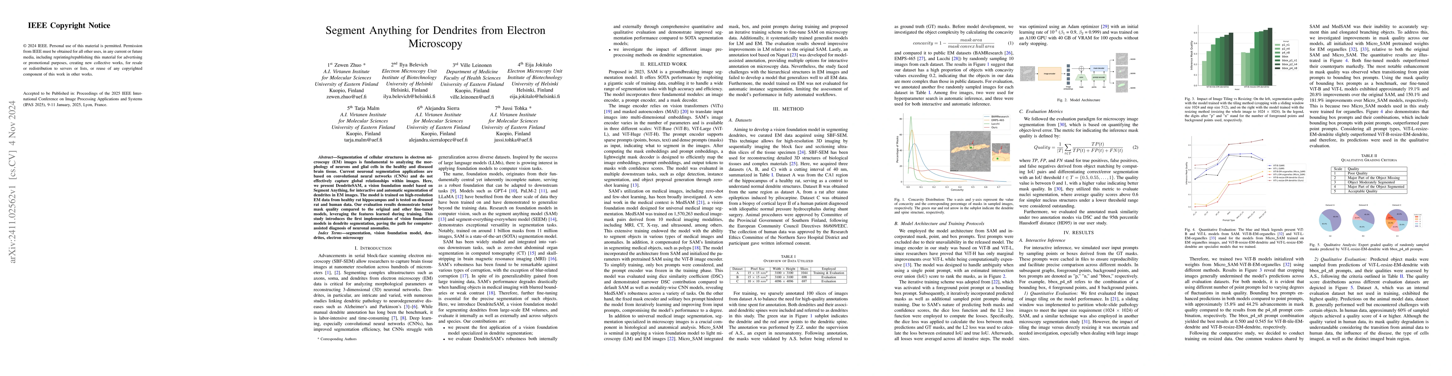
Segmentation of cellular structures in electron microscopy (EM) images is fundamental to analyzing the morphology of neurons and glial cells in the healthy and diseased brain tissue. Current neuronal ...
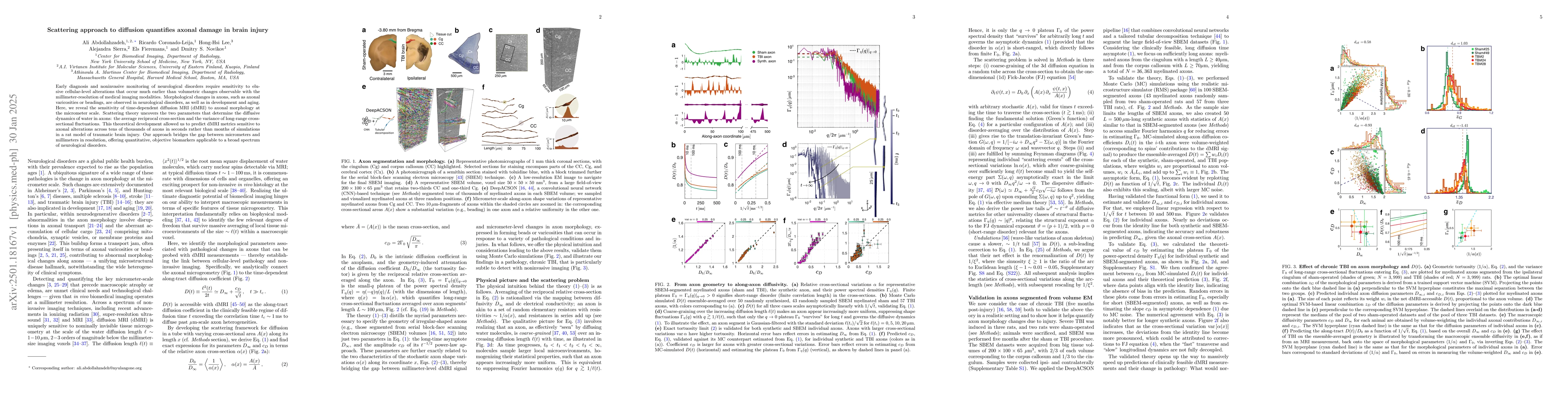
Early diagnosis and noninvasive monitoring of neurological disorders require sensitivity to elusive cellular-level alterations that occur much earlier than volumetric changes observable with the milli...
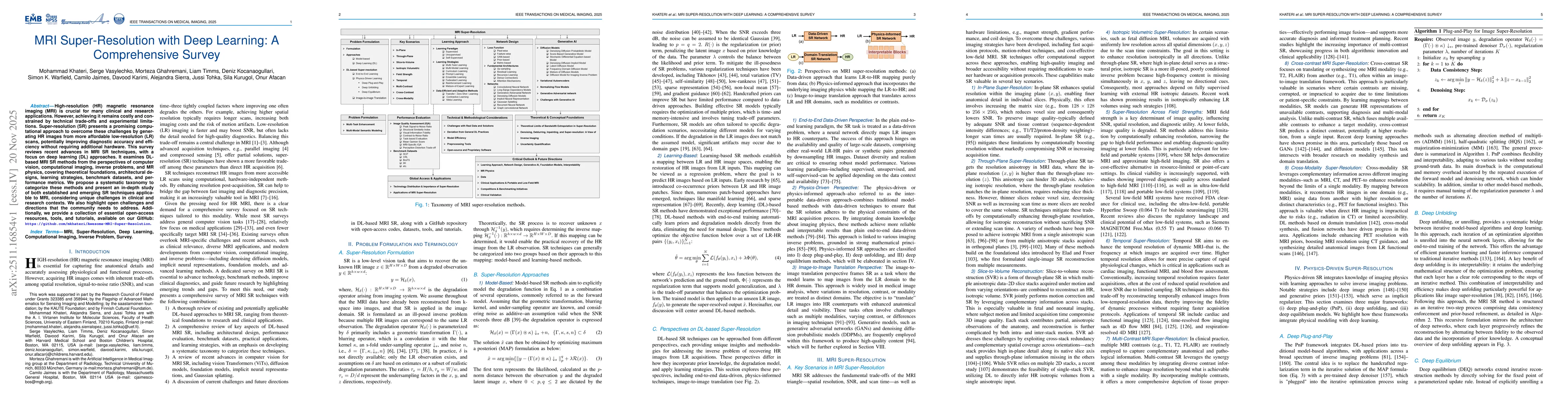
High-resolution (HR) magnetic resonance imaging (MRI) is crucial for many clinical and research applications. However, achieving it remains costly and constrained by technical trade-offs and experimen...
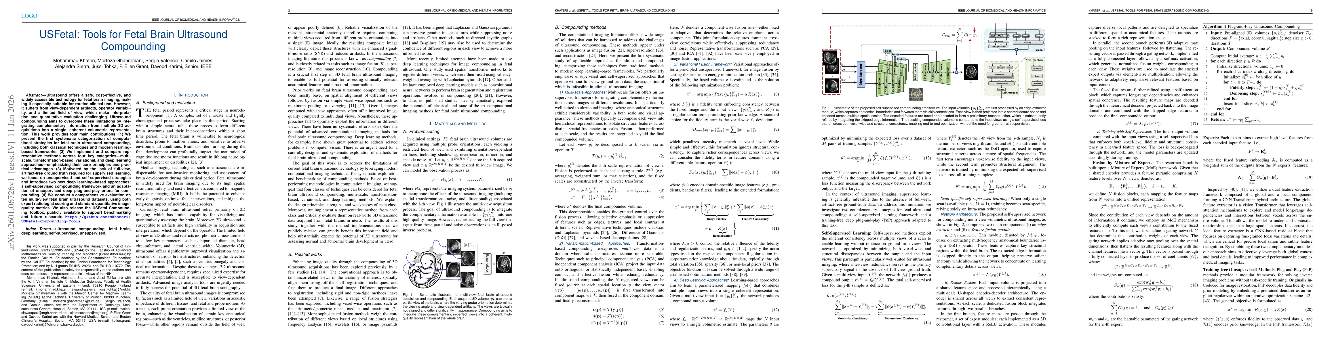
Ultrasound offers a safe, cost-effective, and widely accessible technology for fetal brain imaging, making it especially suitable for routine clinical use. However, it suffers from view-dependent arti...