Volume electron microscopy in injured rat brain validates white matter microstructure metrics from diffusion MRI
Publication
Metrics
AI Quick Summary
This paper validates white matter microstructure metrics derived from diffusion MRI (dMRI) using volume electron microscopy in injured rat brains, demonstrating the sensitivity and specificity of the Standard Model parameters, thus supporting their use as biomarkers for neurological disorders.
Paper Preview
Abstract
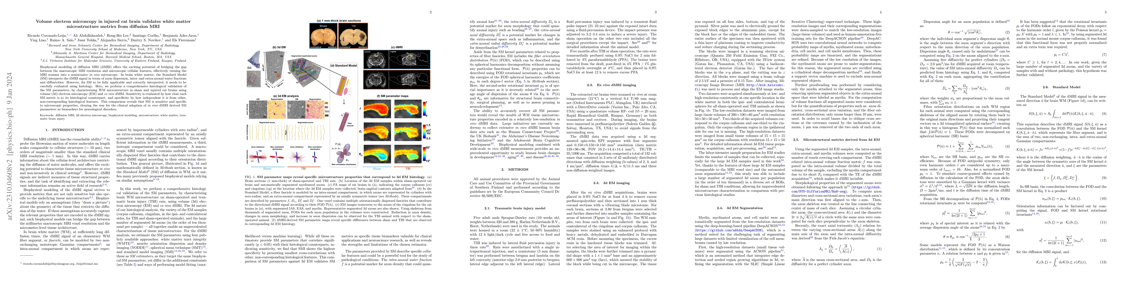
Biophysical modeling of diffusion MRI (dMRI) offers the exciting potential of bridging the gap between the macroscopic MRI resolution and microscopic cellular features, effectively turning the MRI scanner into a noninvasive in vivo microscope. In brain white matter, the Standard Model (SM) interprets the dMRI signal in terms of axon dispersion, intra- and extra-axonal water fractions and diffusivities. However, for SM to be fully applicable and correctly interpreted, it needs to be carefully evaluated using histology. Here, we perform a comprehensive histological validation of the SM parameters, by characterizing WM microstructure in sham and injured rat brains using volume (3d) electron microscopy (EM) and ex vivo dMRI. Sensitivity is evaluated by how close each SM metric is to its histological counterpart, and specificity by how independent it is from other, non-corresponding histological features. This comparison reveals that SM is sensitive and specific to microscopic properties, clearing the way for the clinical adoption of in vivo dMRI derived SM parameters as biomarkers for neurological disorders.
AI Key Findings
Get AI-generated insights about this paper's methodology, results, significance, and more — seven facets brought into focus.
Impact
Paper Details
Authors
PDF Preview
Key Terms
Citation Network
Current paper (gray), citations (green), references (blue)
Display is limited for performance on very large graphs.
Discussion 0