Academic Profile
Statistics
Similar Authors
Papers on arXiv
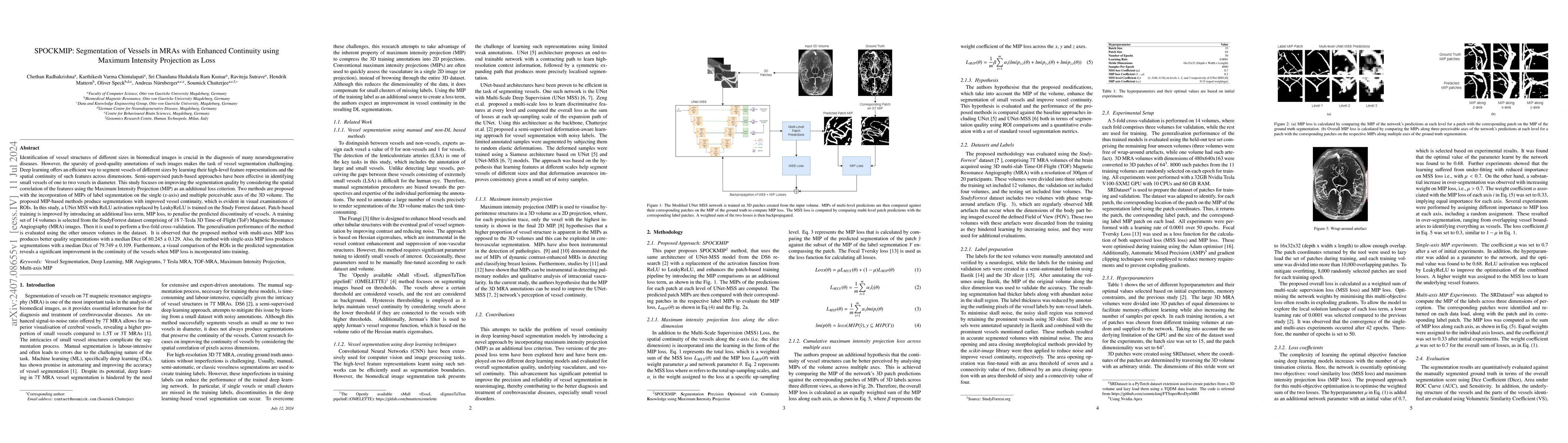
Identification of vessel structures of different sizes in biomedical images is crucial in the diagnosis of many neurodegenerative diseases. However, the sparsity of good-quality annotations of such im...
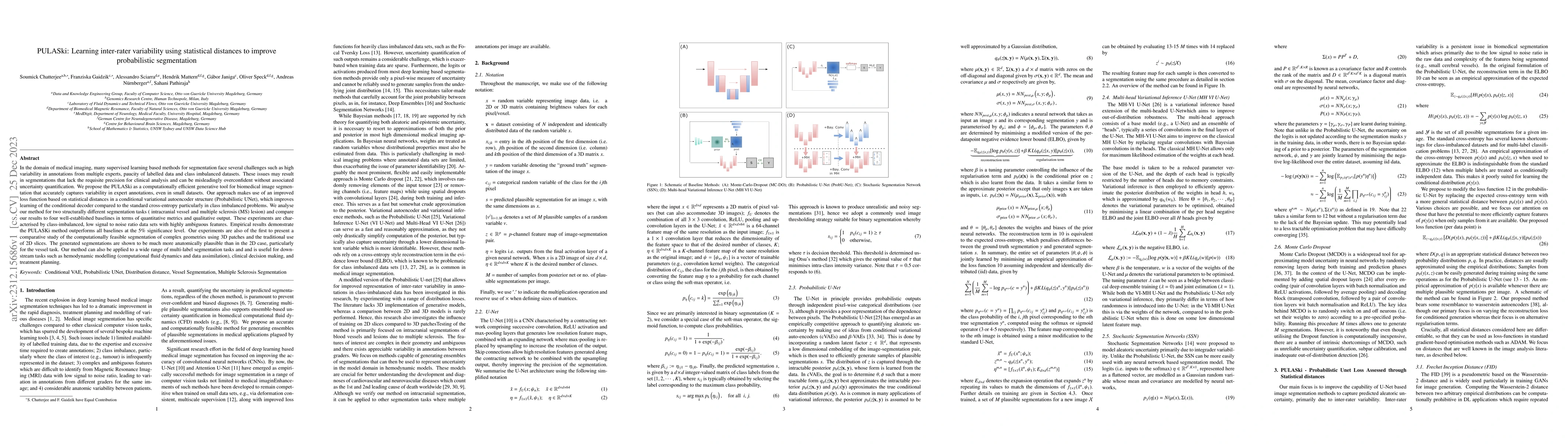
In the domain of medical imaging, many supervised learning based methods for segmentation face several challenges such as high variability in annotations from multiple experts, paucity of labelled d...
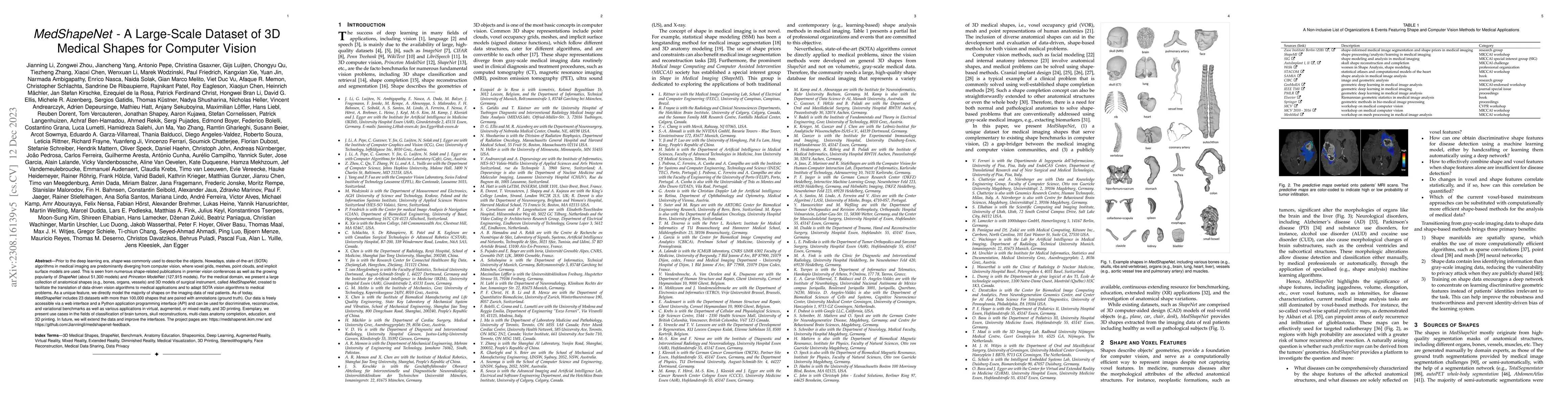
Prior to the deep learning era, shape was commonly used to describe the objects. Nowadays, state-of-the-art (SOTA) algorithms in medical imaging are predominantly diverging from computer vision, whe...
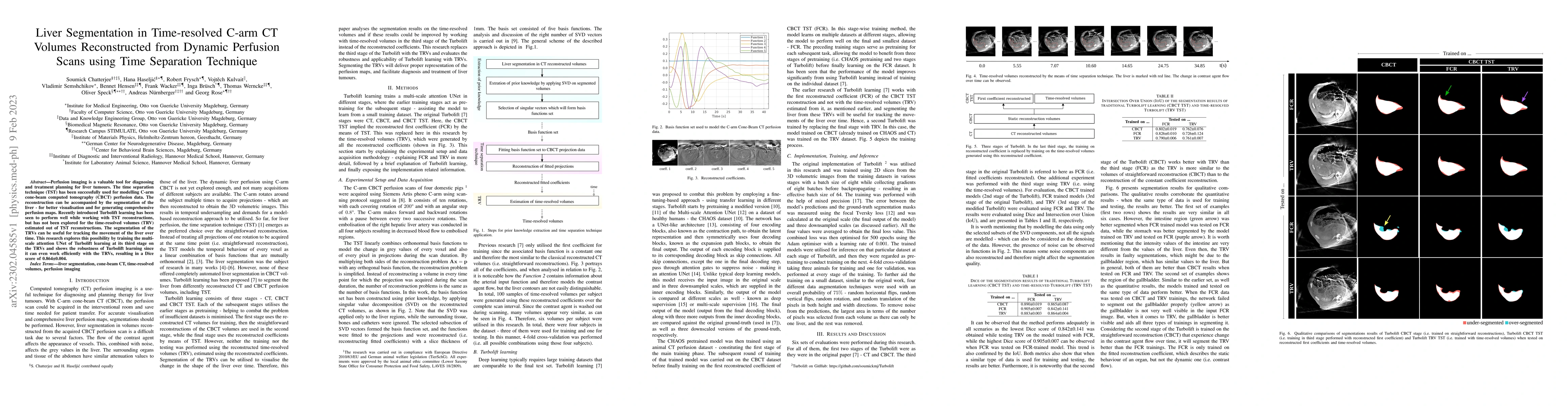
Perfusion imaging is a valuable tool for diagnosing and treatment planning for liver tumours. The time separation technique (TST) has been successfully used for modelling C-arm cone-beam computed to...
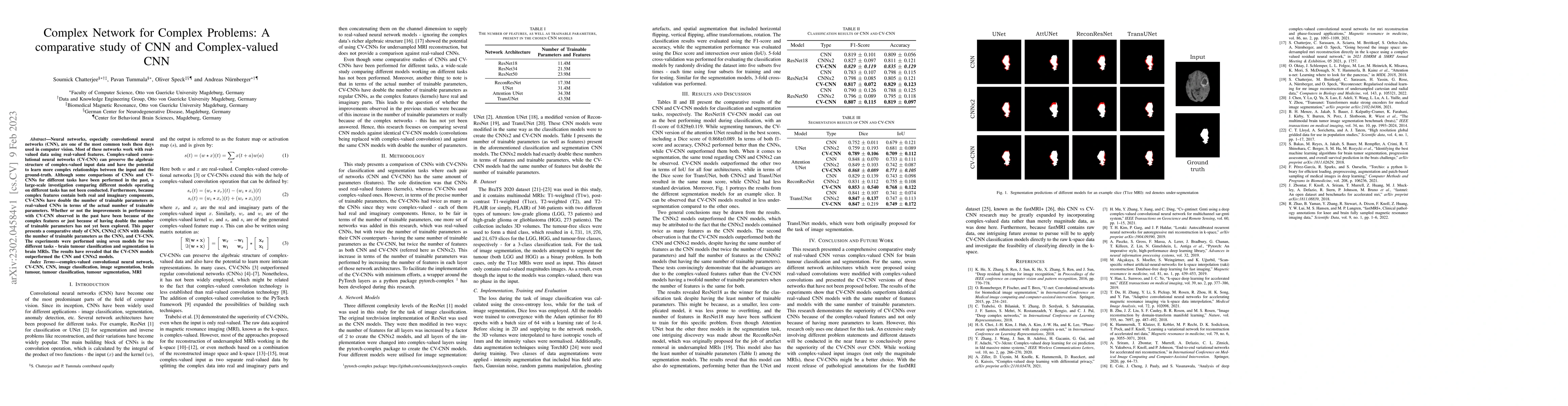
Neural networks, especially convolutional neural networks (CNN), are one of the most common tools these days used in computer vision. Most of these networks work with real-valued data using real-val...
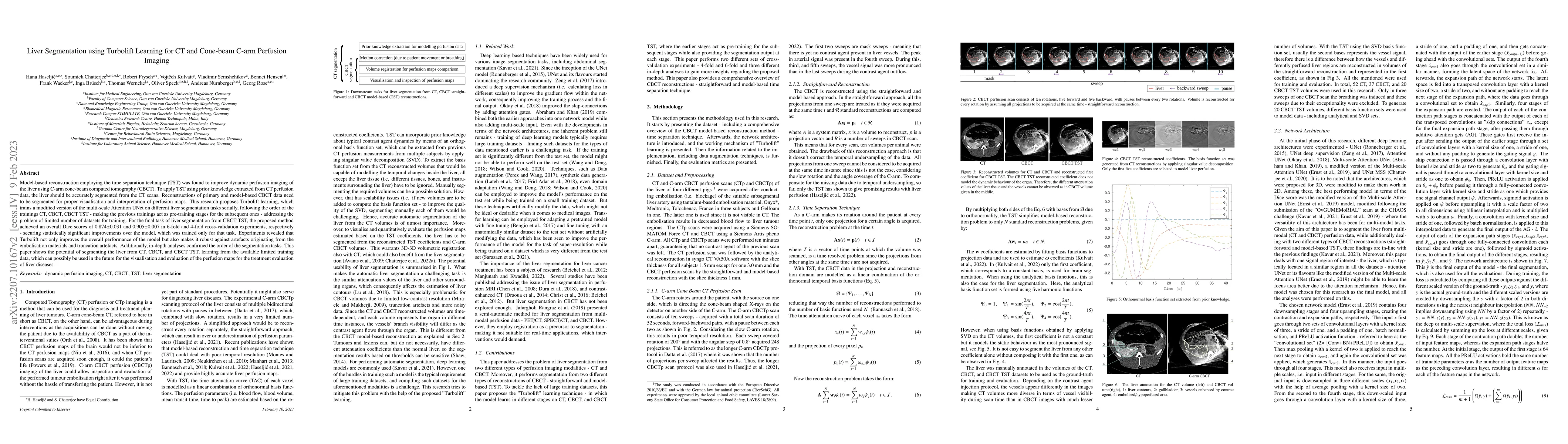
Model-based reconstruction employing the time separation technique (TST) was found to improve dynamic perfusion imaging of the liver using C-arm cone-beam computed tomography (CBCT). To apply TST us...
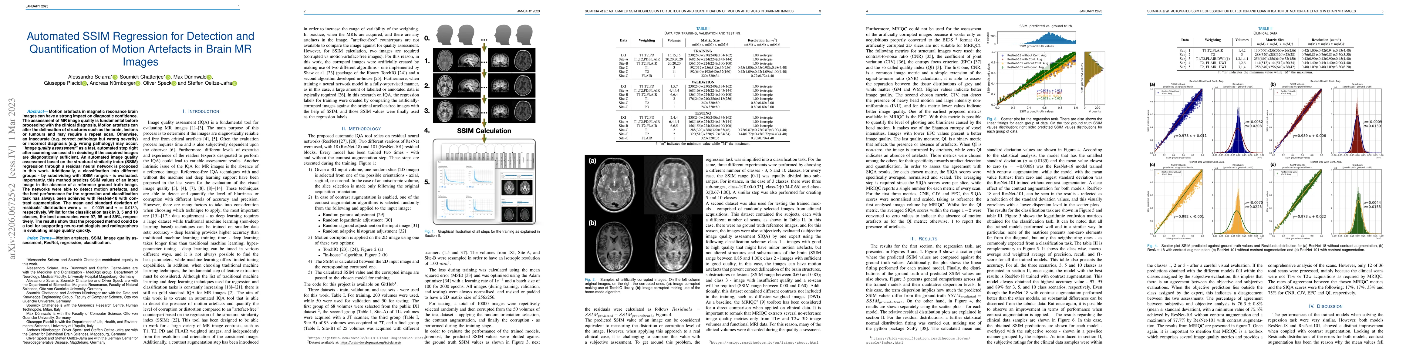
Motion artefacts in magnetic resonance brain images can have a strong impact on diagnostic confidence. The assessment of MR image quality is fundamental before proceeding with the clinical diagnosis...
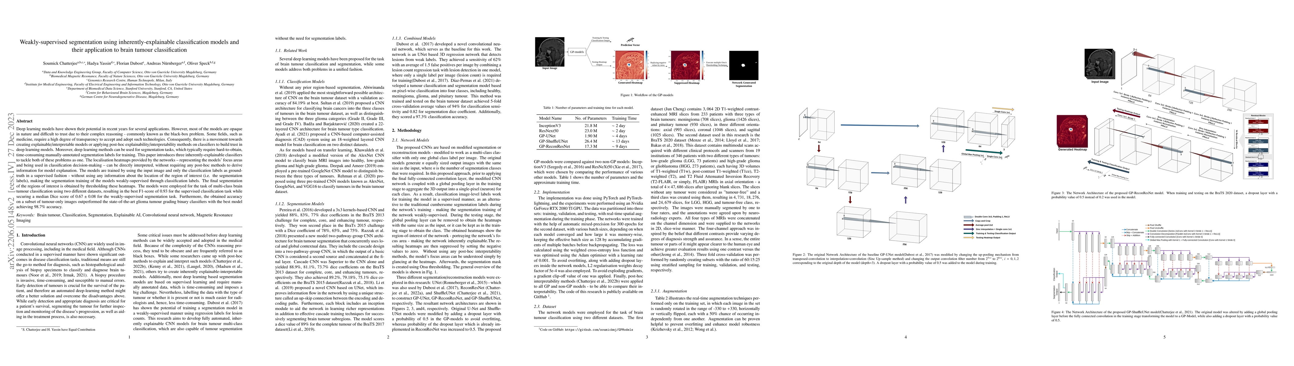
Deep learning models have shown their potential for several applications. However, most of the models are opaque and difficult to trust due to their complex reasoning - commonly known as the black-b...
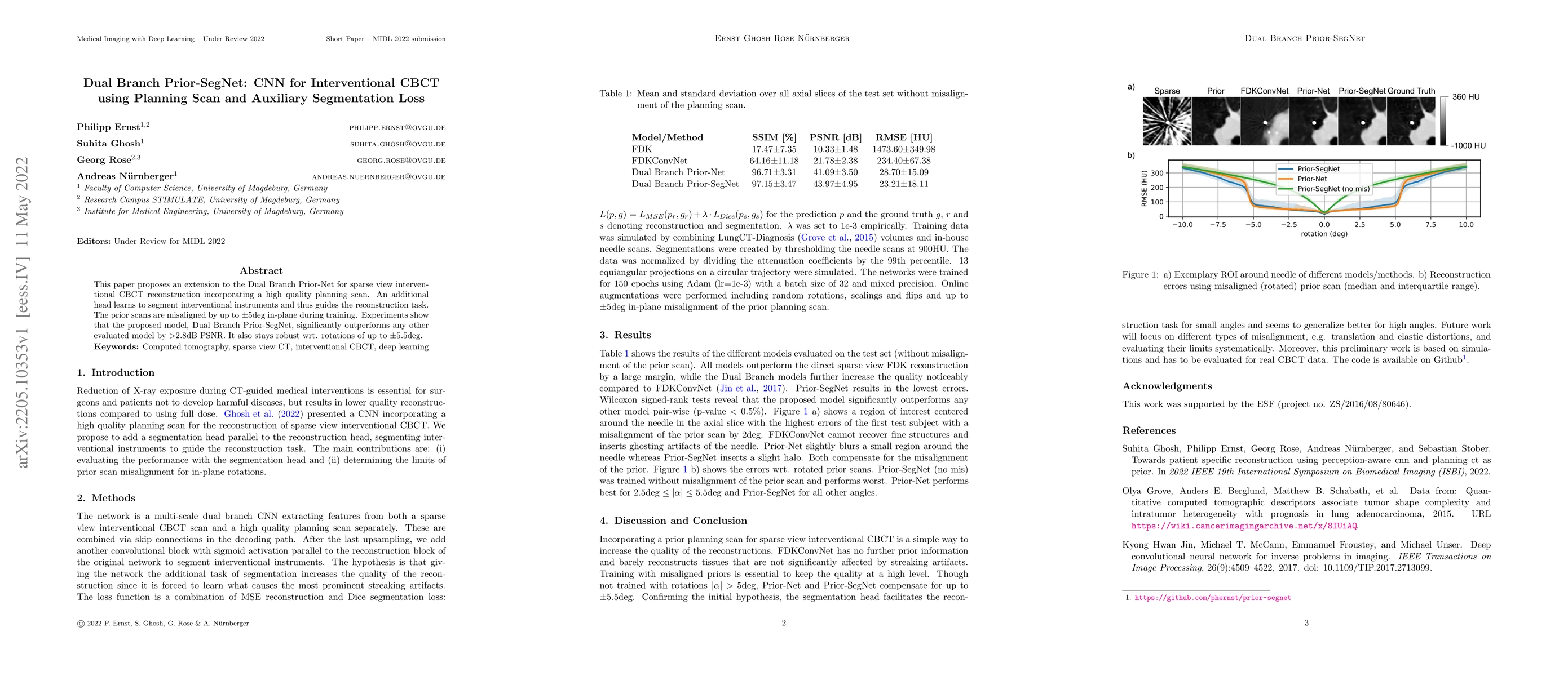
This paper proposes an extension to the Dual Branch Prior-Net for sparse view interventional CBCT reconstruction incorporating a high quality planning scan. An additional head learns to segment inte...
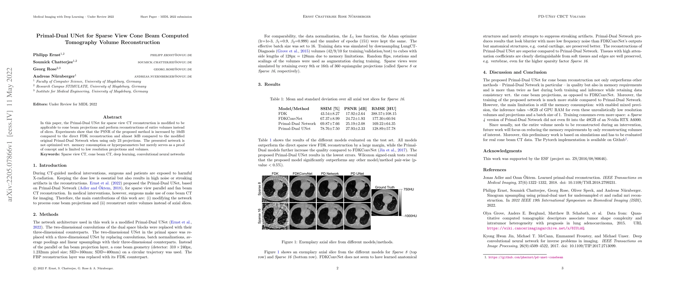
In this paper, the Primal-Dual UNet for sparse view CT reconstruction is modified to be applicable to cone beam projections and perform reconstructions of entire volumes instead of slices. Experimen...
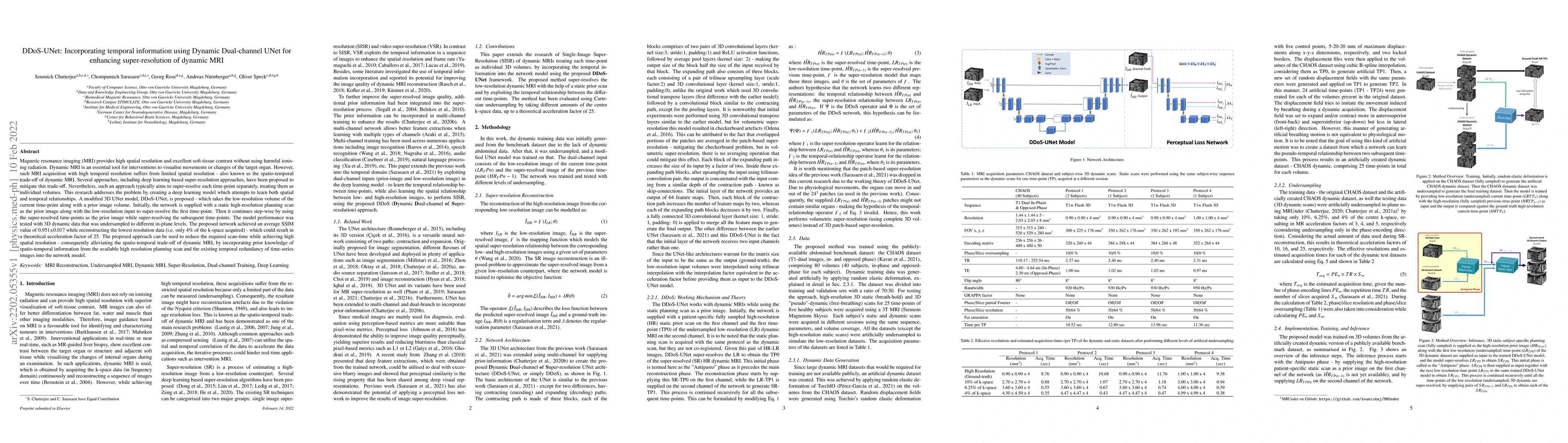
Magnetic resonance imaging (MRI) provides high spatial resolution and excellent soft-tissue contrast without using harmful ionising radiation. Dynamic MRI is an essential tool for interventions to v...
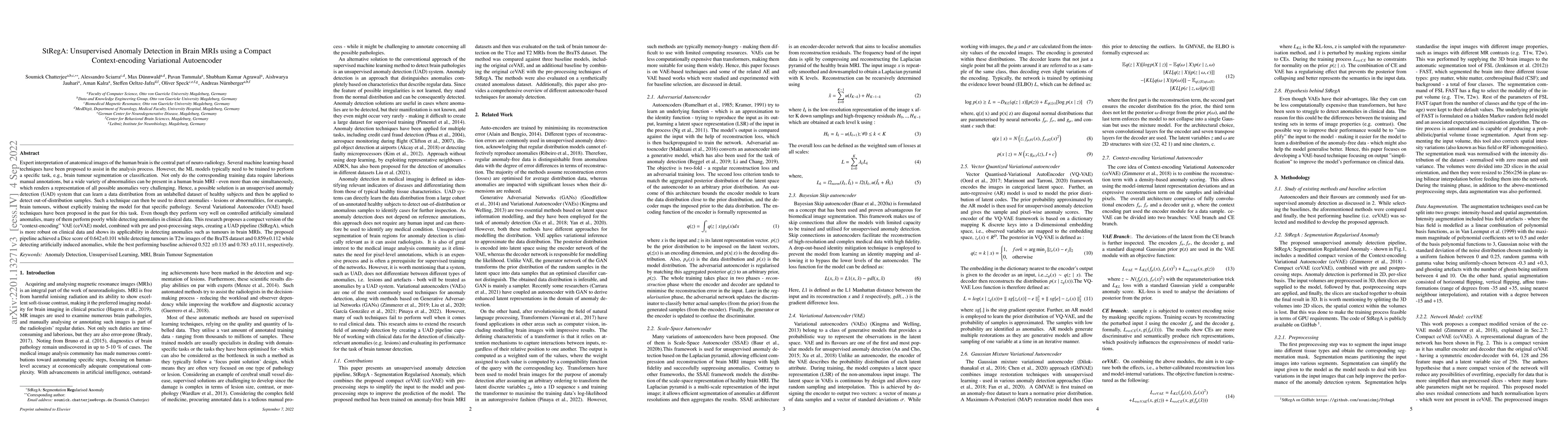
Expert interpretation of anatomical images of the human brain is the central part of neuro-radiology. Several machine learning-based techniques have been proposed to assist in the analysis process. ...
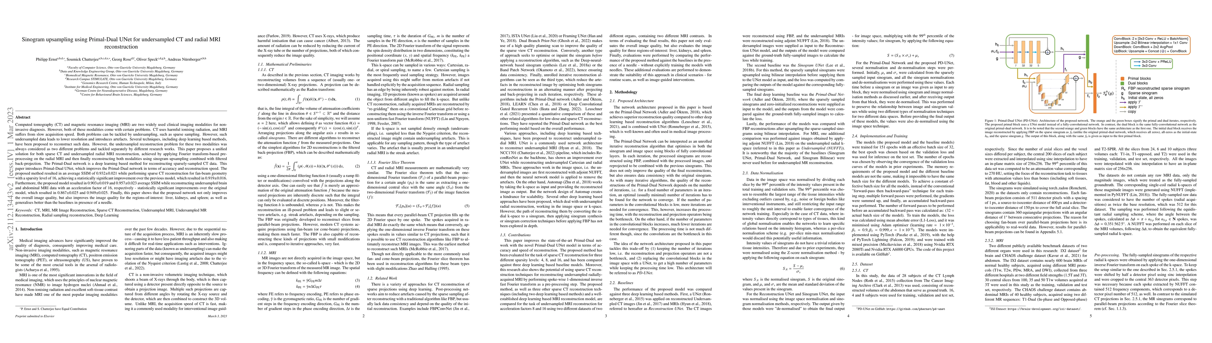
Computed tomography and magnetic resonance imaging are two widely used clinical imaging modalities for non-invasive diagnosis. However, both of these modalities come with certain problems. CT uses h...
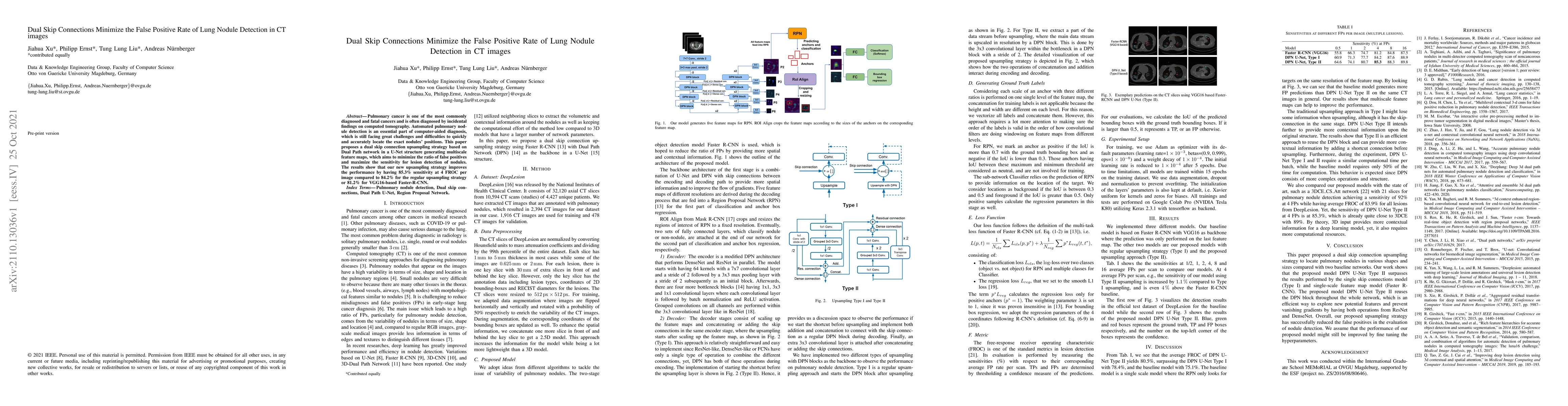
Pulmonary cancer is one of the most commonly diagnosed and fatal cancers and is often diagnosed by incidental findings on computed tomography. Automated pulmonary nodule detection is an essential pa...
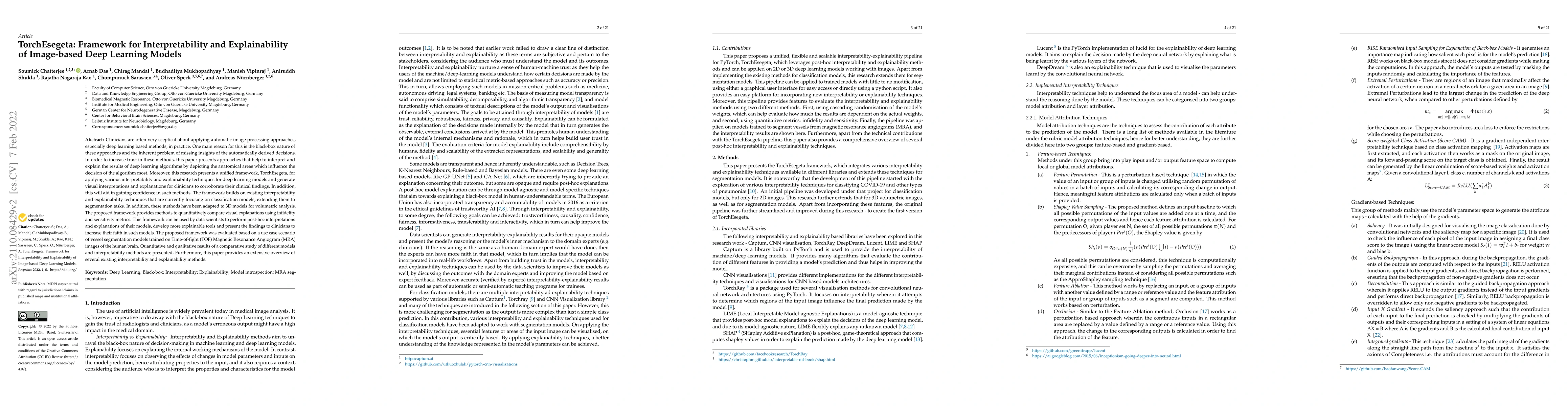
Clinicians are often very sceptical about applying automatic image processing approaches, especially deep learning based methods, in practice. One main reason for this is the black-box nature of the...
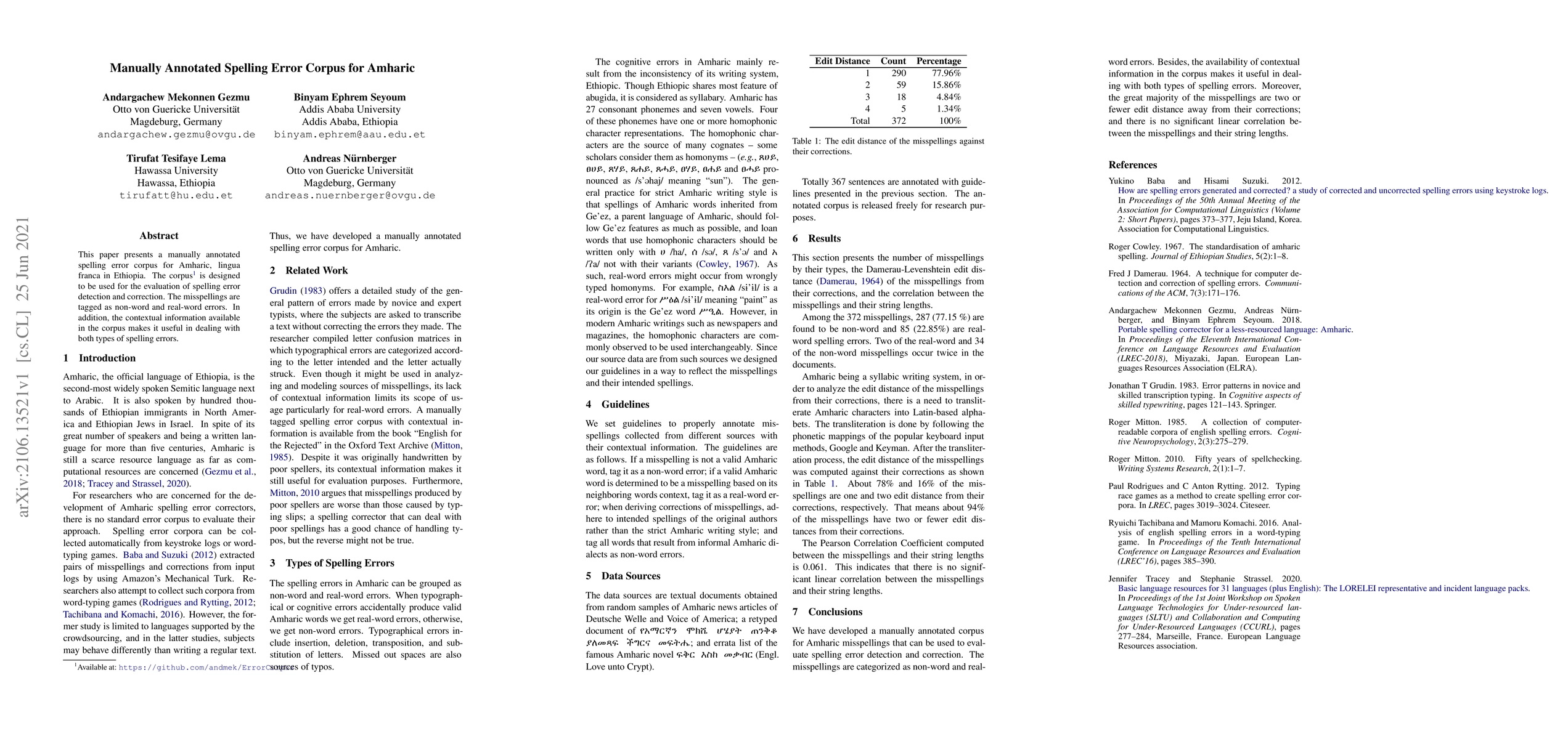
This paper presents a manually annotated spelling error corpus for Amharic, lingua franca in Ethiopia. The corpus is designed to be used for the evaluation of spelling error detection and correction...
We introduced the contemporary Amharic corpus, which is automatically tagged for morpho-syntactic information. Texts are collected from 25,199 documents from different domains and about 24 million o...
A brain tumour is a mass or cluster of abnormal cells in the brain, which has the possibility of becoming life-threatening because of its ability to invade neighbouring tissues and also form metasta...
This paper describes the acquisition, preprocessing, segmentation, and alignment of an Amharic-English parallel corpus. It will be helpful for machine translation of a low-resource language, Amharic...
MRI is an inherently slow process, which leads to long scan time for high-resolution imaging. The speed of acquisition can be increased by ignoring parts of the data (undersampling). Consequently, t...
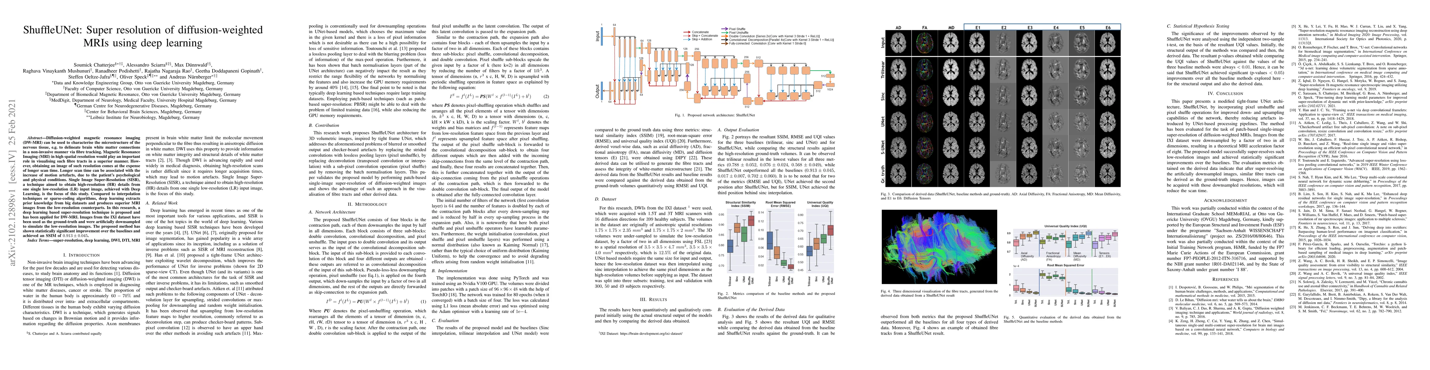
Diffusion-weighted magnetic resonance imaging (DW-MRI) can be used to characterise the microstructure of the nervous tissue, e.g. to delineate brain white matter connections in a non-invasive manner...
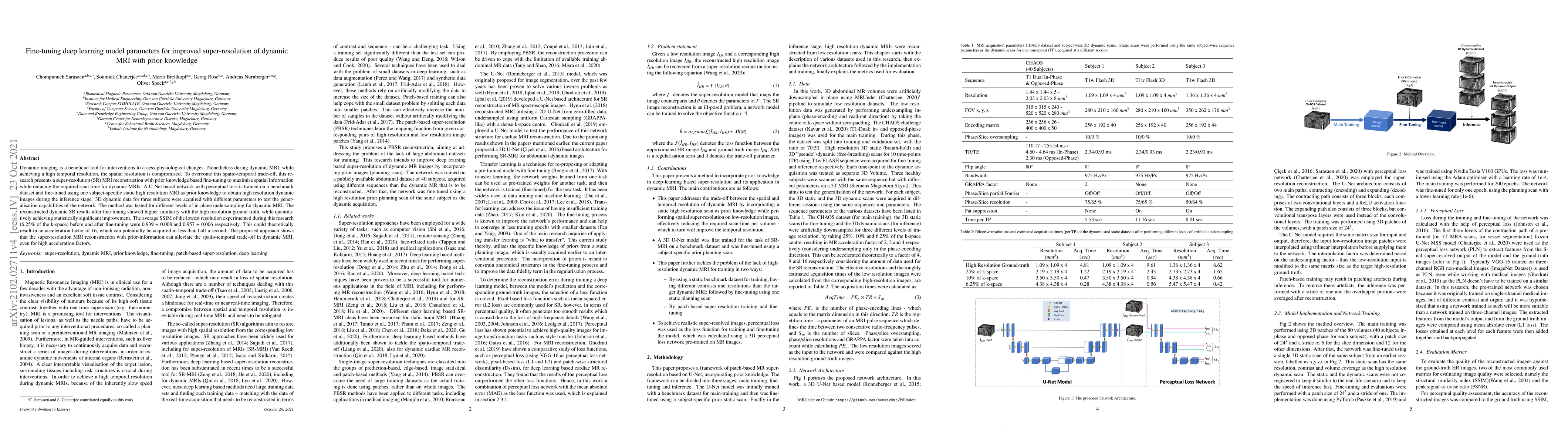
Dynamic imaging is a beneficial tool for interventions to assess physiological changes. Nonetheless during dynamic MRI, while achieving a high temporal resolution, the spatial resolution is compromi...
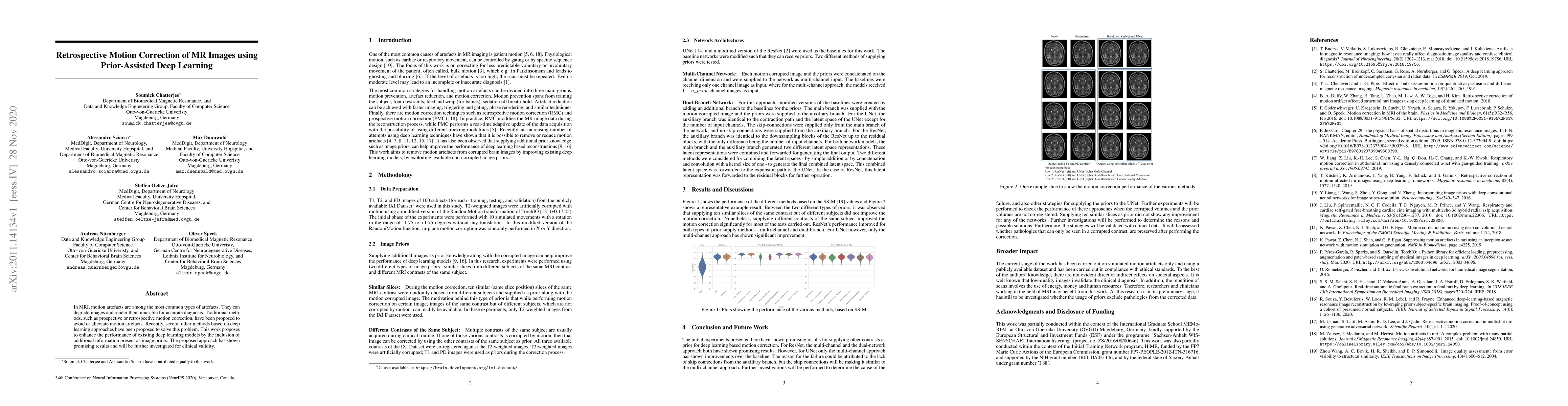
In MRI, motion artefacts are among the most common types of artefacts. They can degrade images and render them unusable for accurate diagnosis. Traditional methods, such as prospective or retrospect...
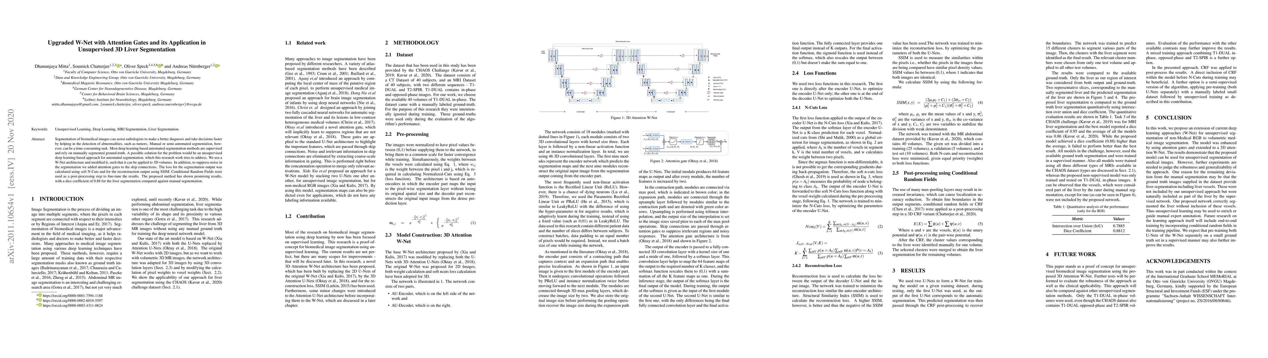
Segmentation of biomedical images can assist radiologists to make a better diagnosis and take decisions faster by helping in the detection of abnormalities, such as tumors. Manual or semi-automated ...
Blood vessels of the brain provide the human brain with the required nutrients and oxygen. As a vulnerable part of the cerebral blood supply, pathology of small vessels can cause serious problems su...
The outbreak of COVID-19 has shocked the entire world with its fairly rapid spread and has challenged different sectors. One of the most effective ways to limit its spread is the early and accurate ...
Segmentation of abdominal organs has been a comprehensive, yet unresolved, research field for many years. In the last decade, intensive developments in deep learning (DL) have introduced new state-o...
Ontologies are formal representations of knowledge in specific domains that provide a structured framework for organizing and understanding complex information. Creating ontologies, however, is a comp...
The human brain receives nutrients and oxygen through an intricate network of blood vessels. Pathology affecting small vessels, at the mesoscopic scale, represents a critical vulnerability within the ...
Liver tumour ablation presents a significant clinical challenge: whilst tumours are clearly visible on pre-operative MRI, they are often effectively invisible on intra-operative CT due to minimal cont...