Academic Profile
Statistics
Similar Authors
Papers on arXiv
Image-based or morphological profiling is a rapidly expanding field wherein cells are "profiled" by extracting hundreds to thousands of unbiased, quantitative features from images of cells that have...
Together with the molecular knowledge of genes and proteins, biological images promise to significantly enhance the scientific understanding of complex cellular systems and to advance predictive and...
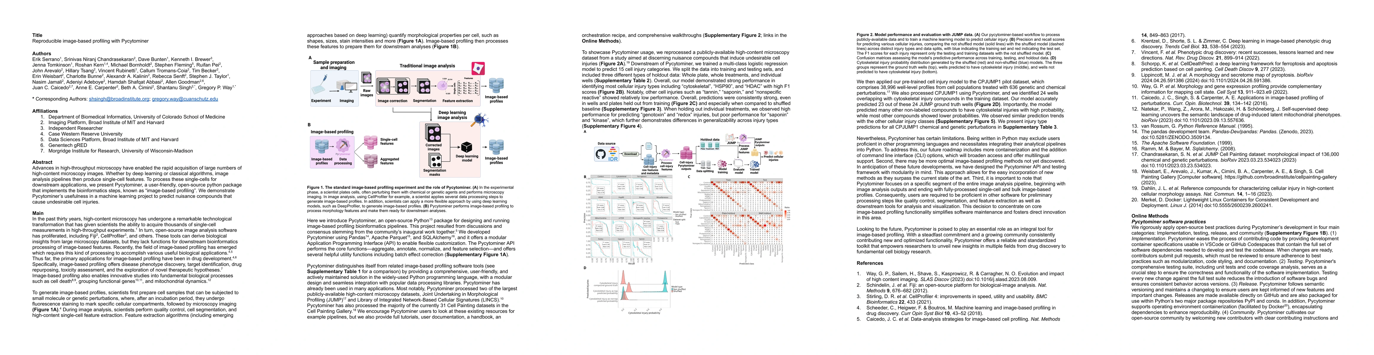
Advances in high-throughput microscopy have enabled the rapid acquisition of large numbers of high-content microscopy images. Whether by deep learning or classical algorithms, image analysis pipelin...
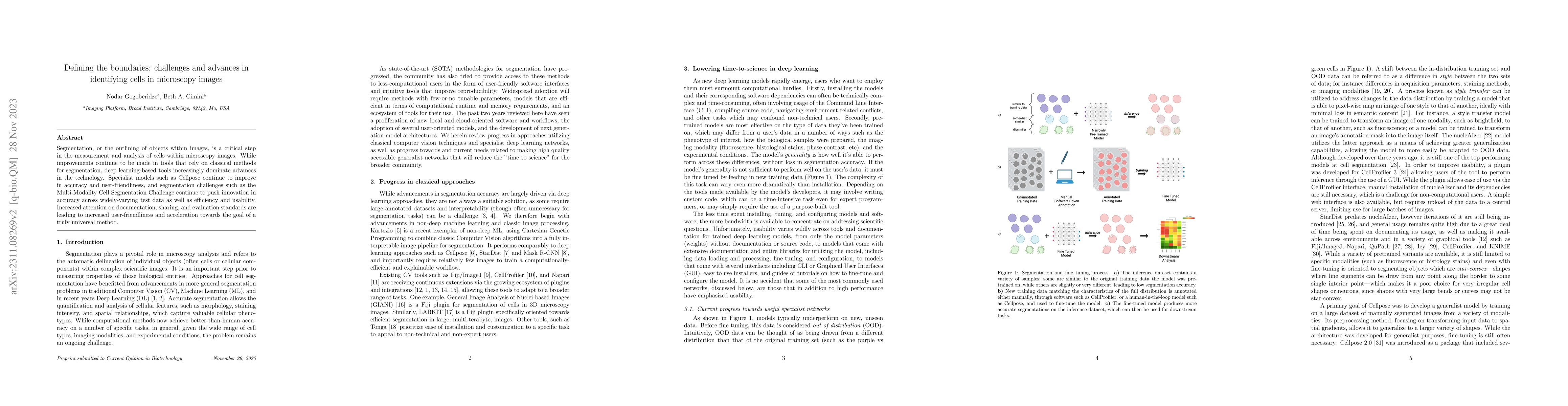
Segmentation, or the outlining of objects within images, is a critical step in the measurement and analysis of cells within microscopy images. While improvements continue to be made in tools that re...
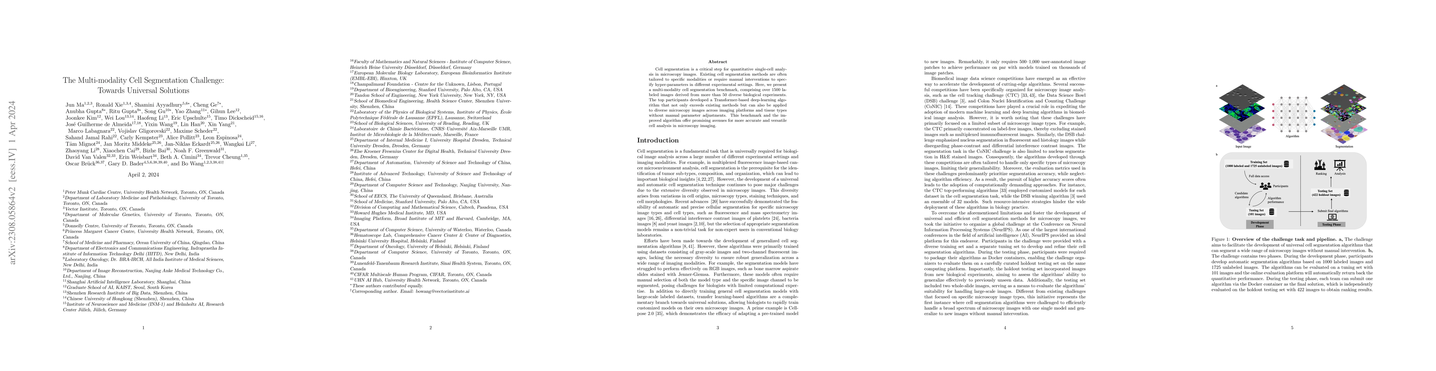
Cell segmentation is a critical step for quantitative single-cell analysis in microscopy images. Existing cell segmentation methods are often tailored to specific modalities or require manual interv...
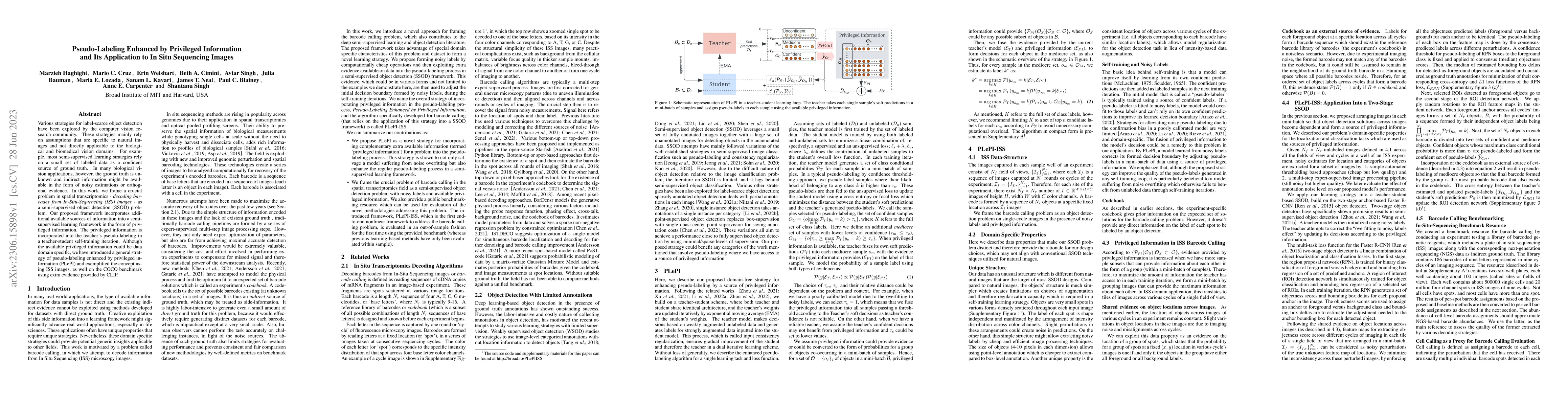
Various strategies for label-scarce object detection have been explored by the computer vision research community. These strategies mainly rely on assumptions that are specific to natural images and...
Validation metrics are key for the reliable tracking of scientific progress and for bridging the current chasm between artificial intelligence (AI) research and its translation into practice. Howeve...
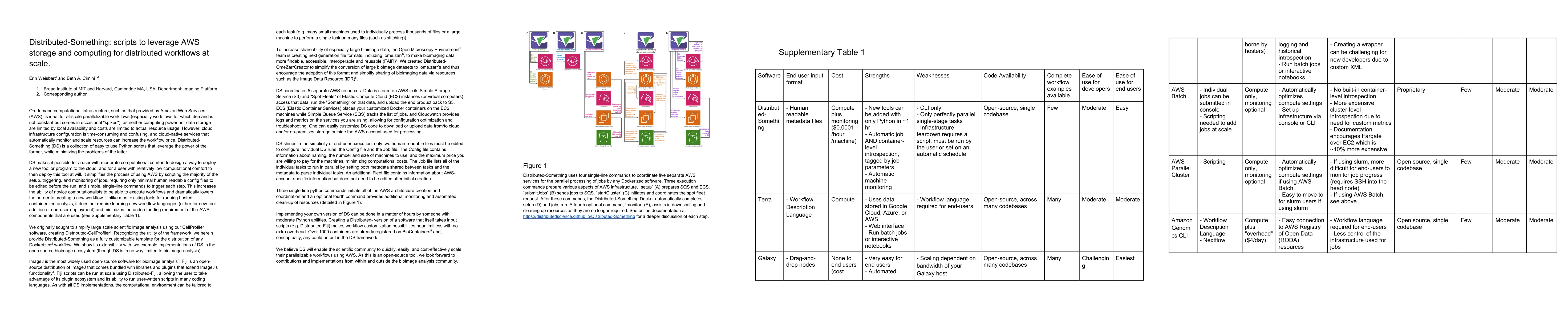
Distributed-Something coordinates the distribution of any Dockerized workflow using on-demand computational infrastructure from Amazon Web Services to enable at-scale workflows where neither computi...
Increasing evidence shows that flaws in machine learning (ML) algorithm validation are an underestimated global problem. Particularly in automatic biomedical image analysis, chosen performance metri...
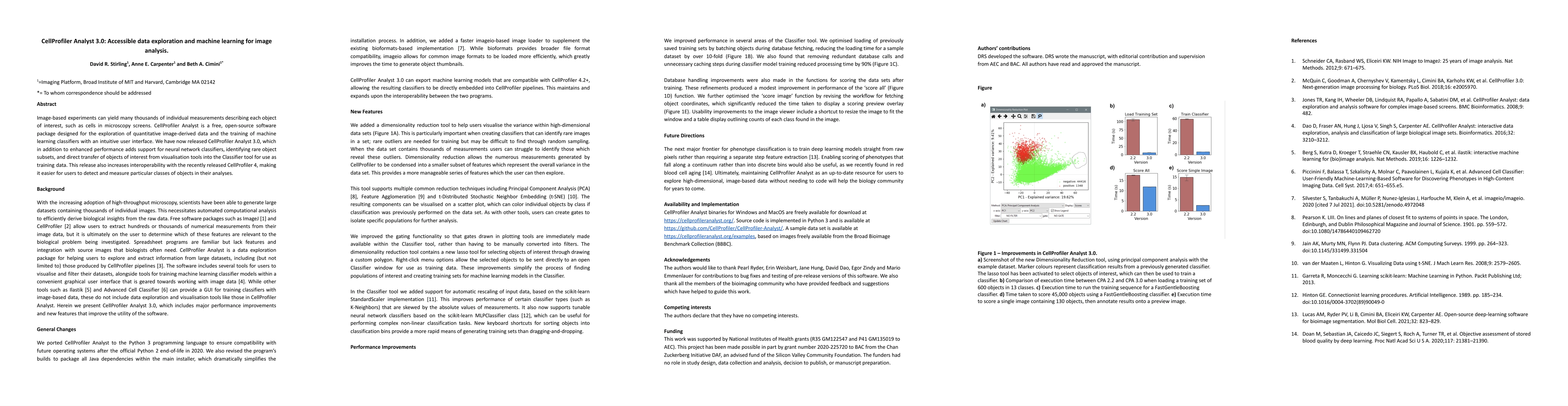
Image-based experiments can yield many thousands of individual measurements describing each object of interest, such as cells in microscopy screens. CellProfiler Analyst is a free, open-source softw...