Quick Summary:

Summary: Pycytominer, an open-source Python package, streamlines image-based profiling in high-content microscopy, enabling reproducible analysis of single-cell features and demonstrating its utility in predicting harmful compounds via machine learning.

Summary

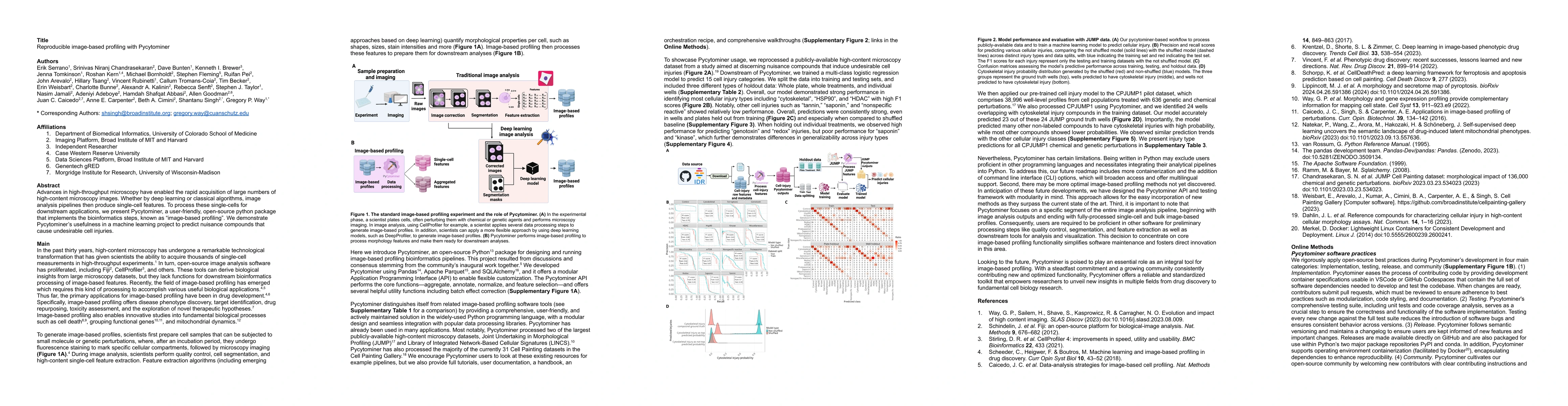
Advances in high-throughput microscopy have enabled the rapid acquisition of large numbers of high-content microscopy images. Whether by deep learning or classical algorithms, image analysis pipelines then produce single-cell features. To process these single-cells for downstream applications, we present Pycytominer, a user-friendly, open-source python package that implements the bioinformatics steps, known as image-based profiling. We demonstrate Pycytominers usefulness in a machine learning project to predict nuisance compounds that cause undesirable cell injuries.

AI Key Findings

Get AI-generated insights about this paper's methodology, results, and significance.

Paper Details

PDF Preview

Download PDF

Key Terms

microscopy (0.282)
cell (0.251)
image (0.203)
python (0.190)
usefulness (0.189)
throughput (0.189)
compounds (0.182)
enabled (0.181)
acquisition (0.172)
package (0.168)
downstream (0.168)
single (0.167)
cells (0.165)
open source (0.165)
learning (0.164)
advances (0.163)
cause (0.161)
project (0.157)
rapid (0.154)
steps (0.152)

Citation Network

Current paper (gray), citations (green), references (blue)

Current Paper
Citations
References
Click to view

Similar Papers

No similar papers found for this research.

Title Authors Year Actions
Cell Painting PLUS: expanding the multiplexing capacity of Cell Painting-based phenotypic profiling using iterative staining-elution cycles
E. von Coburg, M. Wedler, J. M. Muiño, Christopher Wolff, Nils Körber, S. Dunst, Shu Liu
2025
View Paper
High-content microscopy and machine learning characterize a cell morphology signature of NF1 genotype in Schwann cells
Jenna Tomkinson, Cameron Mattson, Michelle Mattson-Hoss, Herb Sarnoff, S. Bouley, James A Walker, Gregory P. Way
2025
View Paper
Drug-induced cytotoxicity prediction in muscle cells, an application of the Cell Painting assay
Roman Lambert, Eva Serrano Candelas, Pablo Aparicio, Aisling Murphy, Rafael Gozalbes, Howard Fearnhead
2025
View Paper
A morphology and secretome map of pyroptosis
Michael J. Lippincott, Jenna Tomkinson, Dave Bunten, Milad Mohammadi, Johanna Kastl, Johannes Knop, Ralf Schwandner, Jiamin Huang, Grant Ongo, Nathaniel Robichaud, M. Dagher, Andrés Mansilla-Soto, Cynthia Saravia-Estrada, Masafumi Tsuboi, Carla Basualto-Alarcón, Gregory P. Way
2025
View Paper
Cell morphological representations of genes enhance prediction of drug targets
Niveditha S. Iyer, Daniel J. Michael, S-Y Gordon Chi, John Arevalo, S. Chandrasekaran, A. Carpenter, P. Rajpurkar, Shantanu Singh
2025
View Paper
Alternate dyes for image-based profiling assays
Suganya Sivagurunathan, Patrick J. Byrne, Alán F. Muñoz, John Arevalo, A. Carpenter, Shantanu Singh, Maria Kost-Alimova, B. Cimini
2025
View Paper
A genome-wide atlas of human cell morphology
Meraj Ramezani, Erin Weisbart, J. Bauman, Avtar Singh, John Yong, Maria Lozada, Gregory P. Way, Sanam L. Kavari, Celeste Diaz, Eddy Leardini, Gunjan Jetley, Jenlu Pagnotta, Marzieh Haghighi, Thiago M. Batista, Joaquín Pérez-Schindler, M. Claussnitzer, Shantanu Singh, B. Cimini, P. Blainey, A. Carpenter, Calvin H Jan, James T Neal
2025
View Paper
MorphoDiff: Cellular Morphology Painting with Diffusion Models
Zeinab Navidi, Jun Ma, Esteban A. Miglietta, Le Liu, A. Carpenter, B. Cimini, Benjamin Haibe-Kains, Bo Wang
2024
View Paper
Morphological Profiling Dataset of EU-OPENSCREEN Bioactive Compounds Over Multiple Imaging Sites and Cell Lines
Christopher Wolff, Martin Neuenschwander, C. Beese, Divya Sitani, María C. Ramos, Alžběta Srovnalova, M. J. Varela, Pavel G. Polishchuk, Katholiki E. Skopelitou, C. Škuta, Bahne Stechmann, J. Brea, Mads Hartvig Clausen, P. Džubák, Rosario Fernández-Godino, O. Genilloud, Marián Hajdúch, M. I. Loza, Martin Lehmann, J. V. von Kries, Han Sun, Christopher Schmied
2024
View Paper
Building, benchmarking, and exploring perturbative maps of transcriptional and morphological data
Safiye Celik, Jan-Christian Hütter, Sandra Melo Carlos, N. Lazar, Ramkumar Veppathur Mohan, Conor A. Tillinghast, Tommaso Biancalani, Marta M. Fay, Berton A. Earnshaw, I. Haque
2024
View Paper
Evaluating batch correction methods for image-based cell profiling
John Arevalo, Ellen Su, Jessica D. Ewald, Robert van Dijk, A. Carpenter, Shantanu Singh
2024
View Paper
PostFocus: automated selective post-acquisition high-throughput focus restoration using diffusion model for label-free time-lapse microscopy
Kwan-Ling Wu, Melisa J Montalvo, Prashant Menon, Badrinath Roysam, N. Varadarajan
2024
View Paper
Anomaly detection for high-content image-based phenotypic cell profiling
Alon Shpigler, Naor Kolet, Shahar Golan, Erin Weisbart, A. Zaritsky
2024
View Paper
Morphological profiling for drug discovery in the era of deep learning
Qiaosi Tang, R. Ratnayake, Gustavo Seabra, Zhe Jiang, Ruogu Fang, Lina Cui, Yousong Ding, Tamer Kahveci, Jiang Bian, Chenglong Li, Hendrik Luesch, Yanjun Li
2024
View Paper
A versatile information retrieval framework for evaluating profile strength and similarity
Alexandr A. Kalinin, John Arevalo, Loan Vulliard, Erik Serrano, Hillary Tsang, Michael Bornholdt, Bartek Rajwa, A. Carpenter, Gregory P. Way, Shantanu Singh
2024
View Paper
Toward generalizable phenotype prediction from single-cell morphology representations
Jenna Tomkinson, Roshan Kern, Cameron Mattson, Gregory P. Way
2024
View Paper

Comments (0)

This website uses cookies to ensure you get the best experience. Learn more