Academic Profile
Statistics
Similar Authors
Papers on arXiv
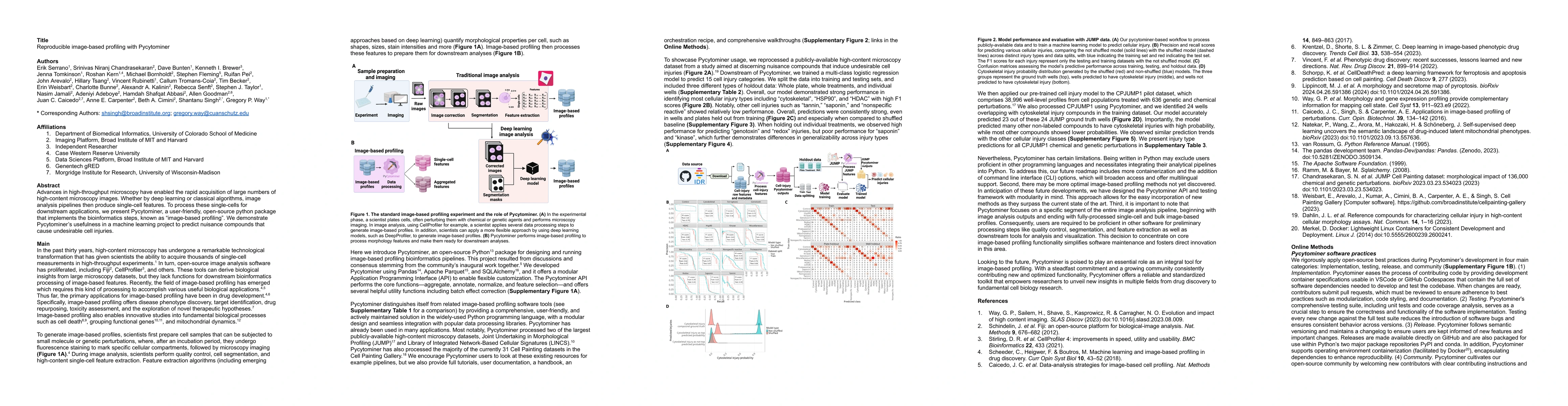
Advances in high-throughput microscopy have enabled the rapid acquisition of large numbers of high-content microscopy images. Whether by deep learning or classical algorithms, image analysis pipelin...
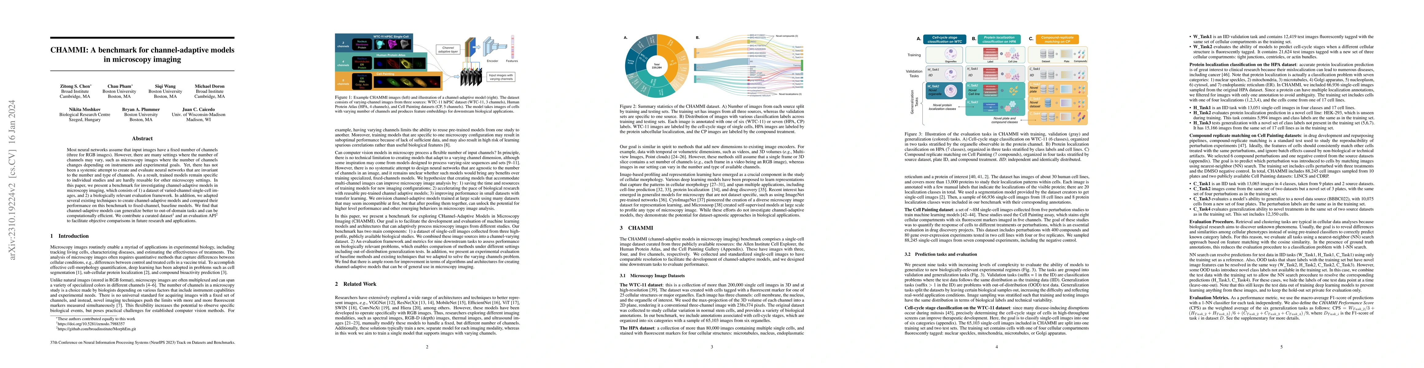
Most neural networks assume that input images have a fixed number of channels (three for RGB images). However, there are many settings where the number of channels may vary, such as microscopy image...
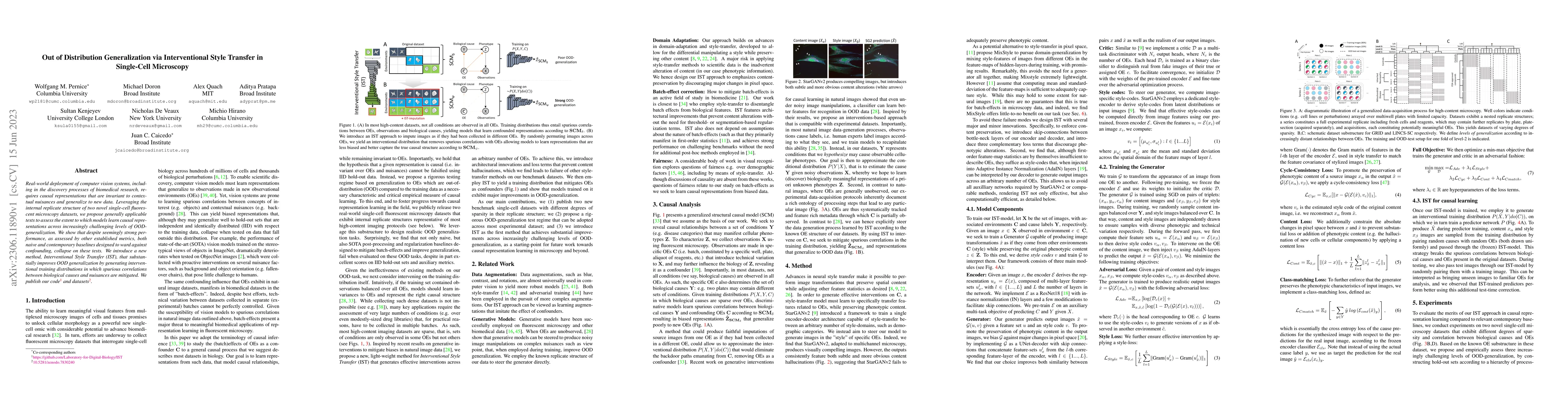
Real-world deployment of computer vision systems, including in the discovery processes of biomedical research, requires causal representations that are invariant to contextual nuisances and generali...
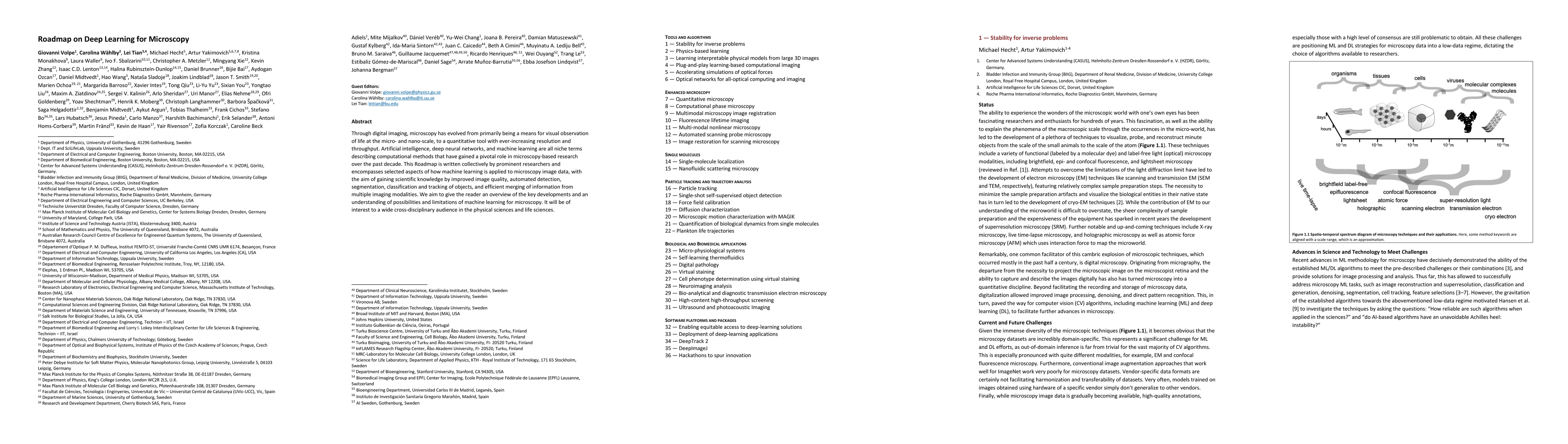
Through digital imaging, microscopy has evolved from primarily being a means for visual observation of life at the micro- and nano-scale, to a quantitative tool with ever-increasing resolution and t...
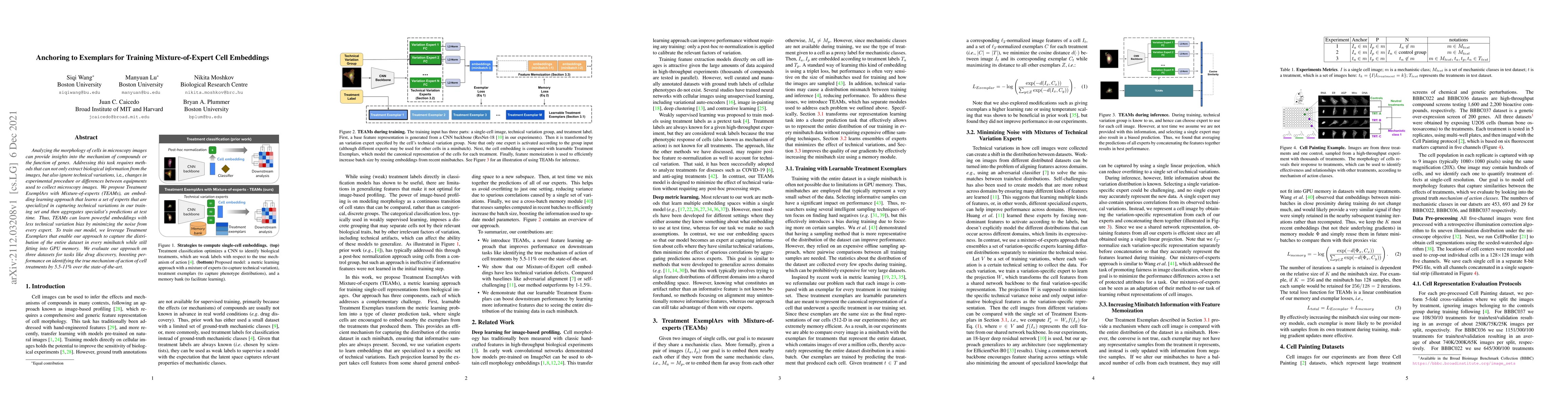
Analyzing the morphology of cells in microscopy images can provide insights into the mechanism of compounds or the function of genes. Addressing this task requires methods that can not only extract ...
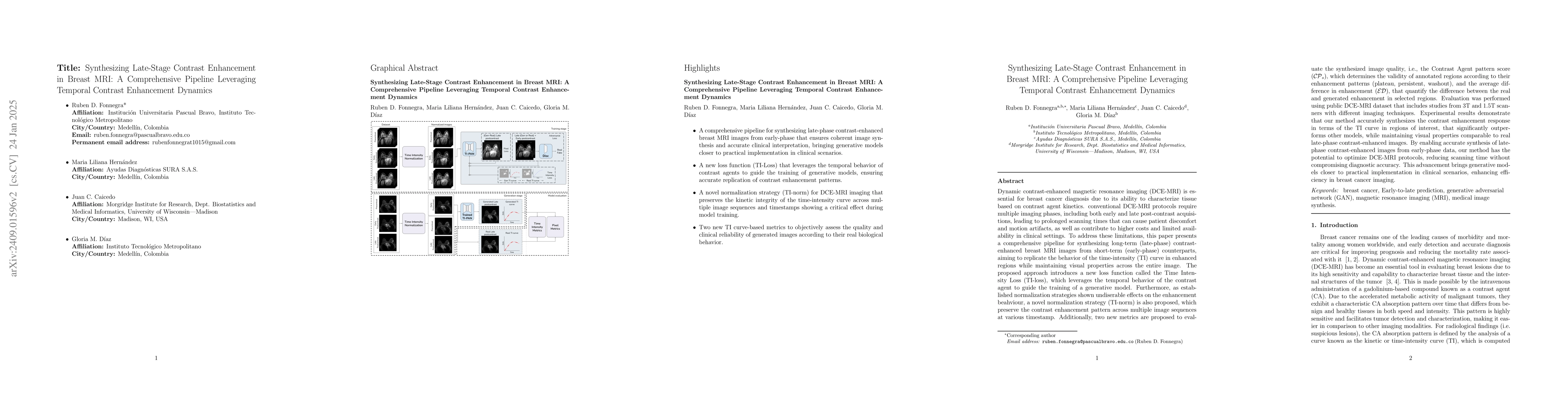
Contrast-enhancement pattern analysis is critical in breast magnetic resonance imaging (MRI) to distinguish benign from probably malignant tumors. However, contrast-enhanced image acquisitions are tim...
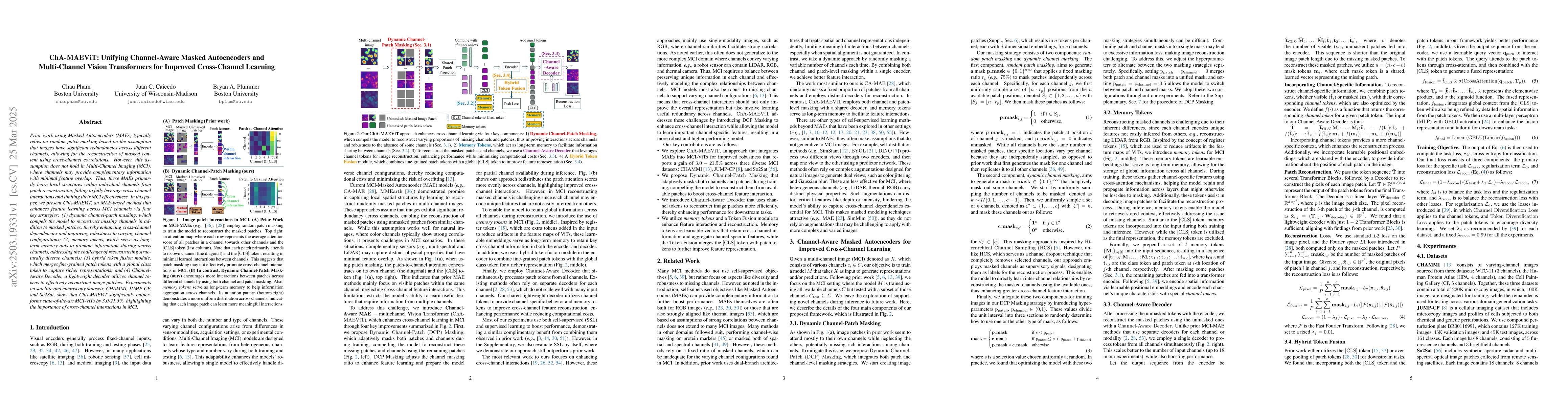
Prior work using Masked Autoencoders (MAEs) typically relies on random patch masking based on the assumption that images have significant redundancies across different channels, allowing for the recon...
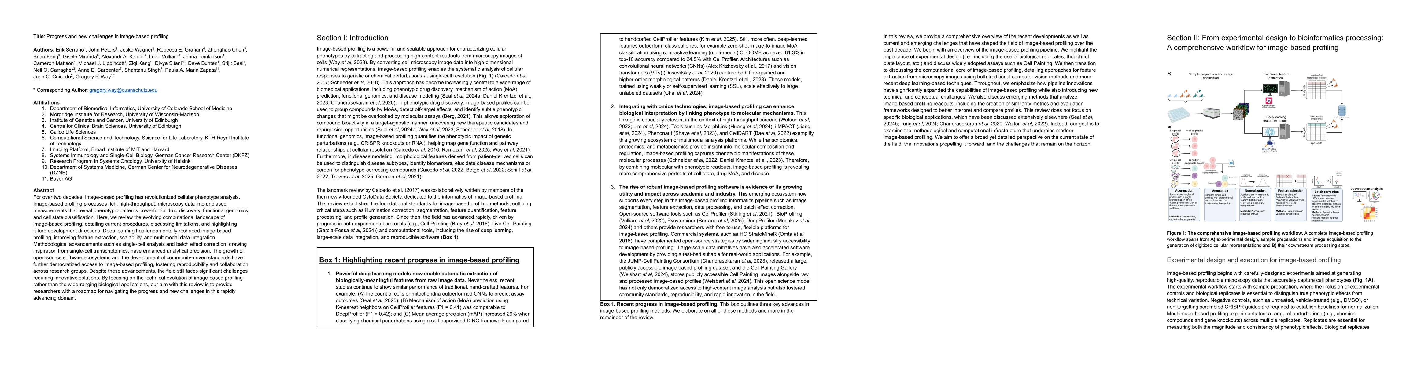
For over two decades, image-based profiling has revolutionized cellular phenotype analysis. Image-based profiling processes rich, high-throughput, microscopy data into unbiased measurements that revea...

Quantifying cell morphology using images and machine learning has proven to be a powerful tool to study the response of cells to treatments. However, models used to quantify cellular morphology are ty...