Academic Profile
Statistics
Similar Authors
Papers on arXiv
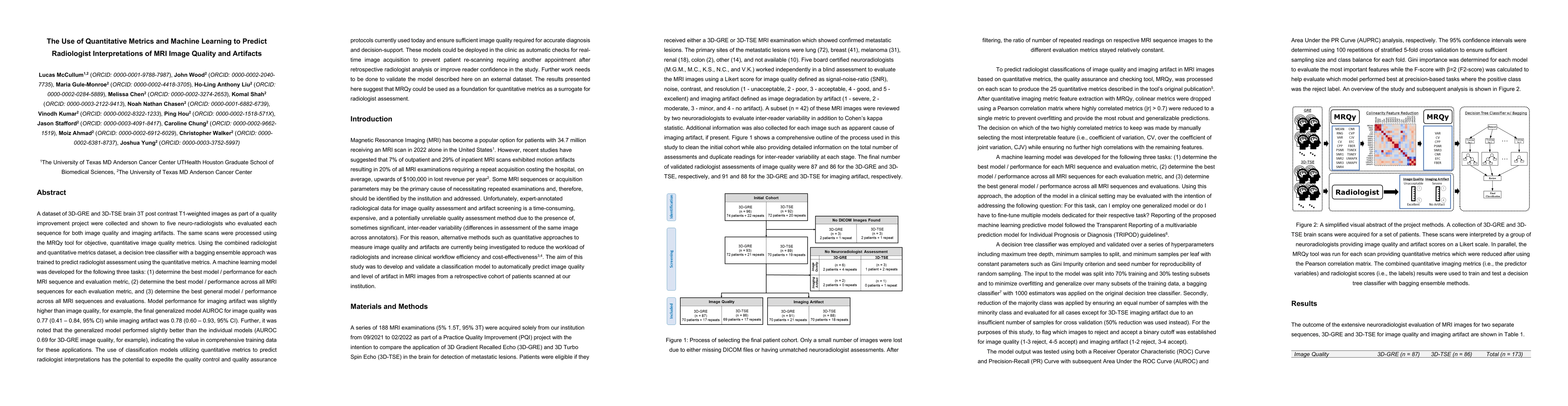
A dataset of 3D-GRE and 3D-TSE brain 3T post contrast T1-weighted images as part of a quality improvement project were collected and shown to five neuro-radiologists who evaluated each sequence for ...
Although generative adversarial networks (GANs) have shown promise in medical imaging, they have four main limitations that impeded their utility: computational cost, data requirements, reliable eva...
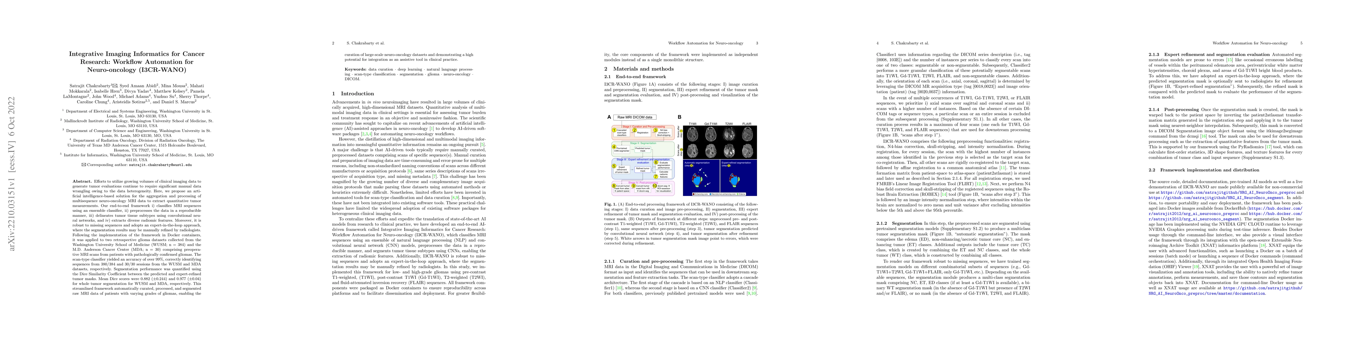
Efforts to utilize growing volumes of clinical imaging data to generate tumor evaluations continue to require significant manual data wrangling owing to the data heterogeneity. Here, we propose an a...
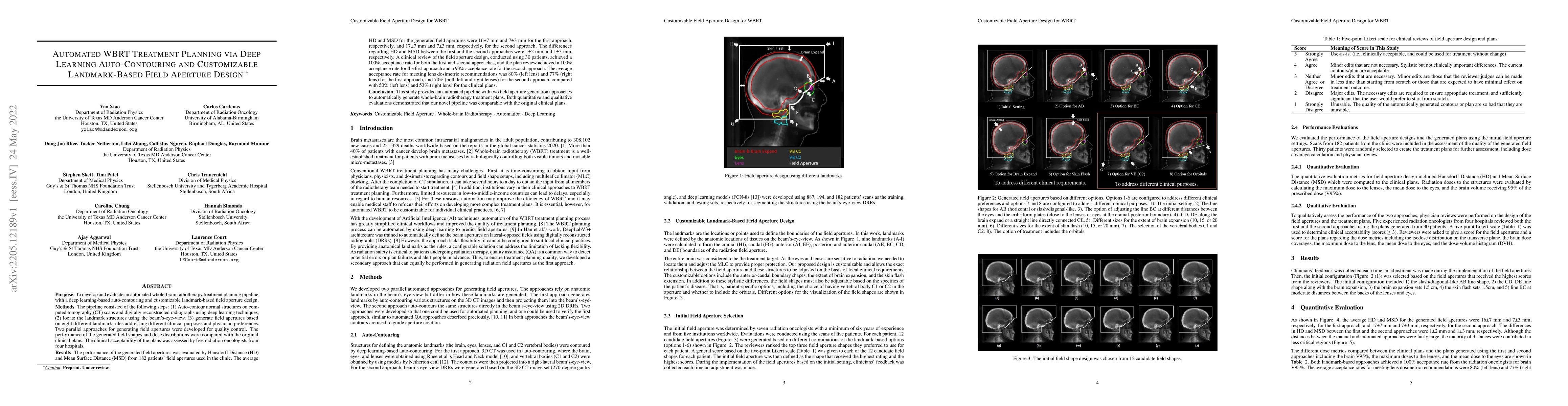
In this work, we developed and evaluated a novel pipeline consisting of two landmark-based field aperture generation approaches for WBRT treatment planning; they are fully automated and customizable...

Deep neural networks with multilevel connections process input data in complex ways to learn the information.A networks learning efficiency depends not only on the complex neural network architectur...
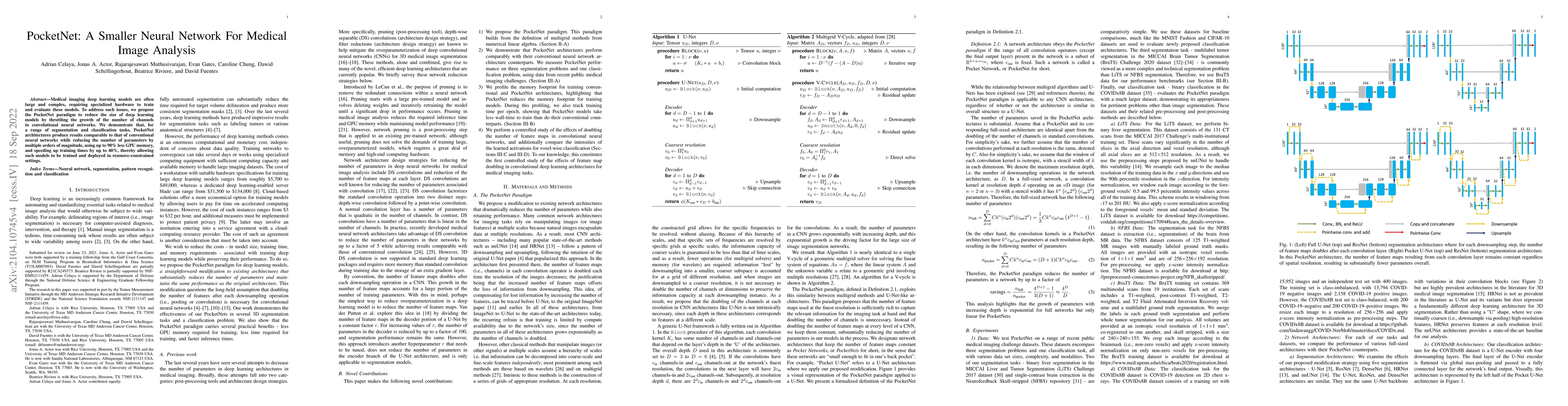
Medical imaging deep learning models are often large and complex, requiring specialized hardware to train and evaluate these models. To address such issues, we propose the PocketNet paradigm to redu...
Medical imaging segmentation is a highly active area of research, with deep learning-based methods achieving state-of-the-art results in several benchmarks. However, the lack of standardized tools for...
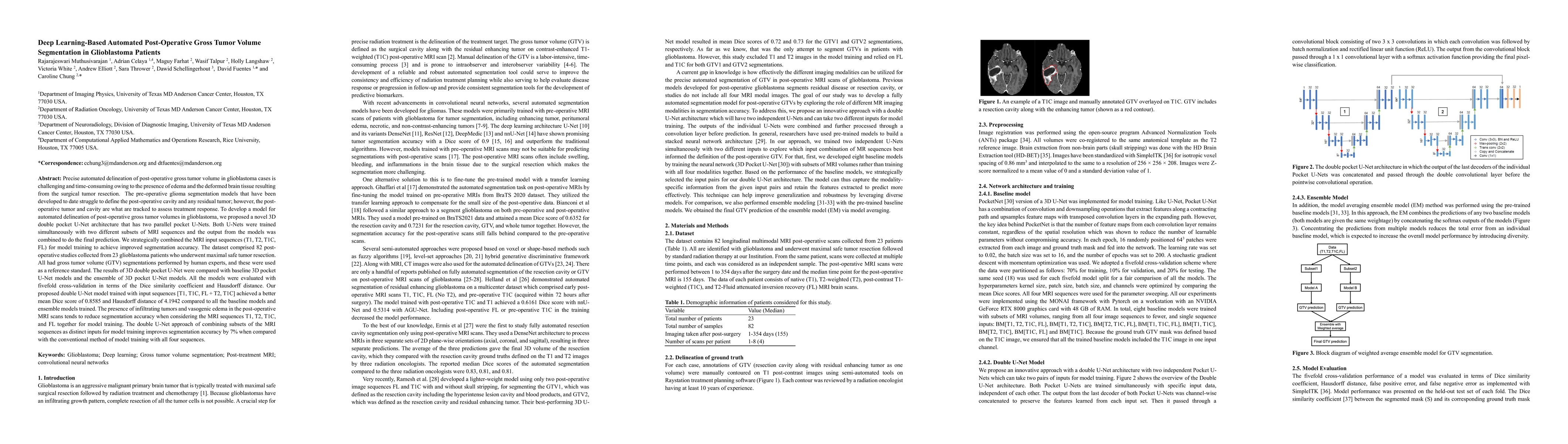
Precise automated delineation of post-operative gross tumor volume in glioblastoma cases is challenging and time-consuming owing to the presence of edema and the deformed brain tissue resulting from t...
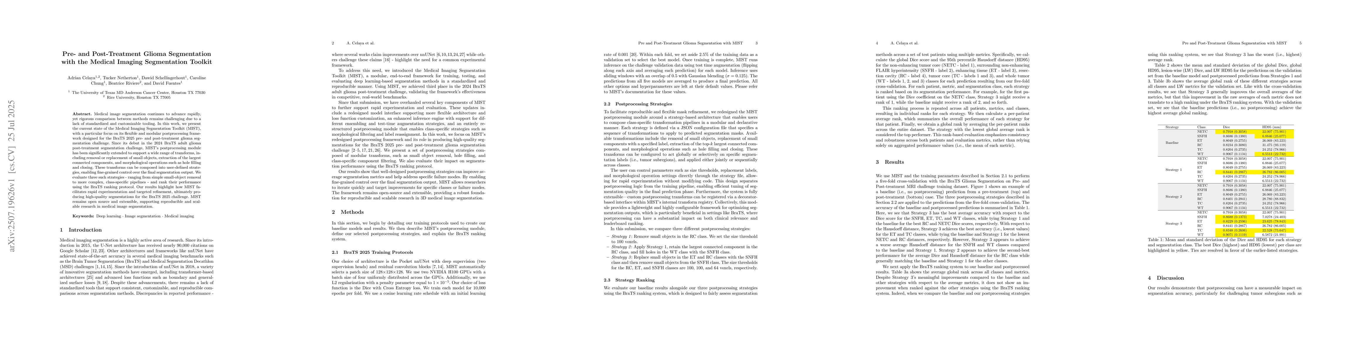
Medical image segmentation continues to advance rapidly, yet rigorous comparison between methods remains challenging due to a lack of standardized and customizable tooling. In this work, we present th...
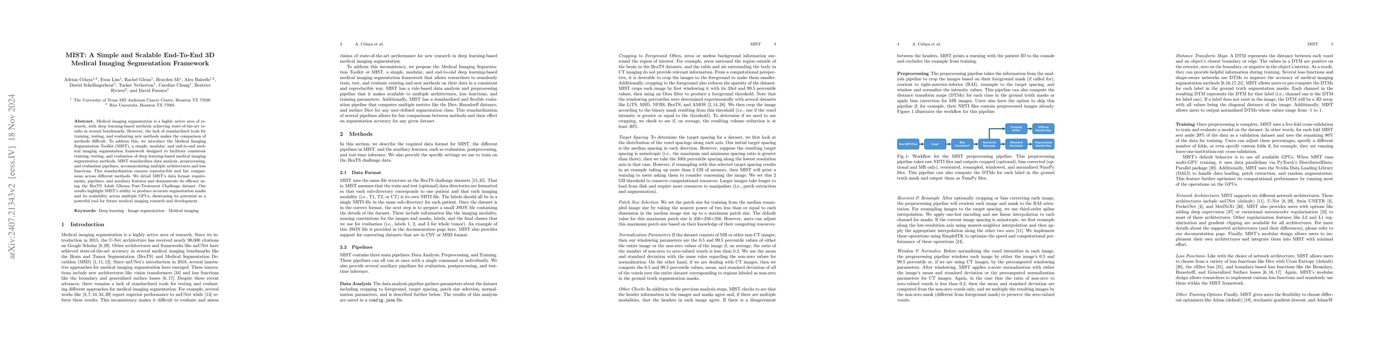
With the recent growth of Deep Learning for AI, there is a need for tools to meet the demand of data flowing into those models. In some cases, source data may exist in multiple formats, and therefore ...