Publication
Metrics
Quick Actions
AI Quick Summary
MIST is a new modular framework for end-to-end medical imaging segmentation that standardizes training, testing, and evaluation processes to facilitate fair comparisons of deep learning methods. It showcases accuracy and scalability using the BraTS dataset, proving its utility for future medical imaging research.
Quick Answers
What is "MIST: A Simple and Scalable End-To-End 3D Medical Imaging Segmentation Framework" about?
MIST is a new modular framework for end-to-end medical imaging segmentation that standardizes training, testing, and evaluation processes to facilitate fair comparisons of deep learning methods. It showcases accuracy and scalability using the BraTS dataset, proving its utility for future medical imaging research.
What methodology did the authors use?
The paper introduces MIST, a simple and scalable end-to-end 3D medical imaging segmentation framework that standardizes data analysis, preprocessing, and evaluation pipelines, accommodating multiple architectures and loss functions. More in Methodology →
What are the key results?
MIST produces accurate segmentation masks for medical imaging. — MIST demonstrates scalability across multiple GPUs, optimizing computational performance with PyTorch's DistributedDataParallel and Nvidia's DALI loader. More in Key Results →
Why is this work significant?
MIST simplifies the comparison of deep learning-based medical imaging segmentation methods by standardizing training, testing, and evaluation processes, which is crucial given the lack of standardized tools in the field. More in Significance →
What are the main limitations?
The paper does not discuss potential limitations of MIST explicitly. — The results are based on a specific dataset (BraTS Adult Glioma Post-Treatment Challenge), so generalizability to other datasets may vary. More in Limitations →
Abstract
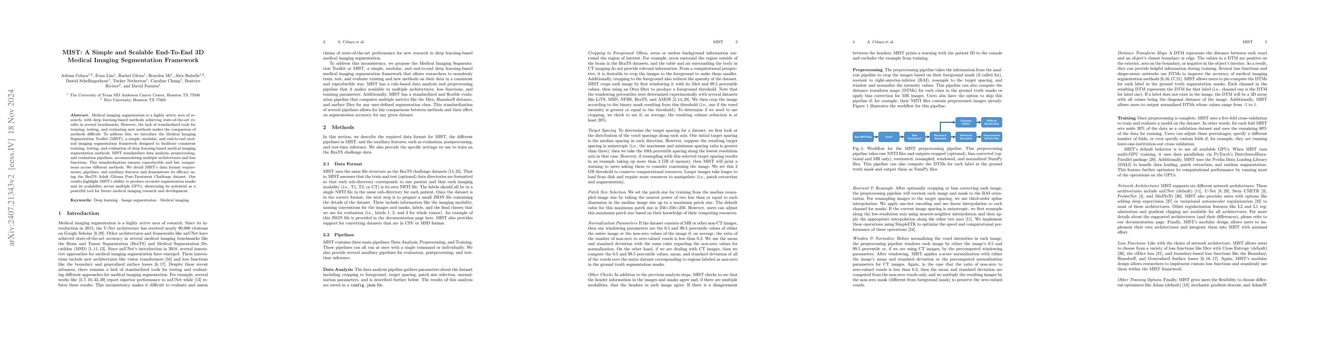
Medical imaging segmentation is a highly active area of research, with deep learning-based methods achieving state-of-the-art results in several benchmarks. However, the lack of standardized tools for training, testing, and evaluating new methods makes the comparison of methods difficult. To address this, we introduce the Medical Imaging Segmentation Toolkit (MIST), a simple, modular, and end-to-end medical imaging segmentation framework designed to facilitate consistent training, testing, and evaluation of deep learning-based medical imaging segmentation methods. MIST standardizes data analysis, preprocessing, and evaluation pipelines, accommodating multiple architectures and loss functions. This standardization ensures reproducible and fair comparisons across different methods. We detail MIST's data format requirements, pipelines, and auxiliary features and demonstrate its efficacy using the BraTS Adult Glioma Post-Treatment Challenge dataset. Our results highlight MIST's ability to produce accurate segmentation masks and its scalability across multiple GPUs, showcasing its potential as a powerful tool for future medical imaging research and development.
AI Key Findings
Generated Sep 02, 2025
Methodology — What approach did the authors take?
The paper introduces MIST, a simple and scalable end-to-end 3D medical imaging segmentation framework that standardizes data analysis, preprocessing, and evaluation pipelines, accommodating multiple architectures and loss functions.
Key Results — What are the main findings?
- MIST produces accurate segmentation masks for medical imaging.
- MIST demonstrates scalability across multiple GPUs, optimizing computational performance with PyTorch's DistributedDataParallel and Nvidia's DALI loader.
- MIST achieves median Dice scores of at least 0.9 for all segmentation classes in a five-fold cross-validation on the BraTS Adult Glioma Post-Treatment Challenge dataset.
- MIST effectively scales for different numbers of A100 and H100 GPUs with multiple batch sizes.
Significance — Why does this research matter?
MIST simplifies the comparison of deep learning-based medical imaging segmentation methods by standardizing training, testing, and evaluation processes, which is crucial given the lack of standardized tools in the field.
Technical Contribution — What is the technical contribution?
MIST offers a modular design that allows users to implement custom architectures and loss functions, along with support for six different network architectures and various loss functions.
Novelty — What is new about this work?
MIST's novelty lies in its standardization of data analysis, preprocessing, and evaluation pipelines, which facilitates consistent and fair comparisons of deep learning-based medical imaging segmentation methods.
Limitations — What are the limitations of this study?
- The paper does not discuss potential limitations of MIST explicitly.
- The results are based on a specific dataset (BraTS Adult Glioma Post-Treatment Challenge), so generalizability to other datasets may vary.
Future Work — What did the authors propose for future work?
- Exploration of different post-processing strategies with MIST's built-in post-processing pipeline.
- Investigation of improvements in accuracy using information from Distance Transform Maps (DTMs) with boundary-based losses.
- Evaluation of other architectures like multi-grid inspired FMG-Net or vision transformer-based architectures like SwinUNETR.
- An ablation study on the selected parameters for the BraTS dataset, including deep supervision, residual convolution blocks, L2 regularization, and pocket architectures.
Paper Details
How to Cite This Paper
@article{celaya2024mist,
title = {MIST: A Simple and Scalable End-To-End 3D Medical Imaging Segmentation
Framework},
author = {Celaya, Adrian and Balsells, Alex and Riviere, Beatrice and others},
year = {2024},
eprint = {2407.21343},
archivePrefix = {arXiv},
primaryClass = {eess.IV},
}
Celaya, A., Balsells, A., Riviere, B., Mi, B., Chung, C., Fuentes, D., Lim, E., Glenn, R., & Netherton, T. (2024). MIST: A Simple and Scalable End-To-End 3D Medical Imaging Segmentation
Framework. arXiv. https://arxiv.org/abs/2407.21343
Celaya, Adrian, et al. "MIST: A Simple and Scalable End-To-End 3D Medical Imaging Segmentation
Framework." arXiv, 2024, arxiv.org/abs/2407.21343.
PDF Preview
Key Terms
Citation Network
Current paper (gray), citations (green), references (blue)
Display is limited for performance on very large graphs.
Similar Papers
Found 4 papersGaNDLF: A Generally Nuanced Deep Learning Framework for Scalable End-to-End Clinical Workflows in Medical Imaging
Sarthak Pati, Siddhesh P. Thakur, İbrahim Ethem Hamamcı et al.
End-to-End Learning of Multi-Organ Implicit Surfaces from 3D Medical Imaging Data
Farahdiba Zarin, Nicolas Padoy, Jérémy Dana et al.
ForestFormer3D: A Unified Framework for End-to-End Segmentation of Forest LiDAR 3D Point Clouds
Binbin Xiang, Maciej Wielgosz, Stefano Puliti et al.
No citations found for this paper.
Comments (0)