Academic Profile
Statistics
Similar Authors
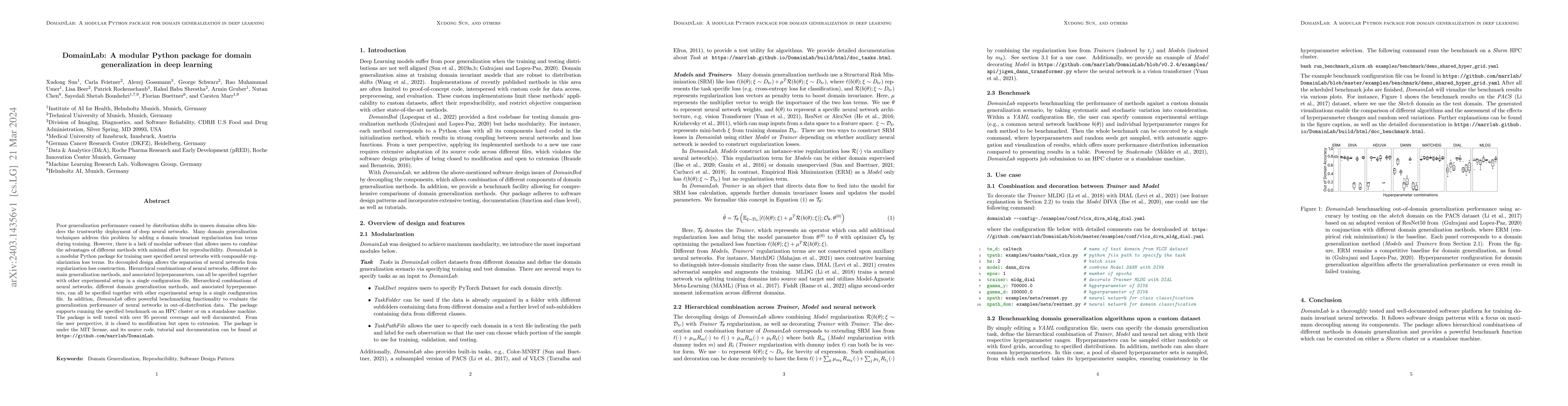
Papers on arXiv
Diagnosis of hematological malignancies depends on accurate identification of white blood cells in peripheral blood smears. Deep learning techniques are emerging as a viable solution to scale and op...
In hematology, computational models offer significant potential to improve diagnostic accuracy, streamline workflows, and reduce the tedious work of analyzing single cells in peripheral blood or bon...
Poor generalization performance caused by distribution shifts in unseen domains often hinders the trustworthy deployment of deep neural networks. Many domain generalization techniques address this p...
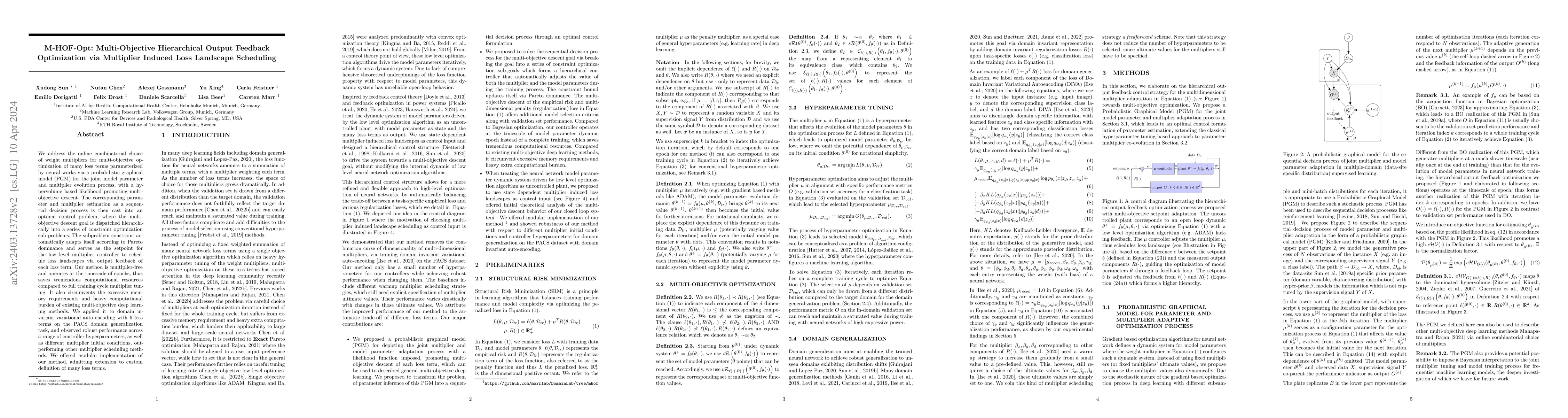
We address the online combinatorial choice of weight multipliers for multi-objective optimization of many loss terms parameterized by neural works via a probabilistic graphical model (PGM) for the j...
Automated disease diagnosis using medical image analysis relies on deep learning, often requiring large labeled datasets for supervised model training. Diseases like Acute Myeloid Leukemia (AML) pos...
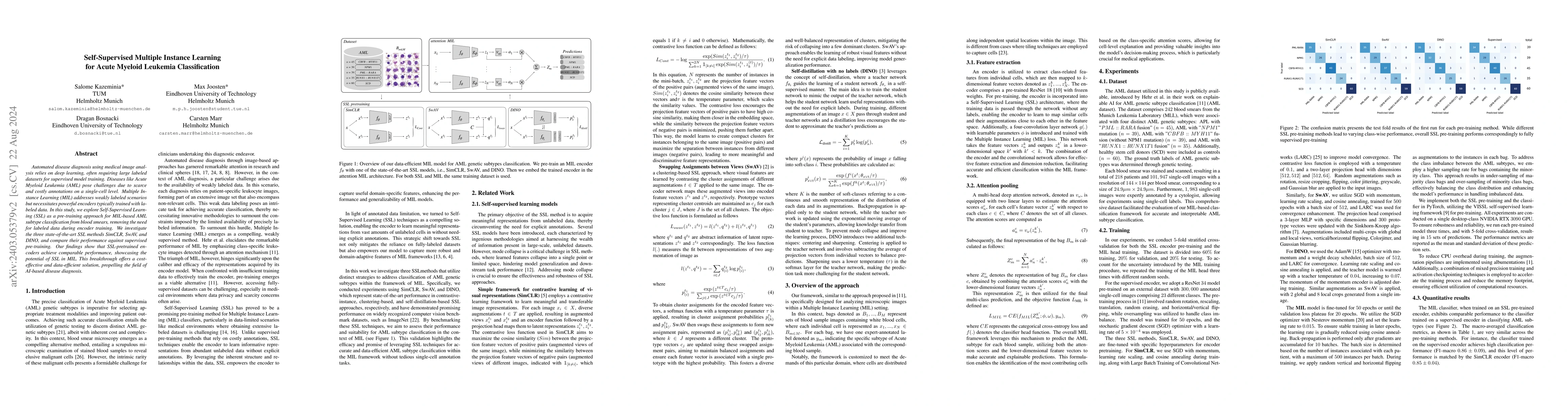
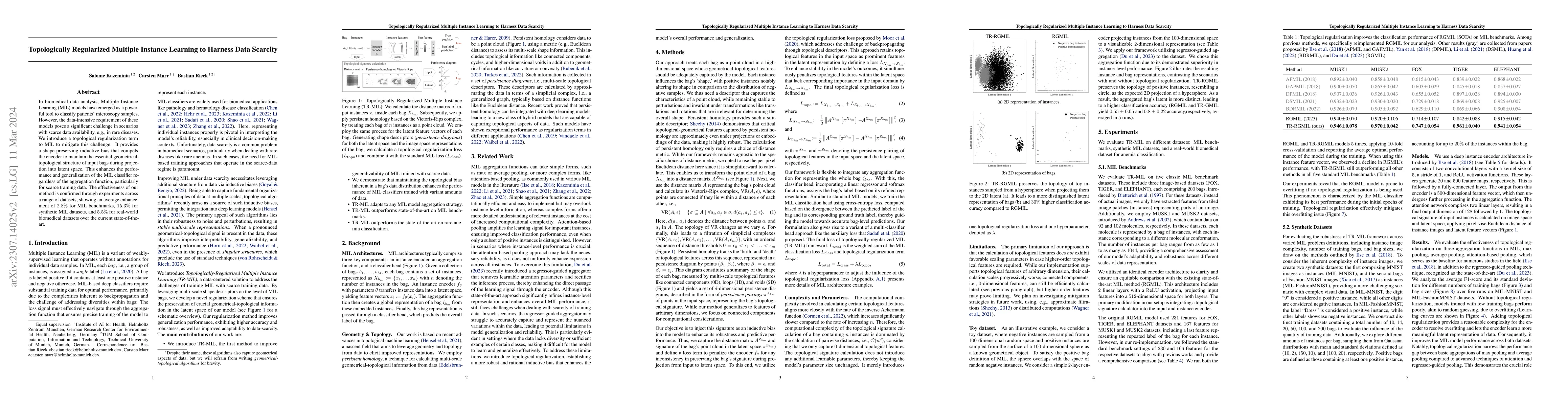
To handle the large scale of whole slide images in computational pathology, most approaches first tessellate the images into smaller patches, extract features from these patches, and finally aggrega...
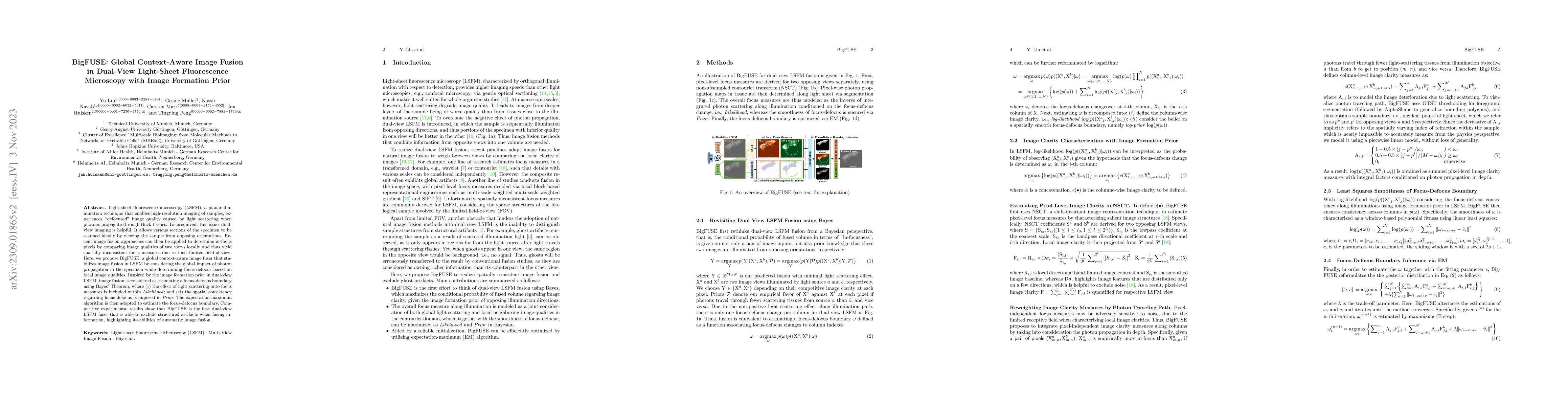
Light-sheet fluorescence microscopy (LSFM), a planar illumination technique that enables high-resolution imaging of samples, experiences defocused image quality caused by light scattering when photo...
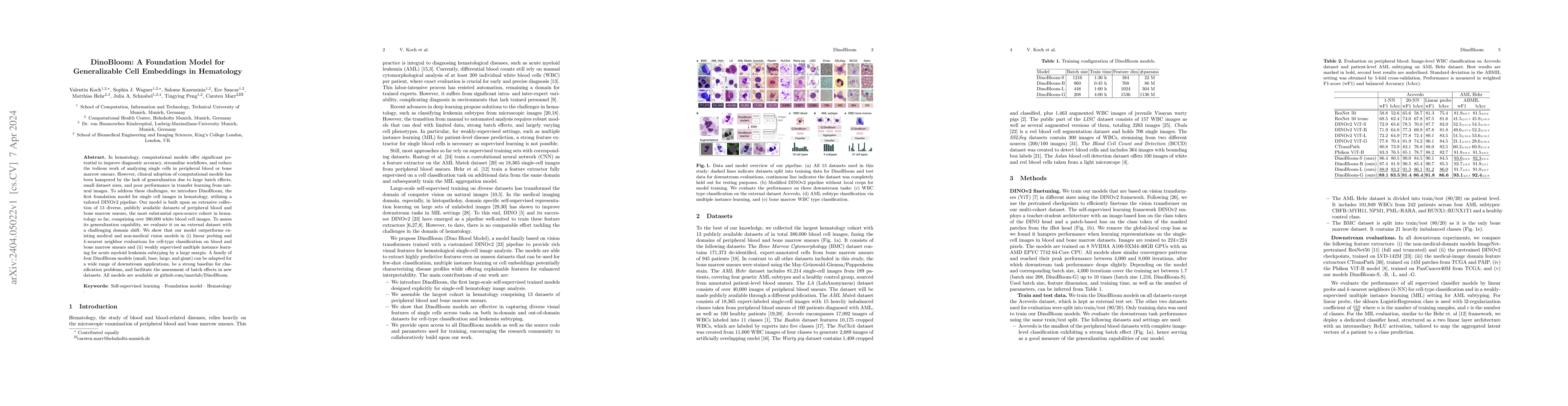
Accurate classification of white blood cells in peripheral blood is essential for diagnosing hematological diseases. Due to constantly evolving clinical settings, data sources, and disease classific...
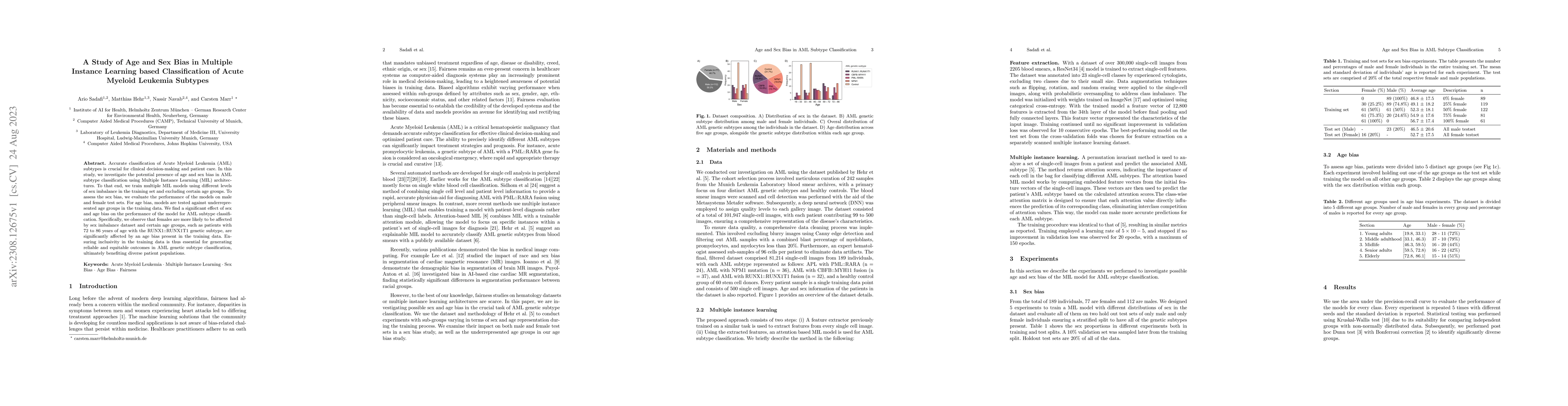
Accurate classification of Acute Myeloid Leukemia (AML) subtypes is crucial for clinical decision-making and patient care. In this study, we investigate the potential presence of age and sex bias in...
In biomedical data analysis, Multiple Instance Learning (MIL) models have emerged as a powerful tool to classify patients' microscopy samples. However, the data-intensive requirement of these models...
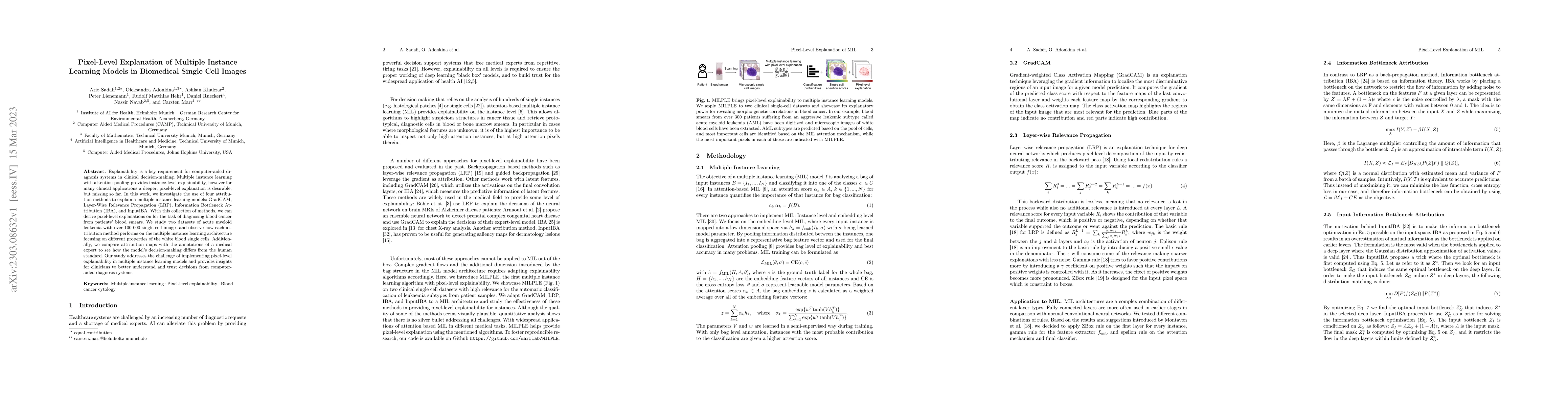
Explainability is a key requirement for computer-aided diagnosis systems in clinical decision-making. Multiple instance learning with attention pooling provides instance-level explainability, howeve...
Accurate morphological classification of white blood cells (WBCs) is an important step in the diagnosis of leukemia, a disease in which nonfunctional blast cells accumulate in the bone marrow. Recen...
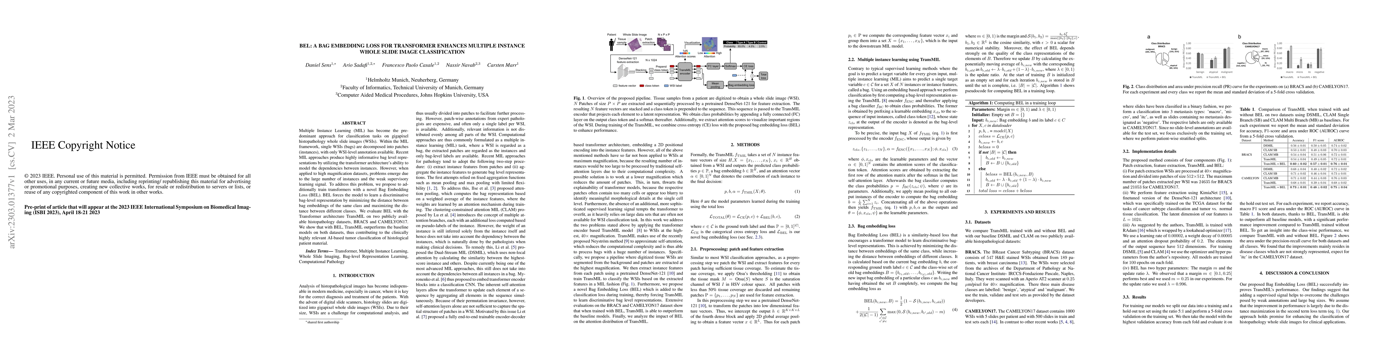
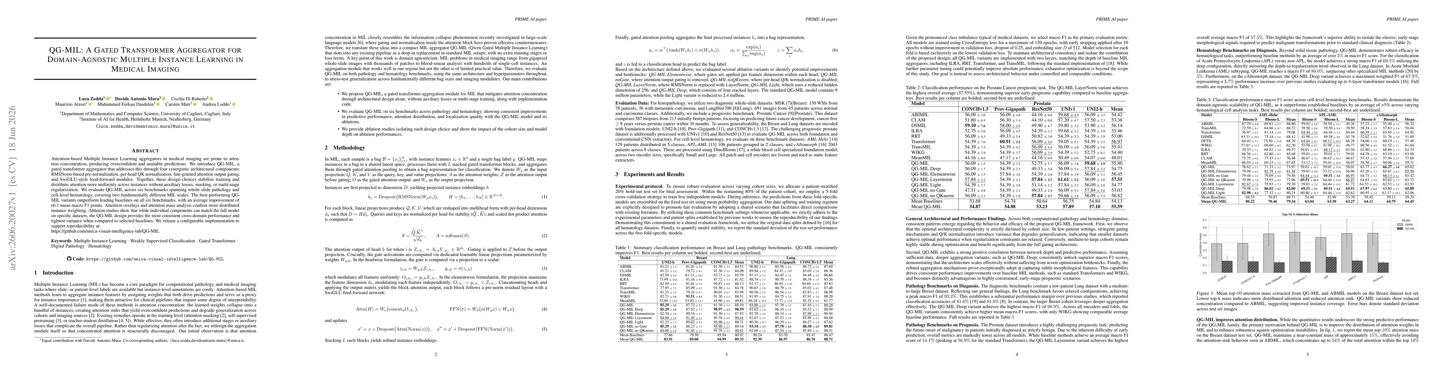
Multiple Instance Learning (MIL) has become the predominant approach for classification tasks on gigapixel histopathology whole slide images (WSIs). Within the MIL framework, single WSIs (bags) are ...
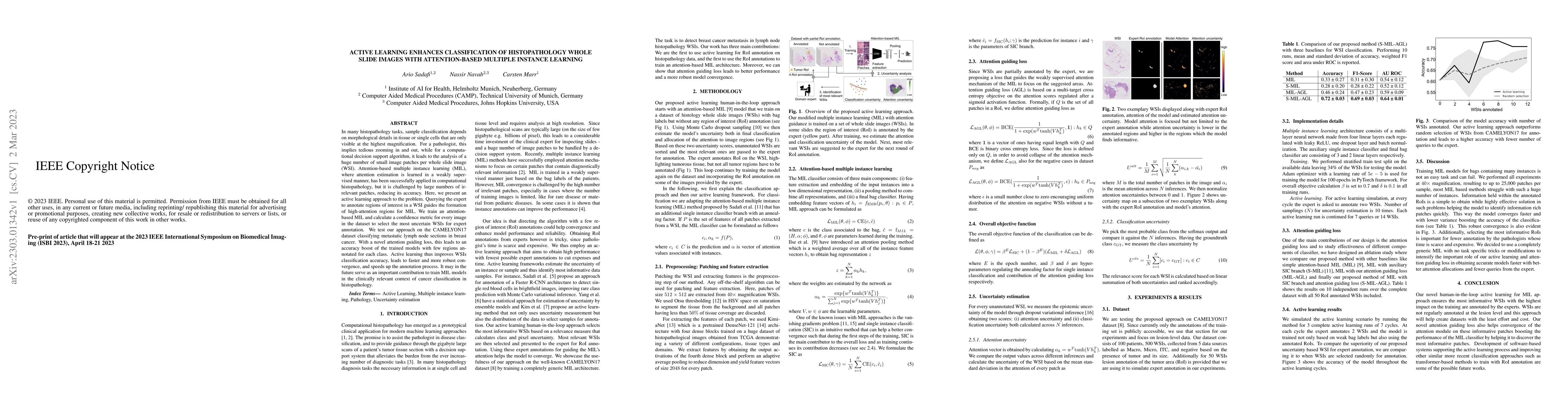
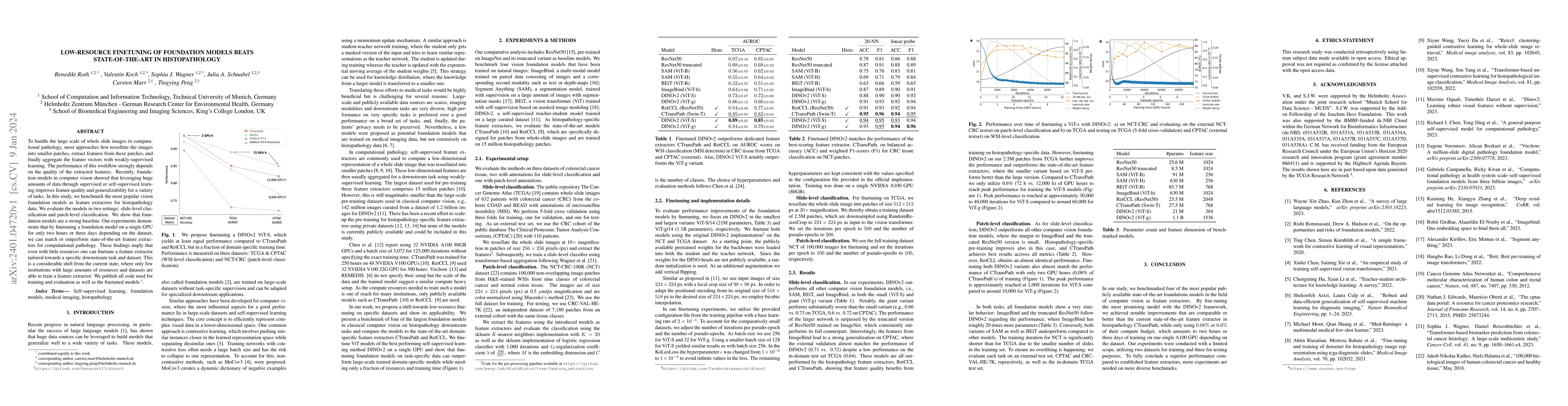
In many histopathology tasks, sample classification depends on morphological details in tissue or single cells that are only visible at the highest magnification. For a pathologist, this implies ted...
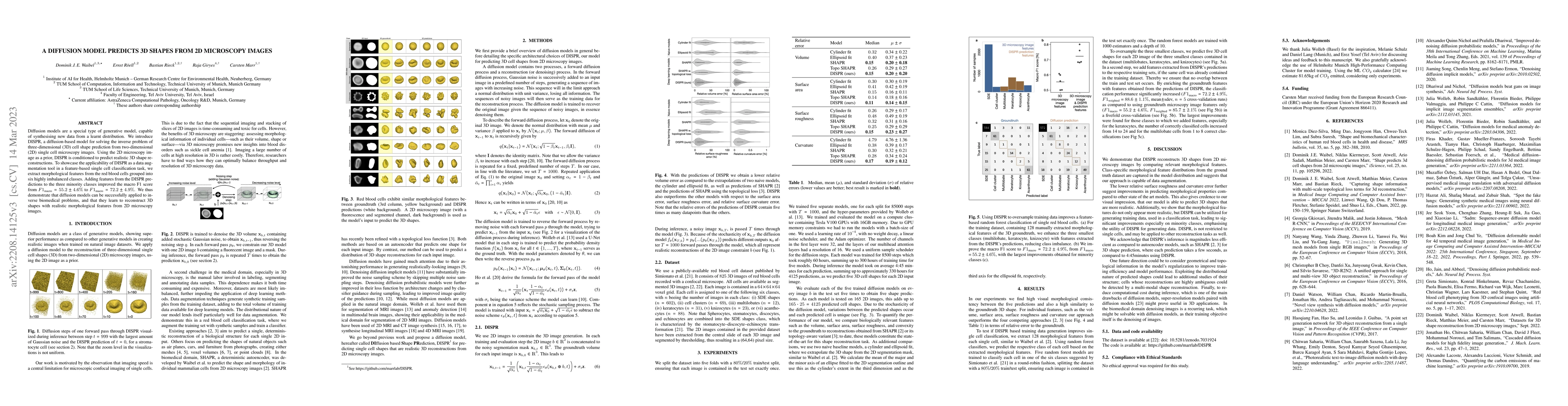
Diffusion models are a special type of generative model, capable of synthesising new data from a learnt distribution. We introduce DISPR, a diffusion-based model for solving the inverse problem of t...
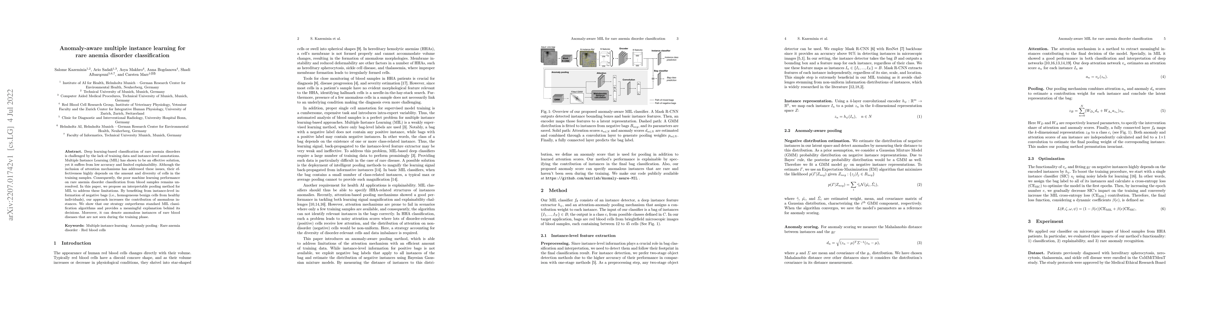
Deep learning-based classification of rare anemia disorders is challenged by the lack of training data and instance-level annotations. Multiple Instance Learning (MIL) has shown to be an effective s...
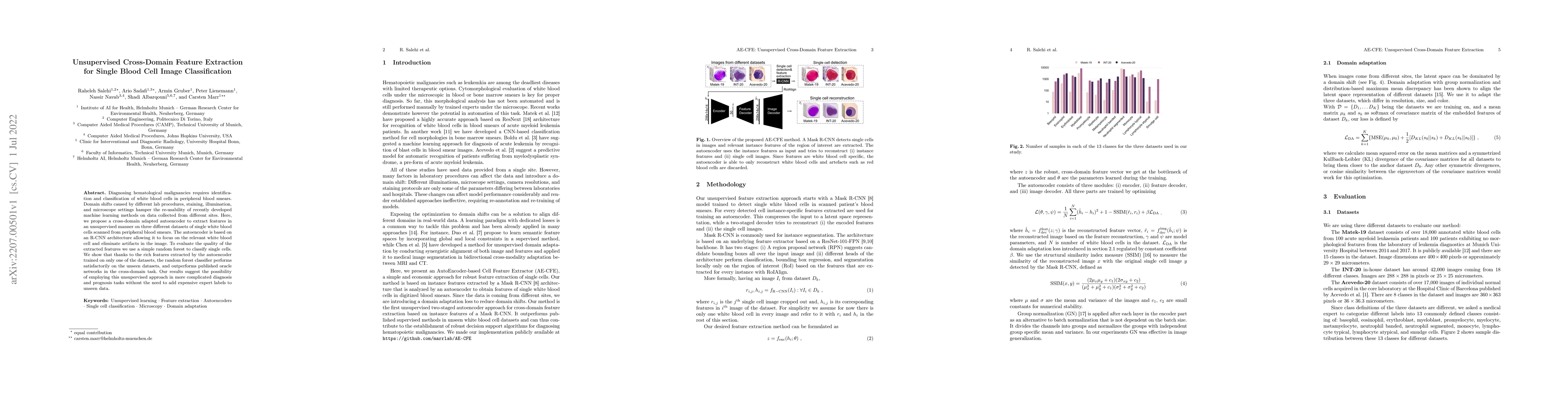
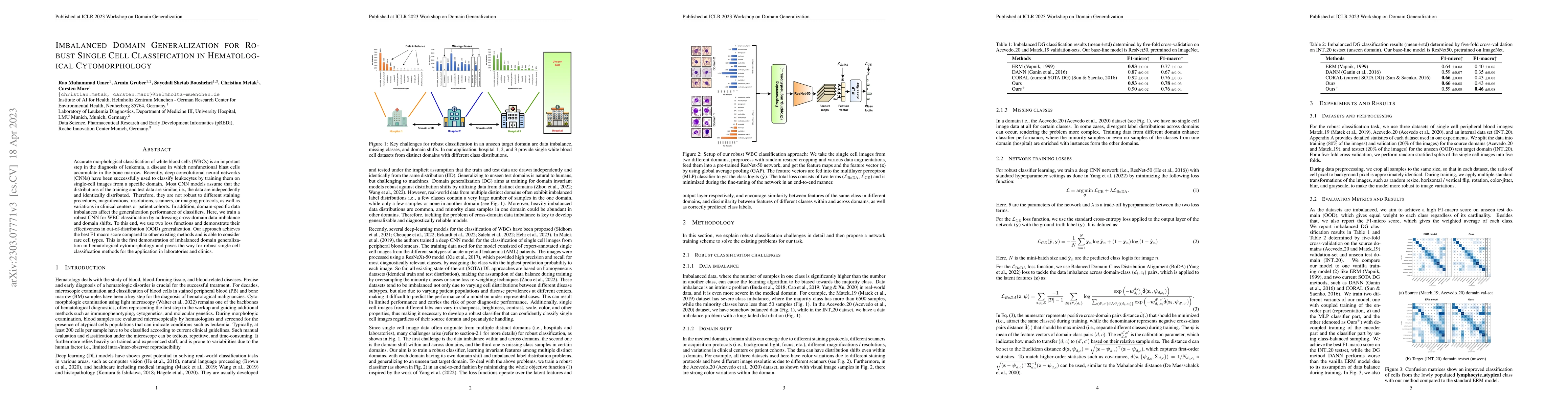
Diagnosing hematological malignancies requires identification and classification of white blood cells in peripheral blood smears. Domain shifts caused by different lab procedures, staining, illumina...
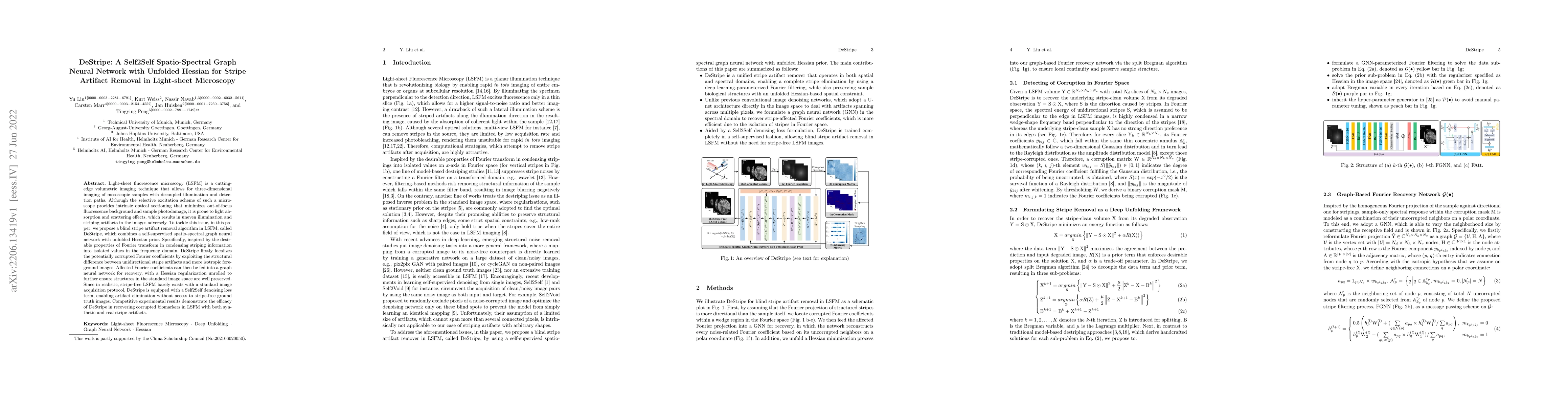
Light-sheet fluorescence microscopy (LSFM) is a cutting-edge volumetric imaging technique that allows for three-dimensional imaging of mesoscopic samples with decoupled illumination and detection pa...
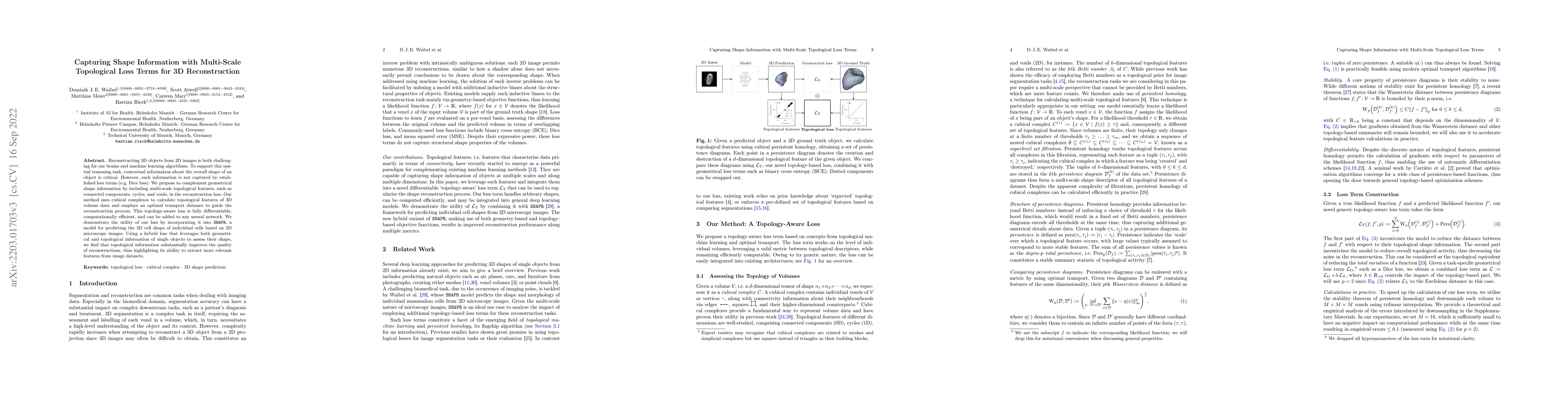
Reconstructing 3D objects from 2D images is both challenging for our brains and machine learning algorithms. To support this spatial reasoning task, contextual information about the overall shape of...
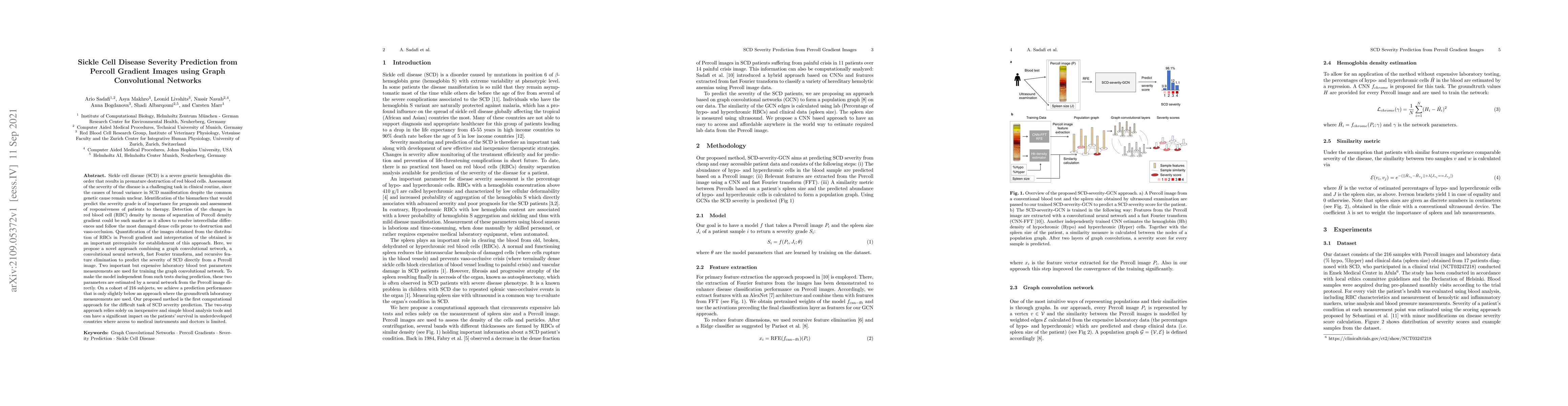
Sickle cell disease (SCD) is a severe genetic hemoglobin disorder that results in premature destruction of red blood cells. Assessment of the severity of the disease is a challenging task in clinica...
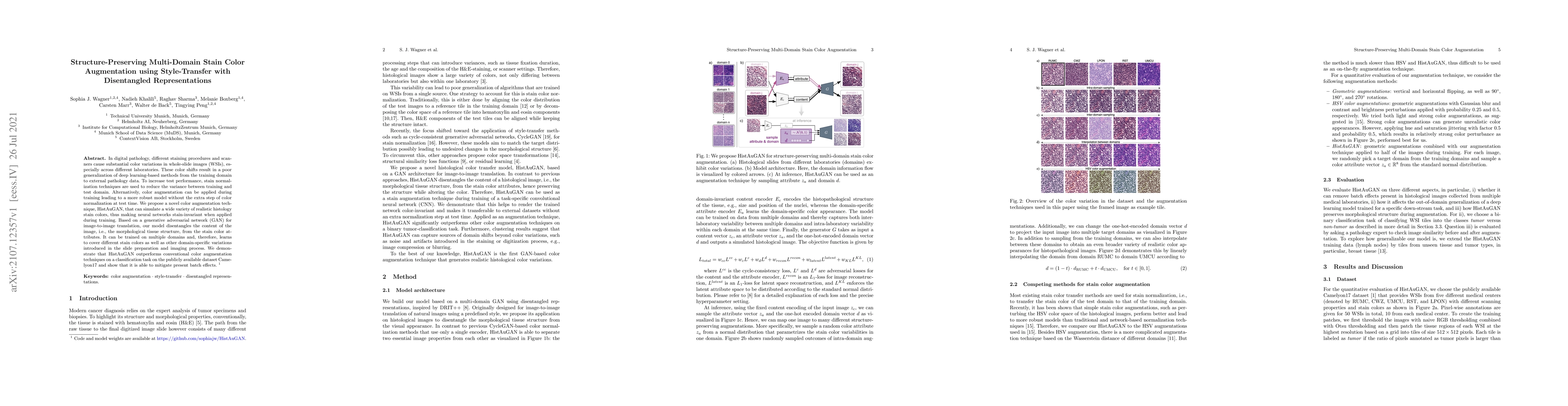
In digital pathology, different staining procedures and scanners cause substantial color variations in whole-slide images (WSIs), especially across different laboratories. These color shifts result ...
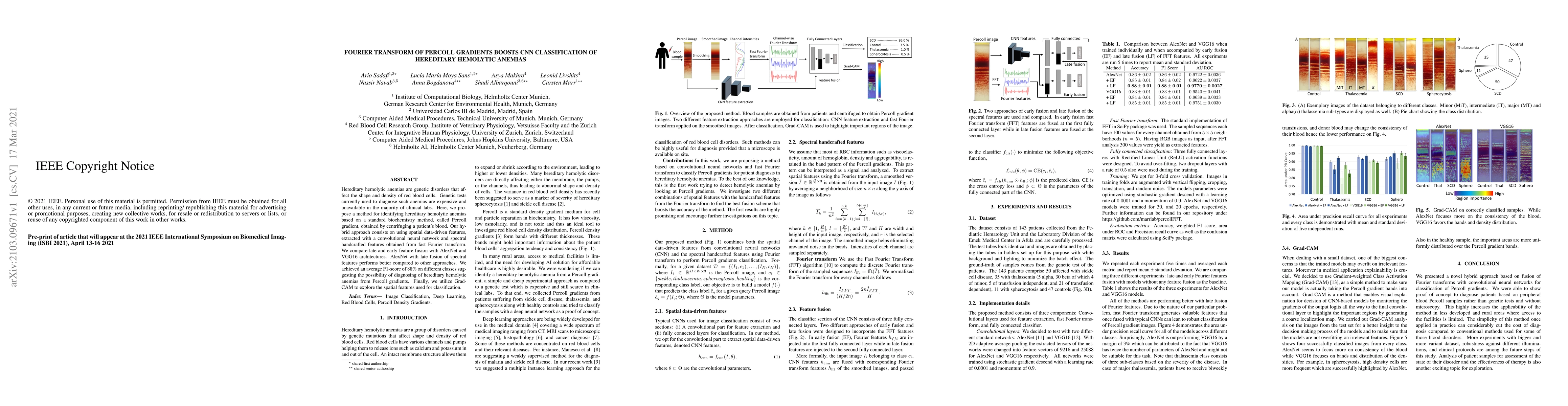
Hereditary hemolytic anemias are genetic disorders that affect the shape and density of red blood cells. Genetic tests currently used to diagnose such anemias are expensive and unavailable in the ma...
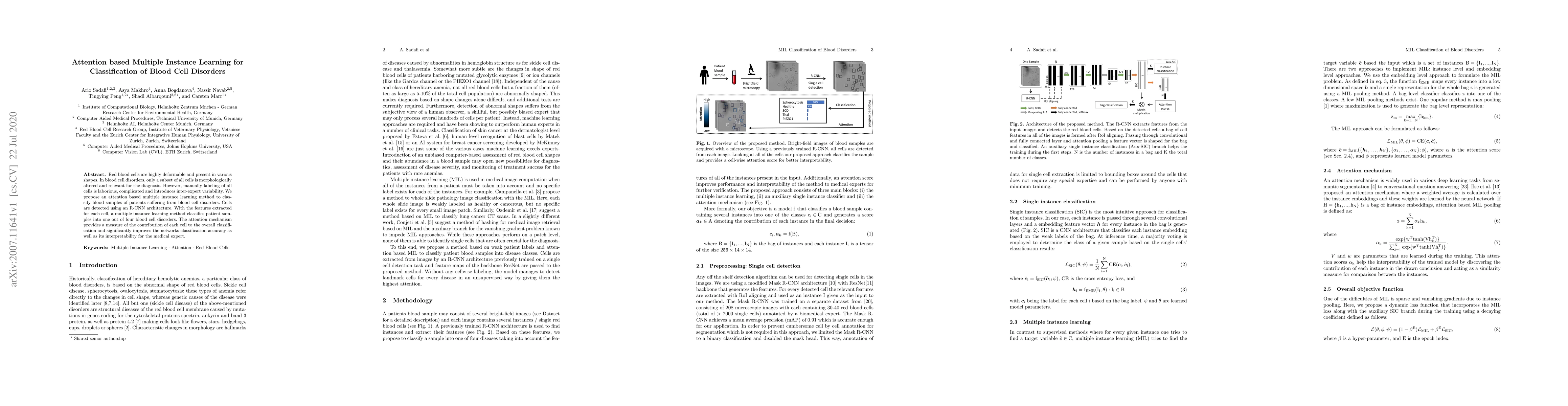
Red blood cells are highly deformable and present in various shapes. In blood cell disorders, only a subset of all cells is morphologically altered and relevant for the diagnosis. However, manually ...
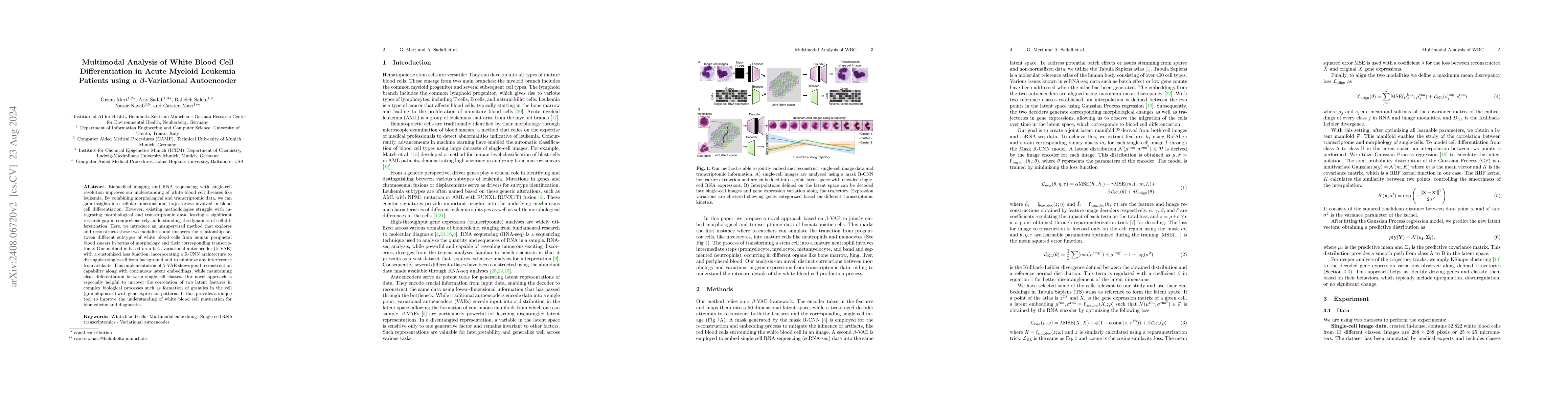
Biomedical imaging and RNA sequencing with single-cell resolution improves our understanding of white blood cell diseases like leukemia. By combining morphological and transcriptomic data, we can gain...
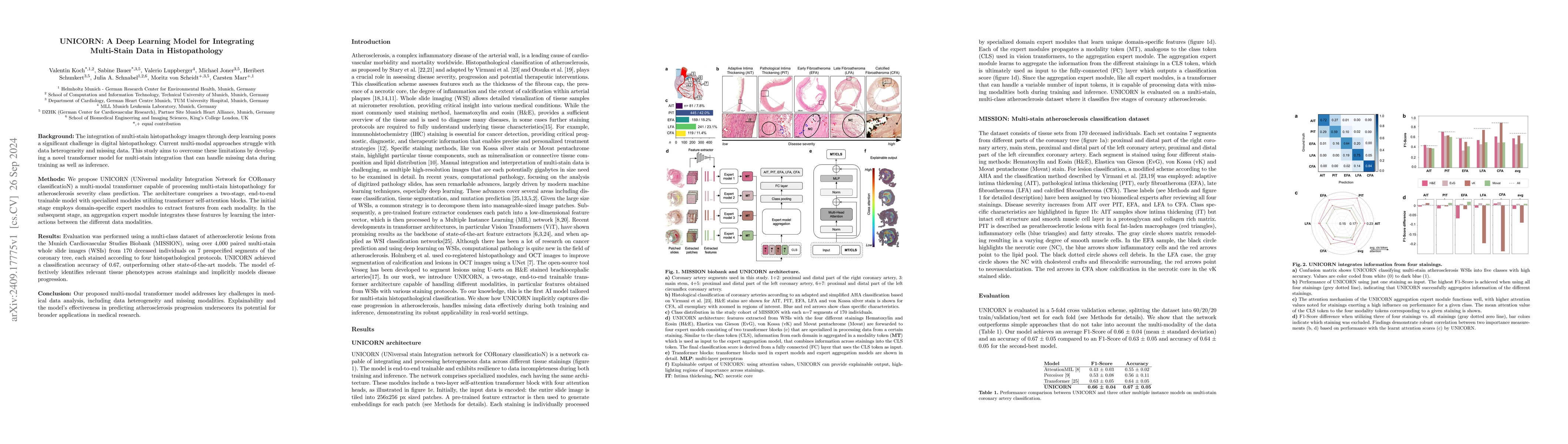
Background: The integration of multi-stain histopathology images through deep learning poses a significant challenge in digital histopathology. Current multi-modal approaches struggle with data hetero...
Histopathology, the microscopic study of diseased tissue, is increasingly digitized, enabling improved visualization and streamlined workflows. An important task in histopathology is the segmentation ...
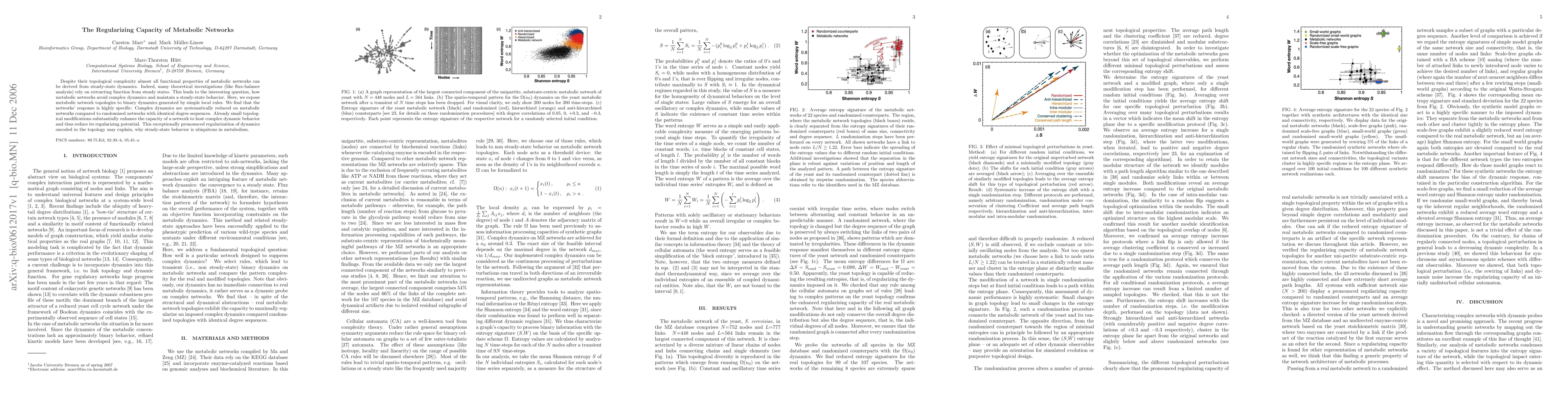
Despite their topological complexity almost all functional properties of metabolic networks can be derived from steady-state dynamics. Indeed, many theoretical investigations (like flux-balance analys...
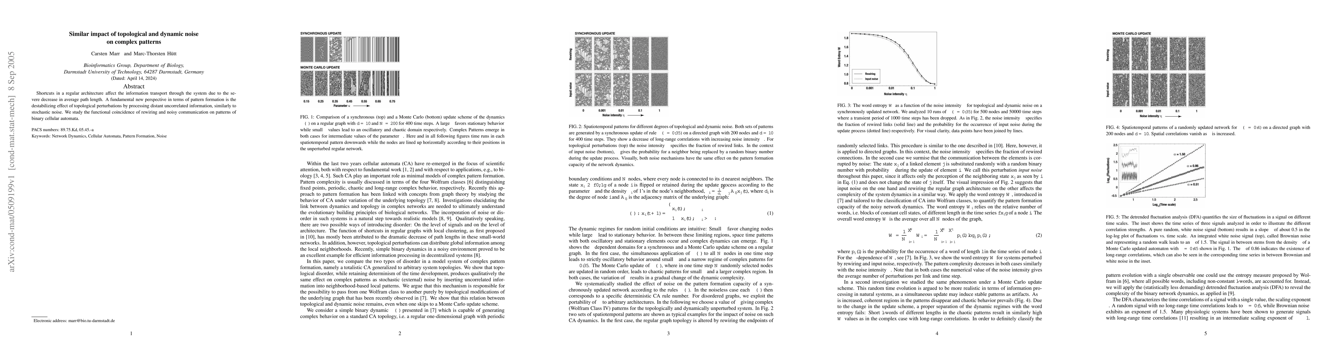
Shortcuts in a regular architecture affect the information transport through the system due to the severe decrease in average path length. A fundamental new perspective in terms of pattern formation i...
We study the effect of topology variation on the dynamic behavior of a system with local update rules. We implement one-dimensional binary cellular automata on graphs with various topologies by formul...
Unbiased data synthesis is crucial for evaluating causal discovery algorithms in the presence of unobserved confounding, given the scarcity of real-world datasets. A common approach, implicit paramete...
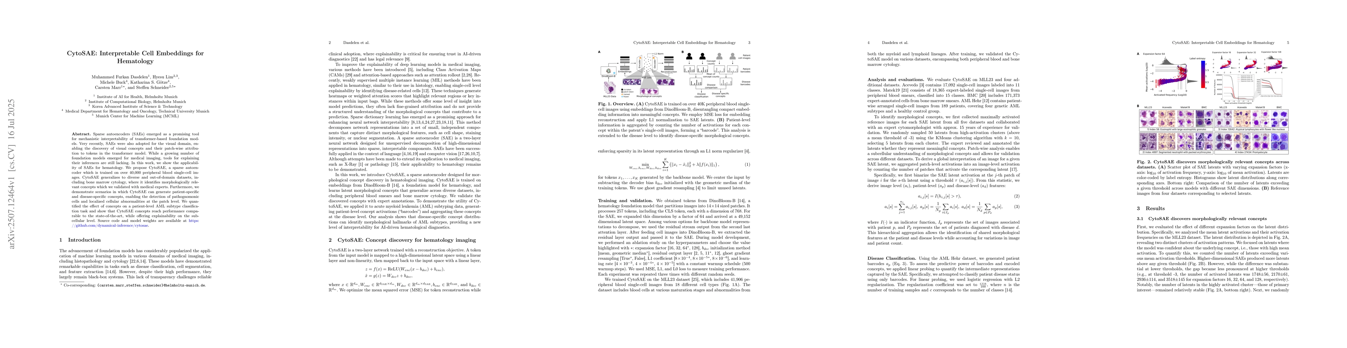
Sparse autoencoders (SAEs) emerged as a promising tool for mechanistic interpretability of transformer-based foundation models. Very recently, SAEs were also adopted for the visual domain, enabling th...
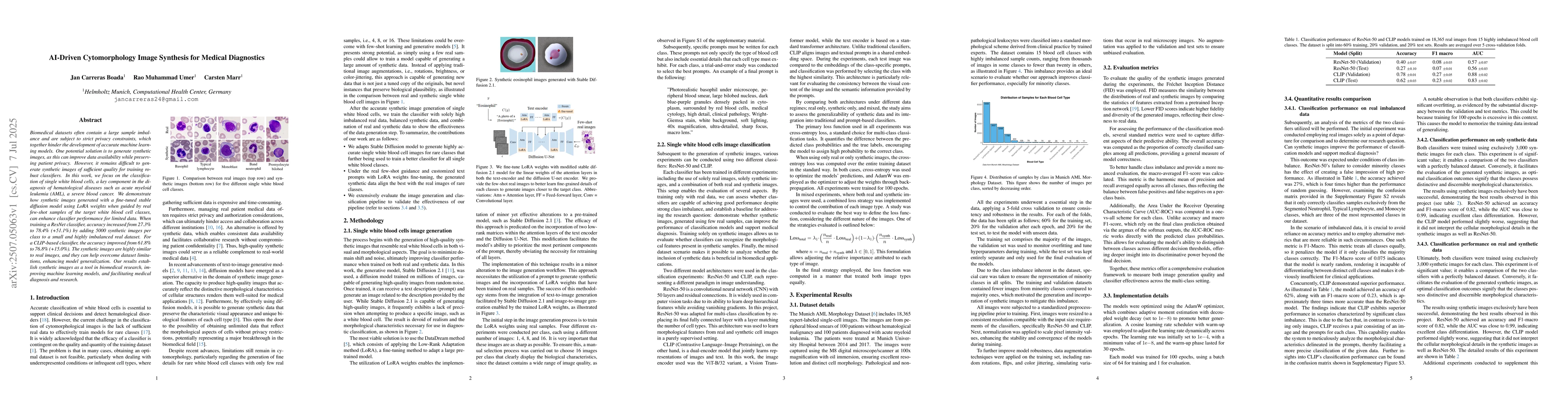
Biomedical datasets often contain a large sample imbalance and are subject to strict privacy constraints, which together hinder the development of accurate machine learning models. One potential solut...
Multiparameter persistence module can capture more topological differences across data instances compared to using a single parameter, where the well-studied matching distance investigates the distanc...
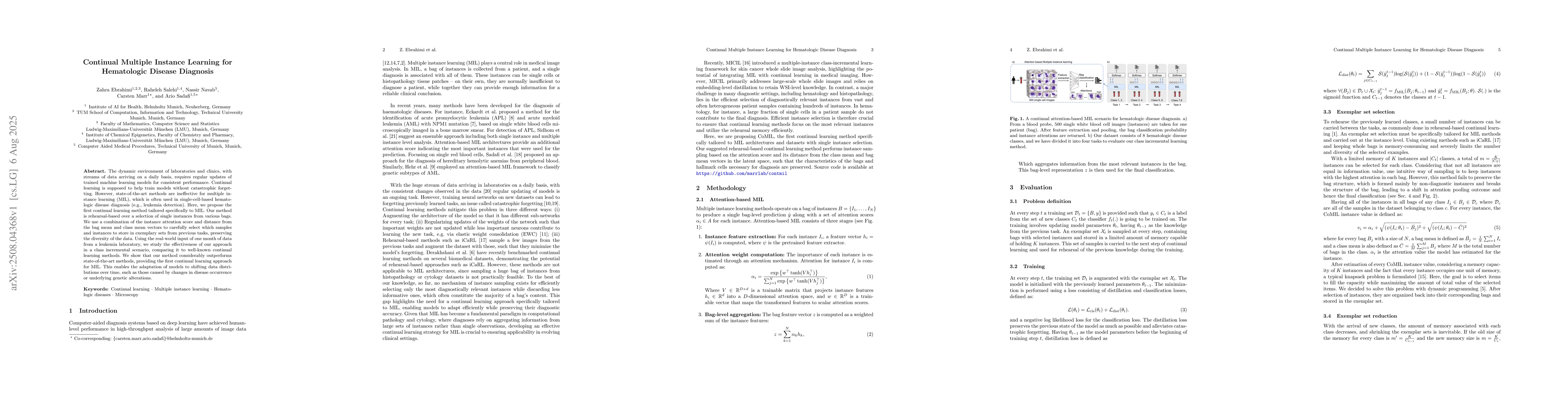
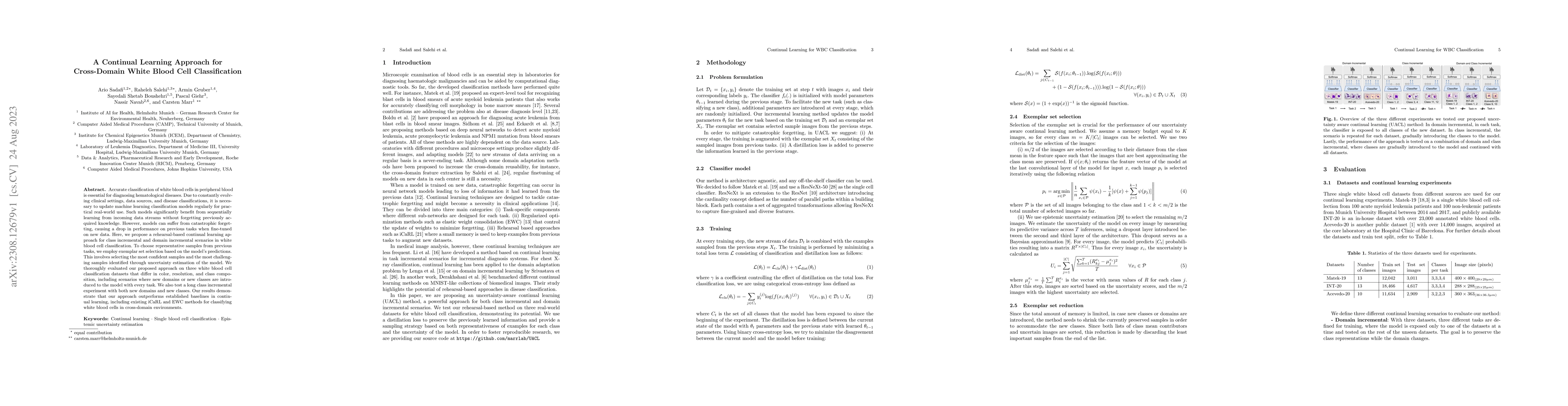
The dynamic environment of laboratories and clinics, with streams of data arriving on a daily basis, requires regular updates of trained machine learning models for consistent performance. Continual l...
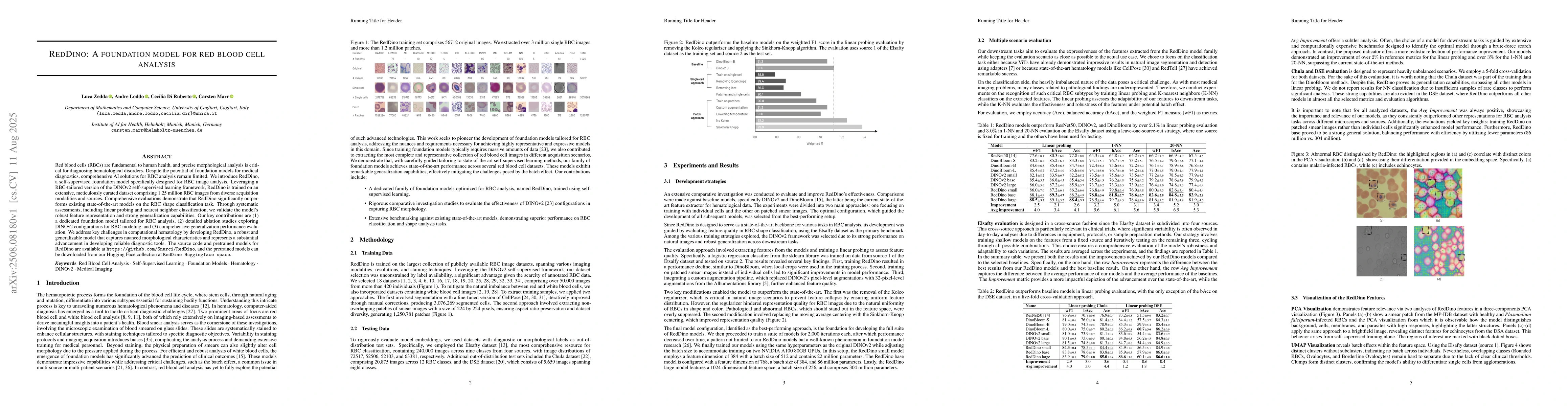
Red blood cells (RBCs) are essential to human health, and their precise morphological analysis is important for diagnosing hematological disorders. Despite the promise of foundation models in medical ...
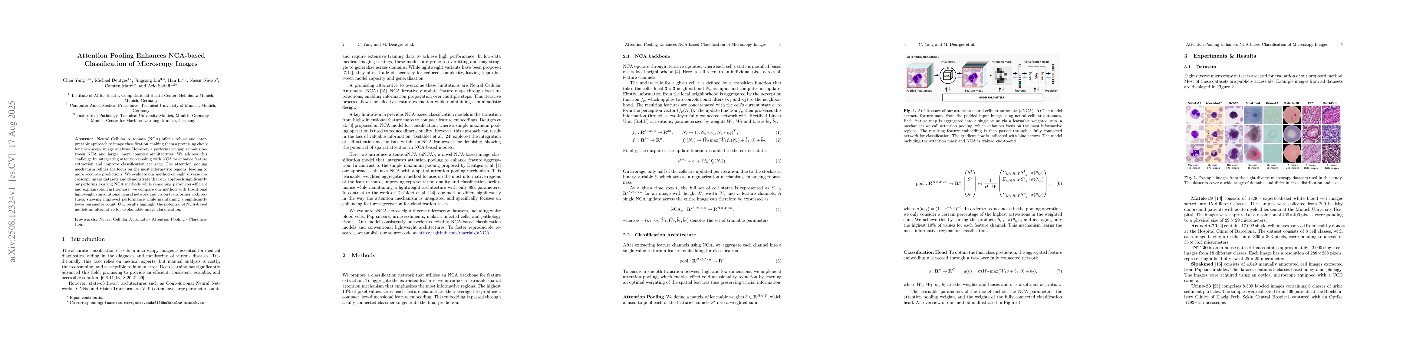
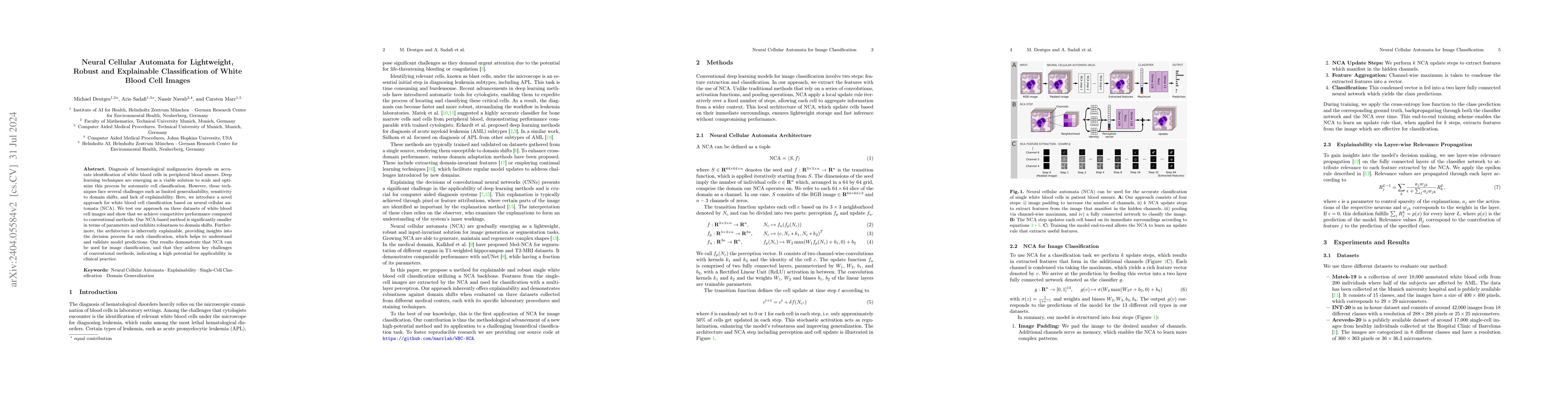
Neural Cellular Automata (NCA) offer a robust and interpretable approach to image classification, making them a promising choice for microscopy image analysis. However, a performance gap remains betwe...
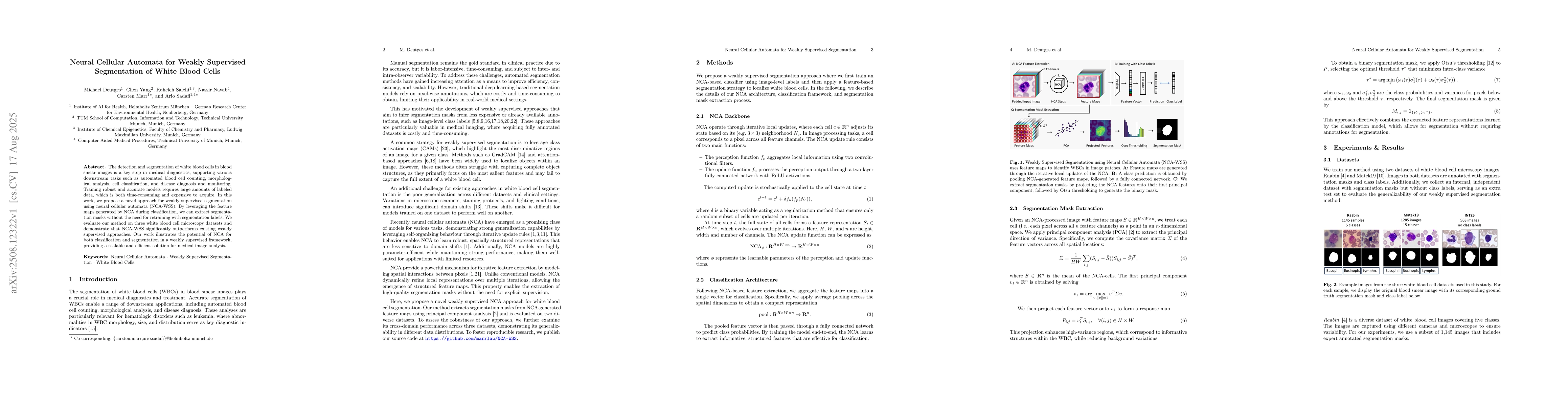
The detection and segmentation of white blood cells in blood smear images is a key step in medical diagnostics, supporting various downstream tasks such as automated blood cell counting, morphological...
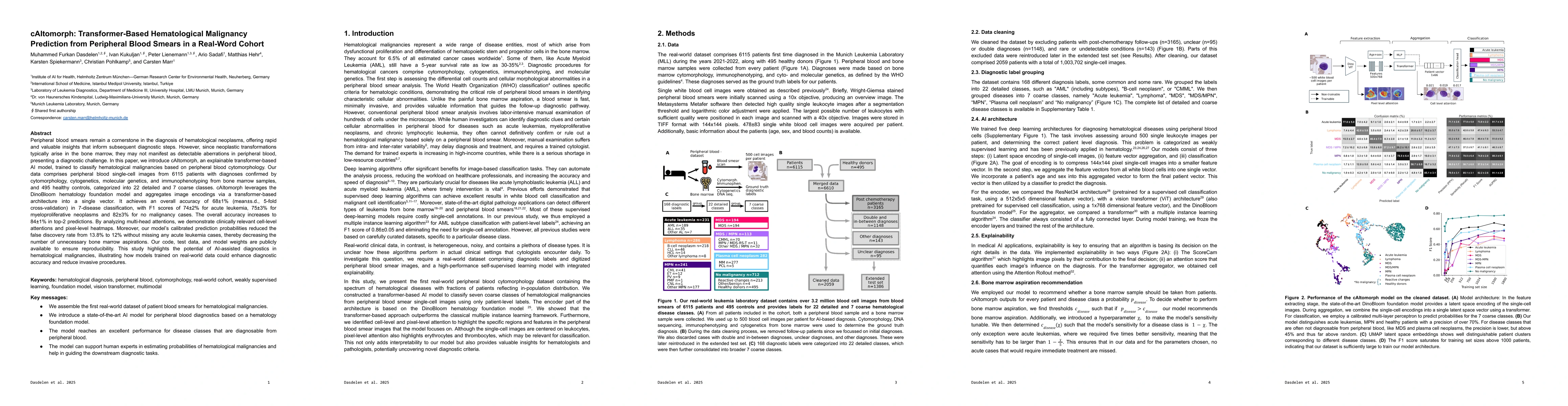
Peripheral blood smears remain a cornerstone in the diagnosis of hematological neoplasms, offering rapid and valuable insights that inform subsequent diagnostic steps. However, since neoplastic transf...
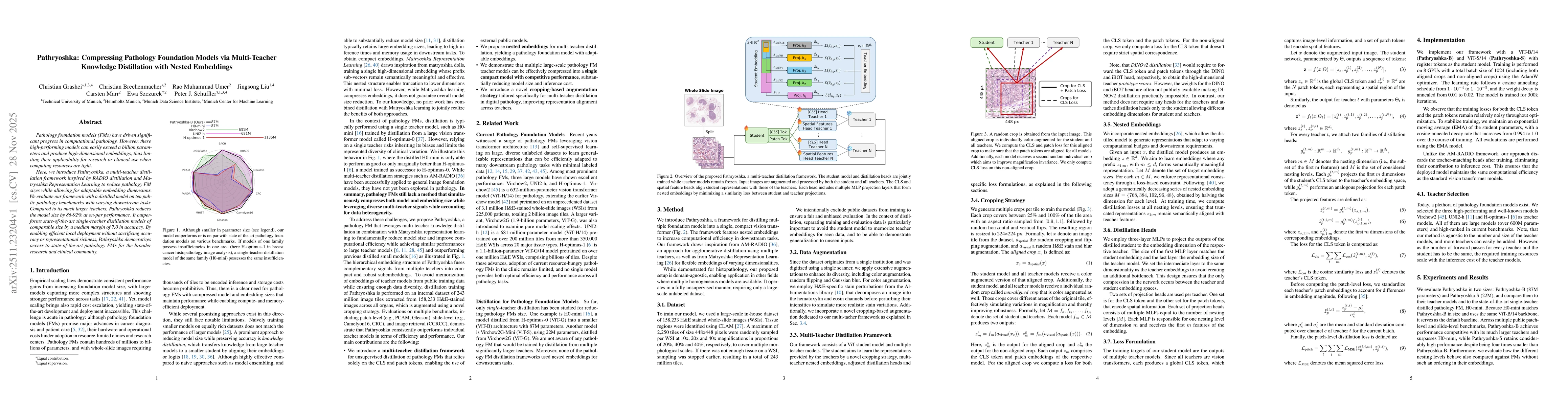
Pathology foundation models (FMs) have driven significant progress in computational pathology. However, these high-performing models can easily exceed a billion parameters and produce high-dimensional...
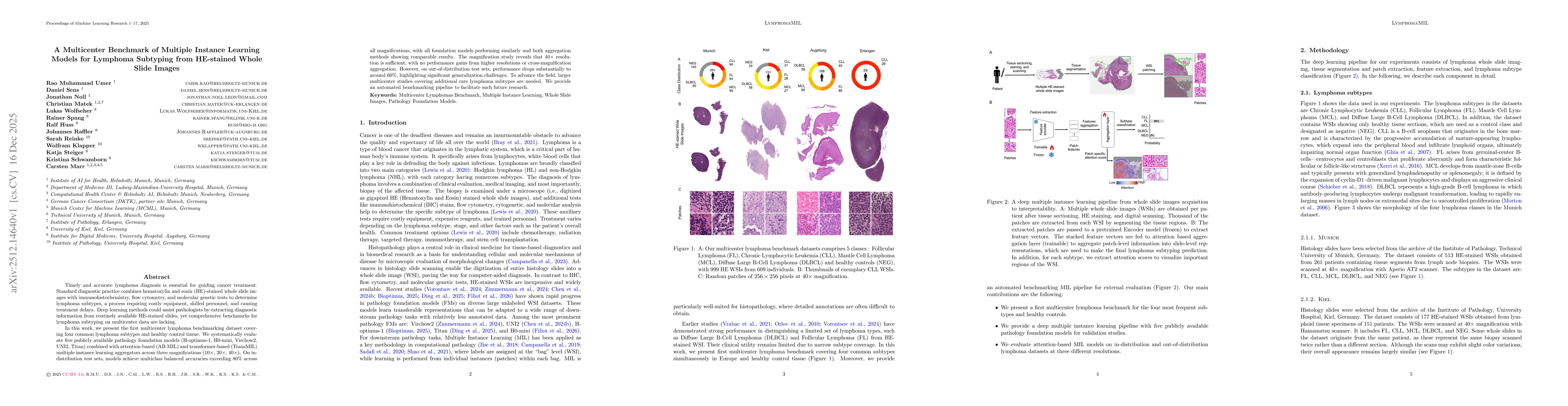
Timely and accurate lymphoma diagnosis is essential for guiding cancer treatment. Standard diagnostic practice combines hematoxylin and eosin (HE)-stained whole slide images with immunohistochemistry,...
Shortcuts in a regular architecture affect the information transport through the system due to the severe decrease in average path length. A fundamental new perspective in terms of pattern formation i...
We study the effect of topology variation on the dynamic behavior of a system with local update rules. We implement one-dimensional binary cellular automata on graphs with various topologies by formul...
Despite their topological complexity almost all functional properties of metabolic networks can be derived from steady-state dynamics. Indeed, many theoretical investigations (like flux-balance analys...
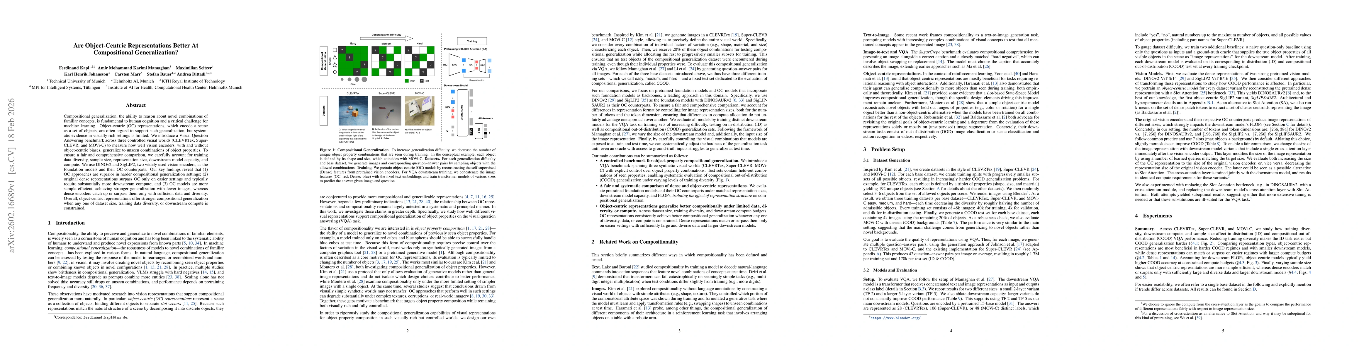
Compositional generalization, the ability to reason about novel combinations of familiar concepts, is fundamental to human cognition and a critical challenge for machine learning. Object-centric (OC) ...
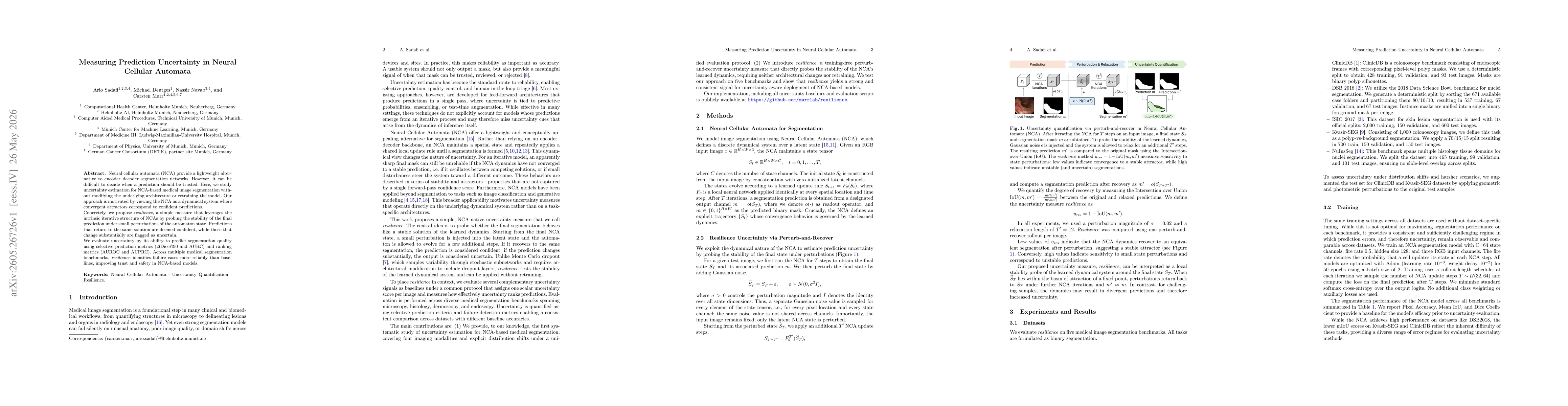
Neural cellular automata (NCA) provide a lightweight alternative to encoder-decoder segmentation networks. However, it can be difficult to decide when a prediction should be trusted. Here, we study un...
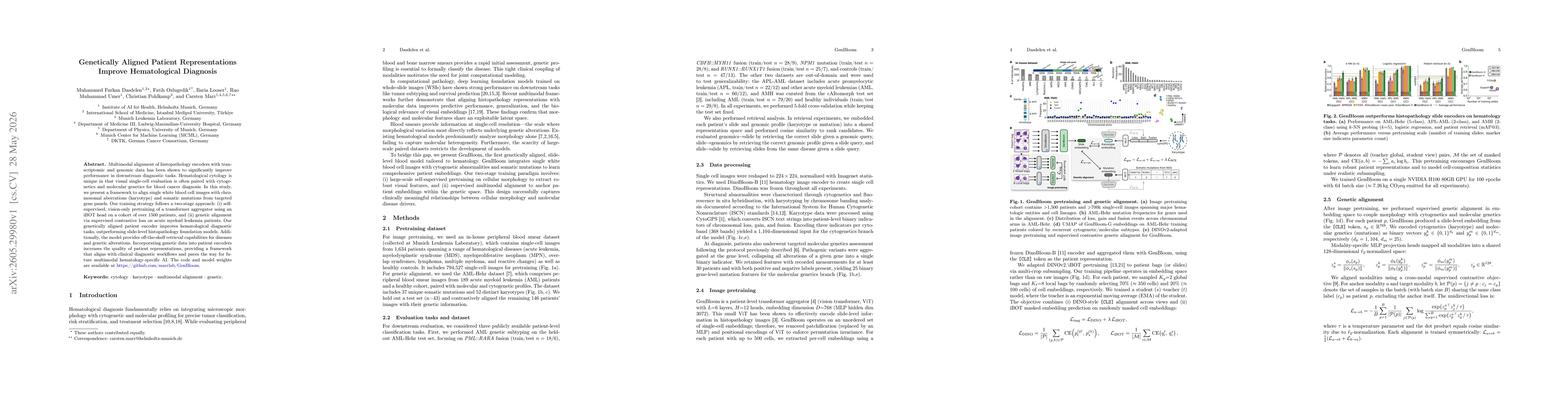
Multimodal alignment of histopathology encoders with transcriptomic and genomic data has been shown to significantly improve performance in downstream diagnostic tasks. Hematological cytology is uniqu...
Attention-based Multiple Instance Learning aggregators in medical imaging are prone to attention concentration, producing overconfident and unstable predictions. We introduce QG-MIL, a gated transform...
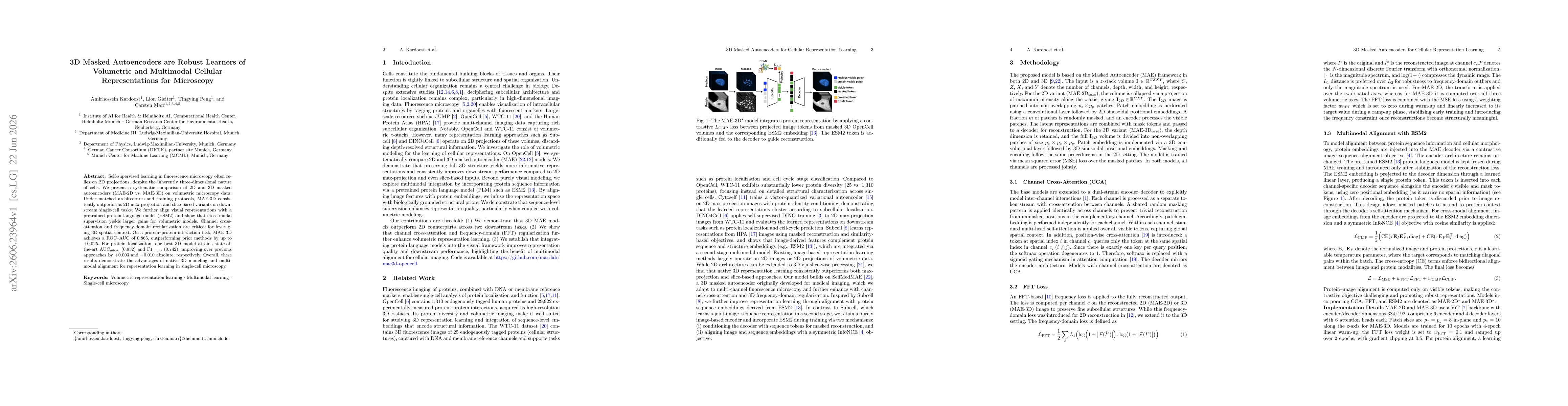
Self-supervised learning in fluorescence microscopy often relies on 2D projections, despite the inherently three-dimensional nature of cells. We present a systematic comparison of 2D and 3D masked aut...
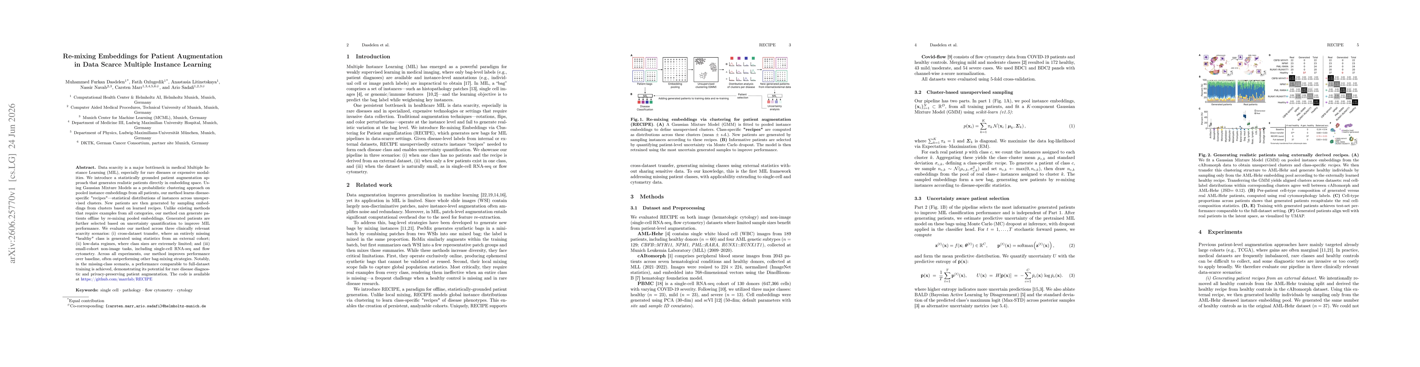
Data scarcity is a major bottleneck in medical Multiple Instance Learning (MIL), especially for rare diseases or expensive modalities. We introduce a statistically grounded patient augmentation approa...
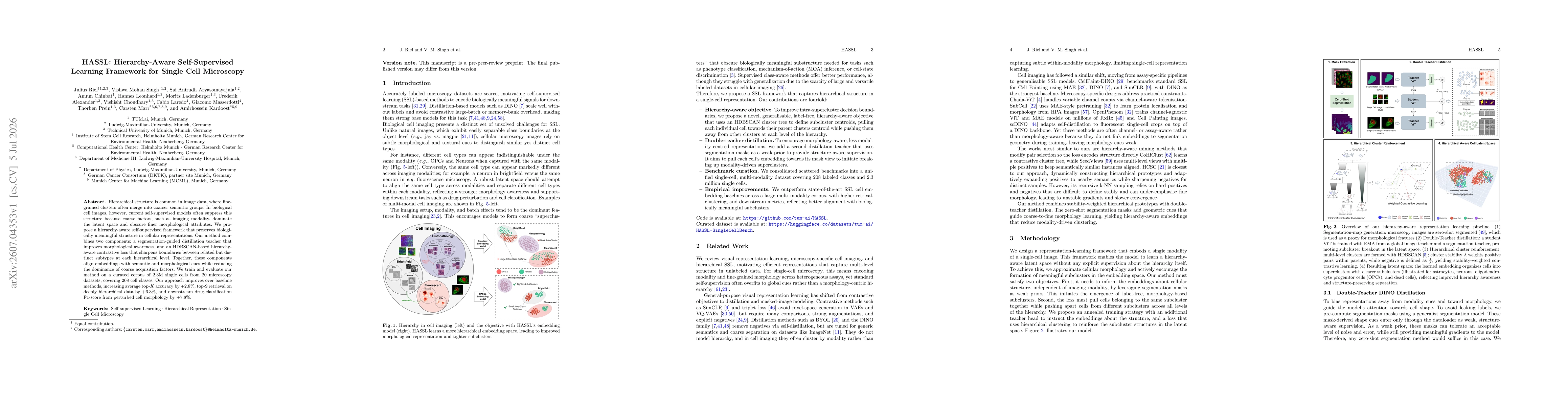
Hierarchical structure is common in image data, where fine-grained clusters often merge into larger, coarser semantic groups. In biological cell images, current self-supervised learning models often s...