A Diffusion Model Predicts 3D Shapes from 2D Microscopy Images
Publication
Metrics
AI Quick Summary
This paper introduces DISPR, a diffusion-based model for predicting 3D cell shapes from 2D microscopy images, demonstrating its effectiveness in reconstructing realistic 3D shapes. The model's features, when added to minority classes, significantly improved macro F1 score in a single cell classification task, showcasing its potential as a data augmentation tool.
Paper Preview
Abstract
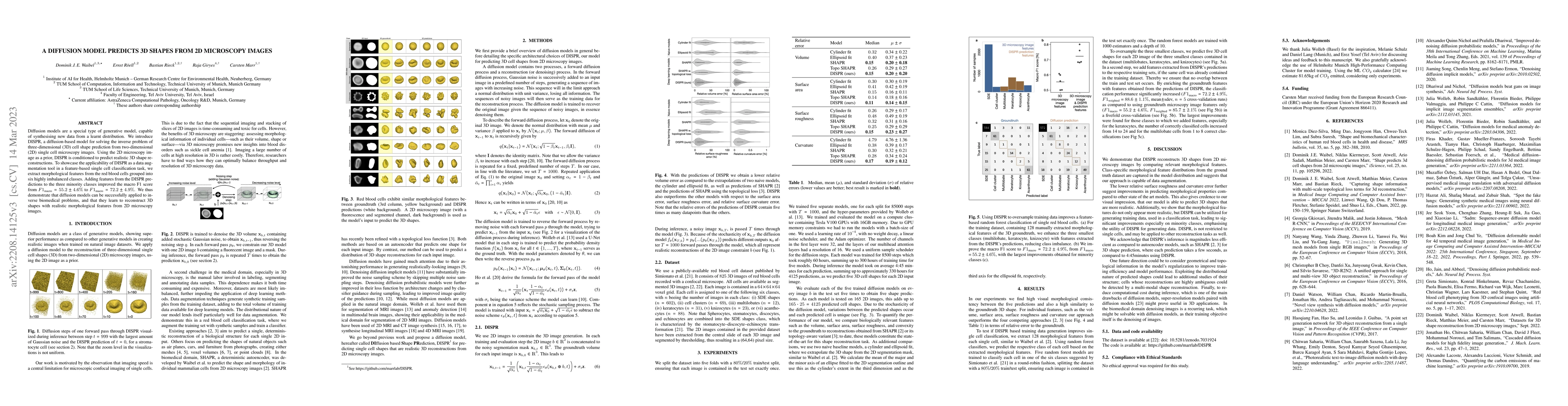
Diffusion models are a special type of generative model, capable of synthesising new data from a learnt distribution. We introduce DISPR, a diffusion-based model for solving the inverse problem of three-dimensional (3D) cell shape prediction from two-dimensional (2D) single cell microscopy images. Using the 2D microscopy image as a prior, DISPR is conditioned to predict realistic 3D shape reconstructions. To showcase the applicability of DISPR as a data augmentation tool in a feature-based single cell classification task, we extract morphological features from the red blood cells grouped into six highly imbalanced classes. Adding features from the DISPR predictions to the three minority classes improved the macro F1 score from $F1_\text{macro} = 55.2 \pm 4.6\%$ to $F1_\text{macro} = 72.2 \pm 4.9\%$. We thus demonstrate that diffusion models can be successfully applied to inverse biomedical problems, and that they learn to reconstruct 3D shapes with realistic morphological features from 2D microscopy images.
AI Key Findings
Get AI-generated insights about this paper's methodology, results, significance, and more — seven facets brought into focus.
Impact
Paper Details
Authors
PDF Preview
Key Terms
Citation Network
Current paper (gray), citations (green), references (blue)
Display is limited for performance on very large graphs.
Discussion 0