Publication
Metrics
AI Quick Summary
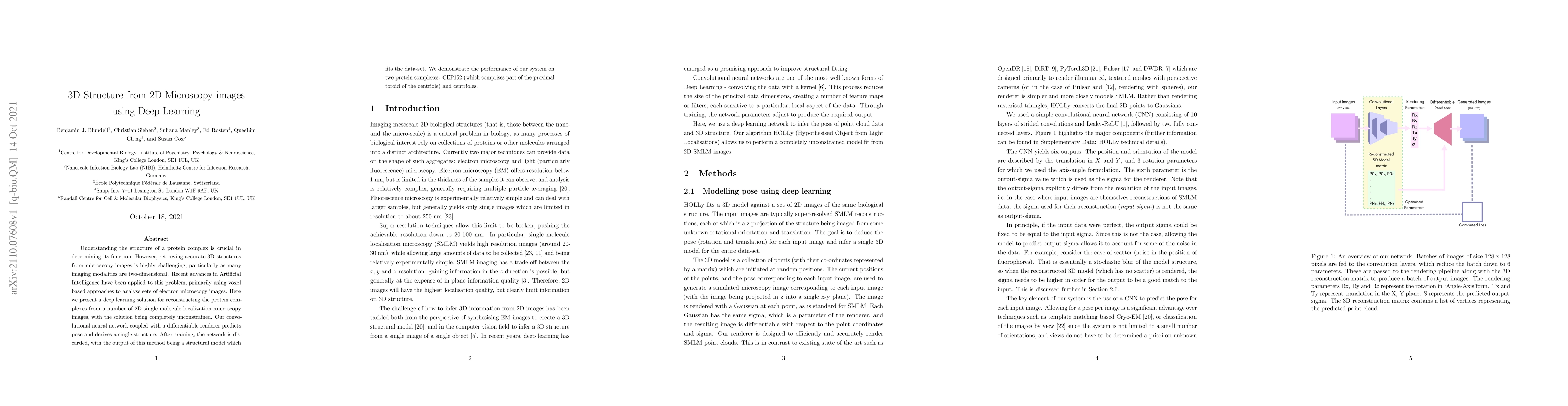
This paper proposes a deep learning method to reconstruct 3D protein complexes from 2D single-molecule localization microscopy images using a convolutional neural network and differentiable renderer. The trained network outputs a structural model that accurately fits the dataset, demonstrated on CEP152 and centrioles.
Paper Preview
Abstract
Understanding the structure of a protein complex is crucial indetermining its function. However, retrieving accurate 3D structures from microscopy images is highly challenging, particularly as many imaging modalities are two-dimensional. Recent advances in Artificial Intelligence have been applied to this problem, primarily using voxel based approaches to analyse sets of electron microscopy images. Herewe present a deep learning solution for reconstructing the protein com-plexes from a number of 2D single molecule localization microscopy images, with the solution being completely unconstrained. Our convolutional neural network coupled with a differentiable renderer predicts pose and derives a single structure. After training, the network is dis-carded, with the output of this method being a structural model which fits the data-set. We demonstrate the performance of our system on two protein complexes: CEP152 (which comprises part of the proximal toroid of the centriole) and centrioles.
AI Key Findings
Get AI-generated insights about this paper's methodology, results, significance, and more — seven facets brought into focus.
Impact
Paper Details
Authors
PDF Preview
Key Terms
Citation Network
Current paper (gray), citations (green), references (blue)
Display is limited for performance on very large graphs.
Discussion 0