Academic Profile
Statistics
Similar Authors
Papers on arXiv
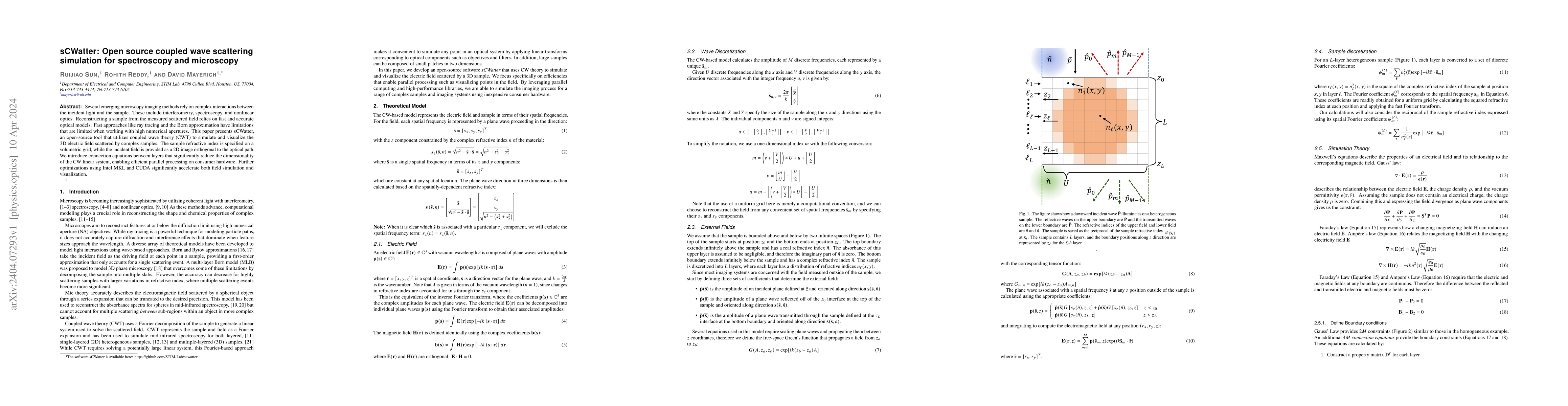
Several emerging microscopy imaging methods rely on complex interactions between the incident light and the sample. These include interferometry, spectroscopy, and nonlinear optics. Reconstructing a...
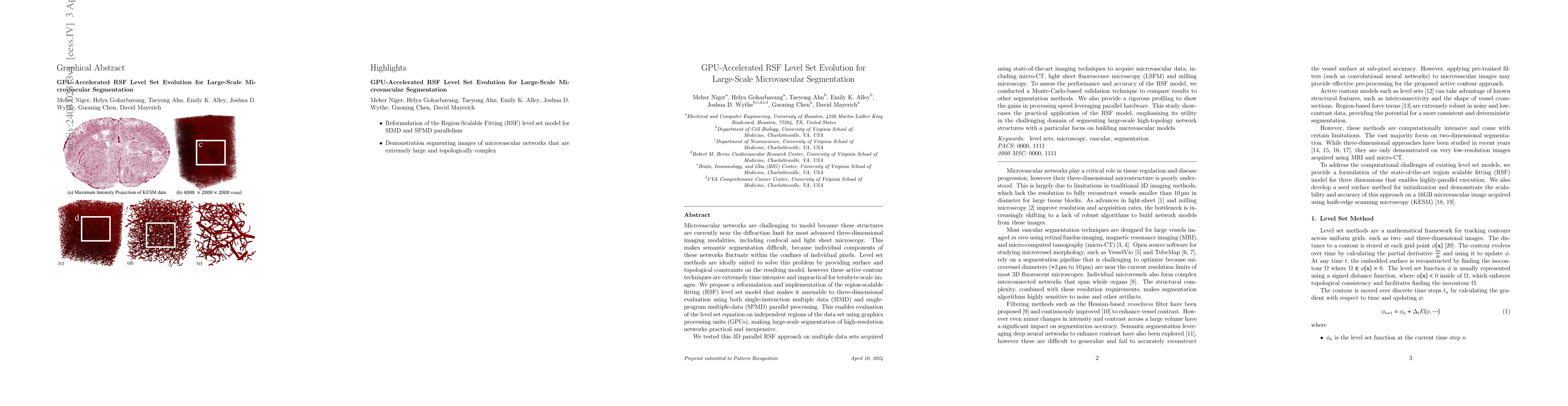
Microvascular networks are challenging to model because these structures are currently near the diffraction limit for most advanced three-dimensional imaging modalities, including confocal and light...
Ovarian cancer detection has traditionally relied on a multi-step process that includes biopsy, tissue staining, and morphological analysis by experienced pathologists. While widely practiced, this ...
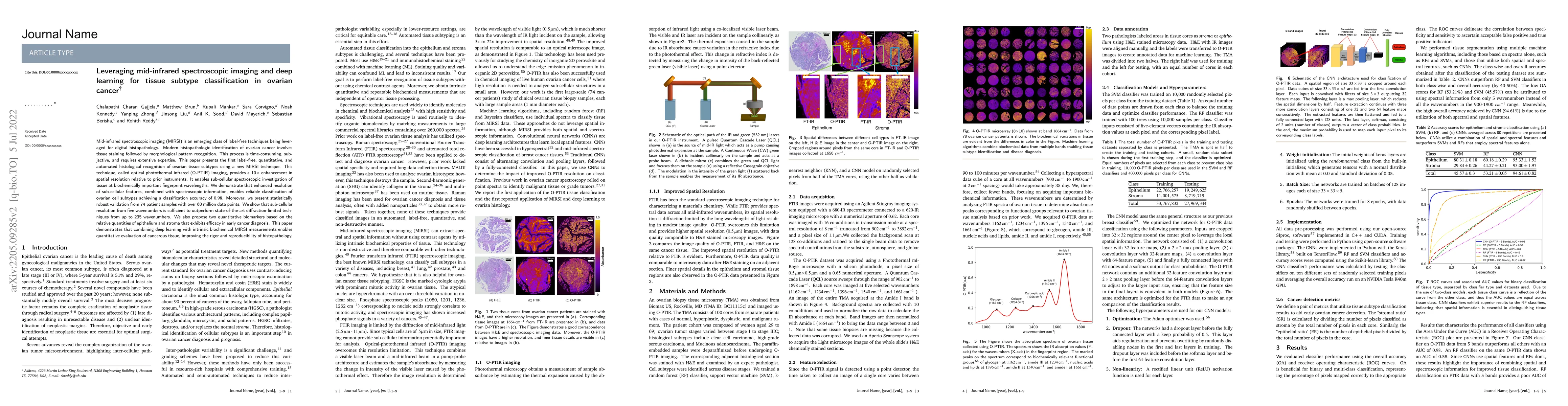
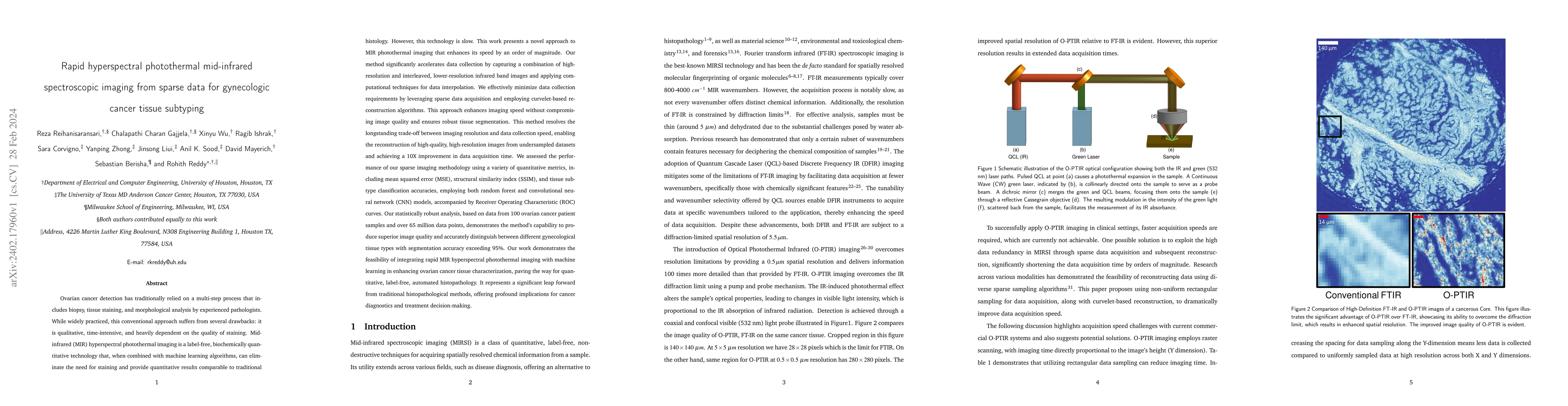
Mid-infrared spectroscopic imaging (MIRSI) is an emerging class of label-free techniques being leveraged for digital histopathology. Modern histopathologic identification of ovarian cancer involves ...
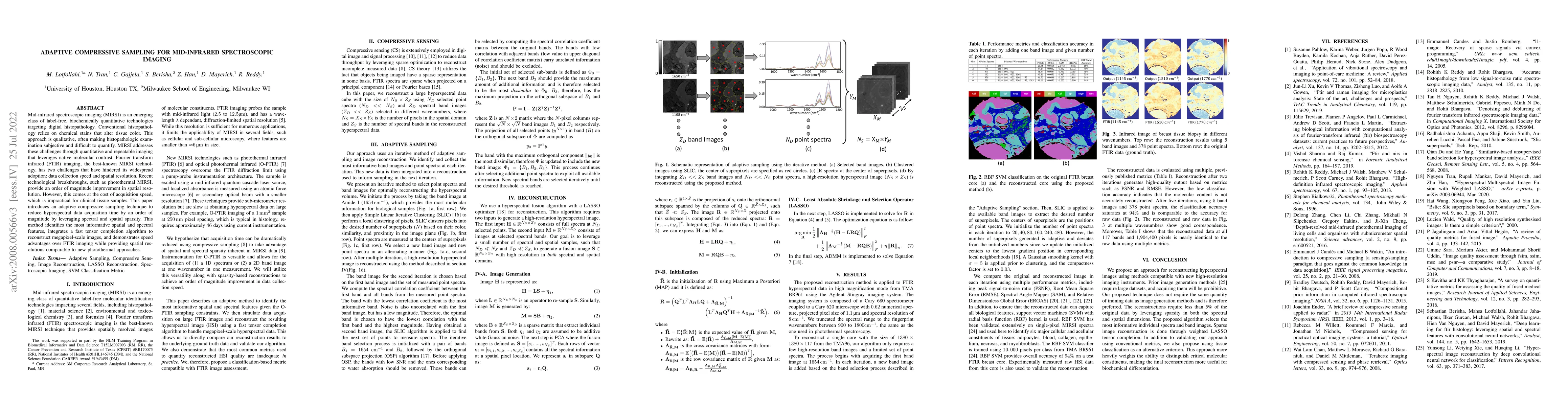
Minfrared spectroscopic imaging (MIRSI) is an emerging class of label-free, biochemically quantitative technologies targeting digital histopathology. Conventional histopathology relies on chemical s...
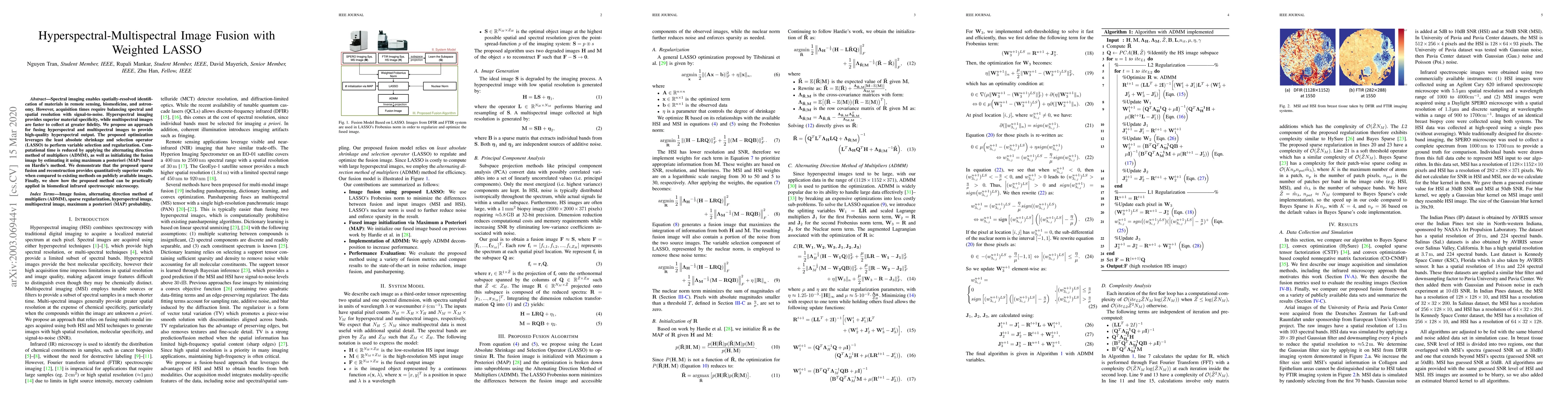
Spectral imaging enables spatially-resolved identification of materials in remote sensing, biomedicine, and astronomy. However, acquisition times require balancing spectral and spatial resolution wi...
Maps of brain microarchitecture are important for understanding neurological function and behavior, including alterations caused by chronic conditions such as neurodegenerative disease. Techniques s...
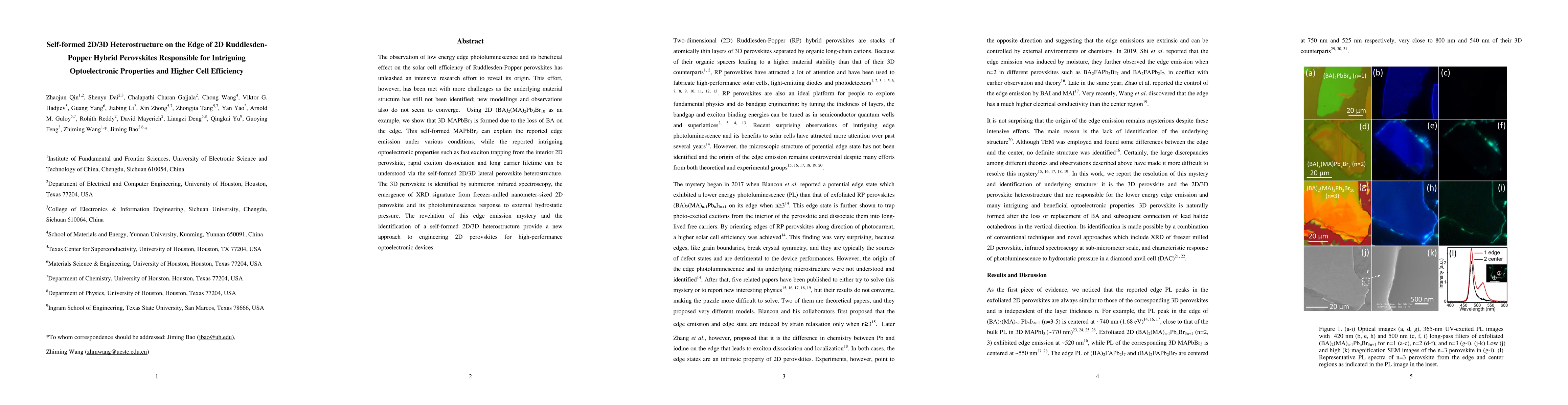
The observation of low energy edge photoluminescence and its beneficial effect on the solar cell efficiency of Ruddlesden-Popper perovskites has unleashed an intensive research effort to reveal its ...
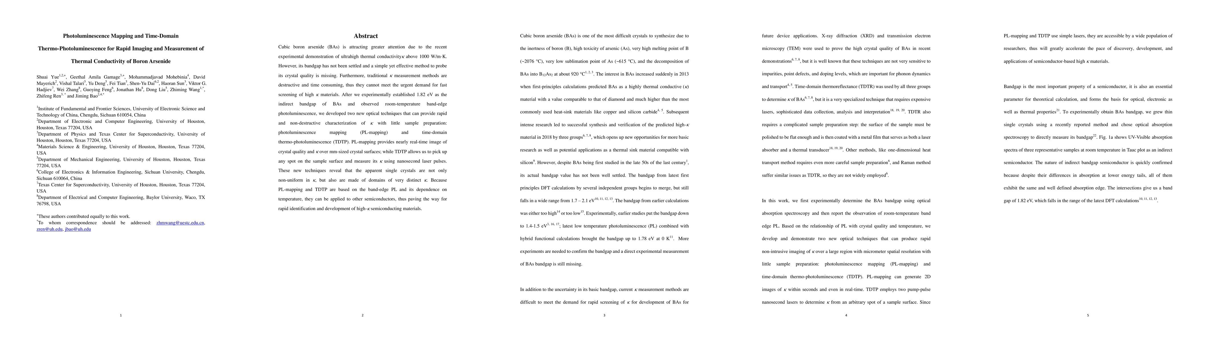
Cubic boron arsenide (BAs) is attracting greater attention due to the recent experimental demonstration of ultrahigh thermal conductivity \k{appa} above 1000 W/mK. However, its bandgap has not been ...
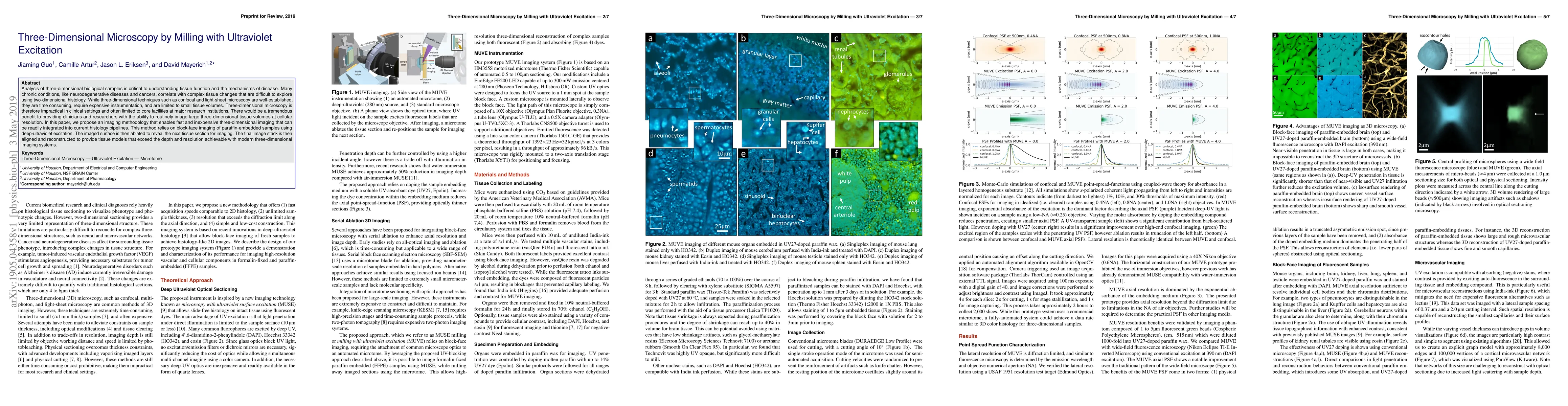
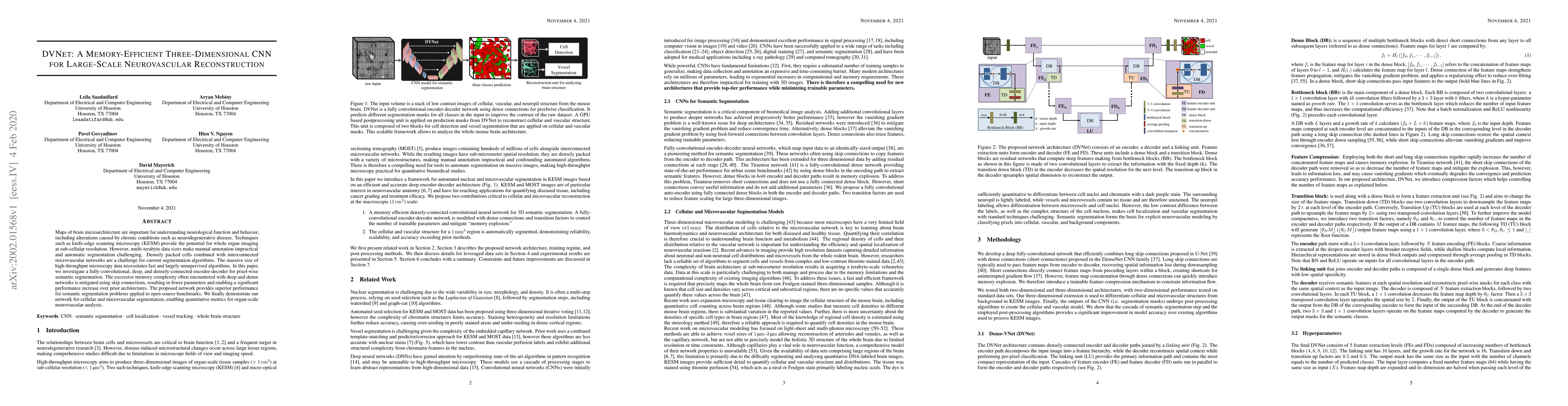
Analysis of three-dimensional biological samples is critical to understanding tissue function and the mechanisms of disease. Many chronic conditions, like neurodegenerative diseases and cancers, cor...
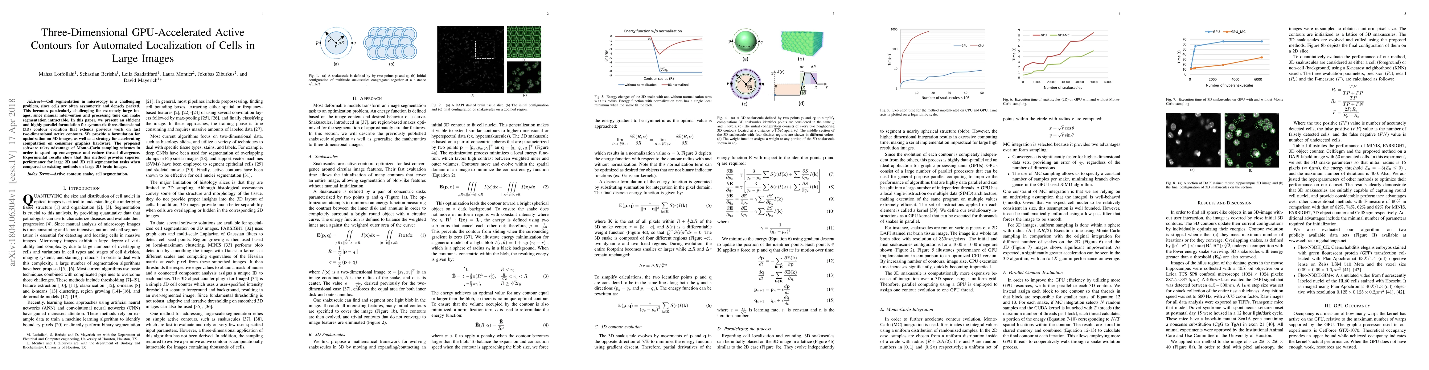
Cell segmentation in microscopy is a challenging problem, since cells are often asymmetric and densely packed. This becomes particularly challenging for extremely large images, since manual interven...