Authors
Publication
Metrics
Quick Actions
AI Quick Summary
This paper presents a GPU-accelerated reformulation of the region-scalable fitting (RSF) level set method for segmenting large-scale microvascular networks, enabling practical and fast processing of high-resolution 3D imaging data. The proposed parallel implementation significantly enhances computational efficiency and accuracy, validated through comparison with other segmentation methods and profiling of parallel hardware performance.
Quick Answers
What methodology did the authors use?
The paper proposes a GPU-accelerated reformulation of the Region-Scalable Fitting (RSF) level set method for large-scale microvascular segmentation. This approach leverages both volume-level and voxel-level parallelism to handle terabyte-scale images efficiently using graphics processing units (GPUs). More in Methodology →
What are the key results?
The proposed 3D parallel RSF approach significantly improves processing speed and practicality for large-scale microvascular segmentation. — Monte-Carlo-based validation demonstrated the accuracy and efficiency of the RSF model compared to other segmentation methods across various microscopy datasets. More in Key Results →
Why is this work significant?
This research is significant as it addresses the challenge of segmenting high-resolution, high-topology microvascular networks, which are difficult to model due to their near-diffraction limit in imaging. The GPU-accelerated RSF method provides a practical and inexpensive solution for large-scale image processing, enabling the creation of detailed microvascular models. More in Significance →
What are the main limitations?
The study did not explore the applicability of the method to other types of biological networks with similar complexity. — Limitations in the validation method, such as relying on Monte-Carlo sampling, might not fully capture the performance across all possible imaging conditions. More in Limitations →
Abstract
Microvascular networks are challenging to model because these structures are currently near the diffraction limit for most advanced three-dimensional imaging modalities, including confocal and light sheet microscopy. This makes semantic segmentation difficult, because individual components of these networks fluctuate within the confines of individual pixels. Level set methods are ideally suited to solve this problem by providing surface and topological constraints on the resulting model, however these active contour techniques are extremely time intensive and impractical for terabyte-scale images. We propose a reformulation and implementation of the region-scalable fitting (RSF) level set model that makes it amenable to three-dimensional evaluation using both single-instruction multiple data (SIMD) and single-program multiple-data (SPMD) parallel processing. This enables evaluation of the level set equation on independent regions of the data set using graphics processing units (GPUs), making large-scale segmentation of high-resolution networks practical and inexpensive. 
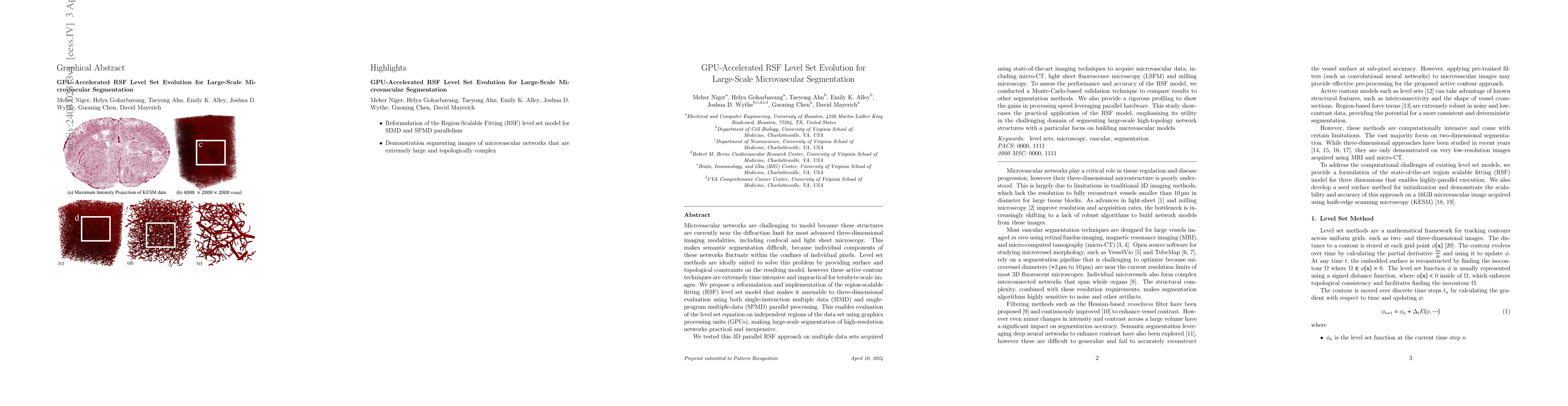
We tested this 3D parallel RSF approach on multiple data sets acquired using state-of-the-art imaging techniques to acquire microvascular data, including micro-CT, light sheet fluorescence microscopy (LSFM) and milling microscopy. To assess the performance and accuracy of the RSF model, we conducted a Monte-Carlo-based validation technique to compare results to other segmentation methods. We also provide a rigorous profiling to show the gains in processing speed leveraging parallel hardware. This study showcases the practical application of the RSF model, emphasizing its utility in the challenging domain of segmenting large-scale high-topology network structures with a particular focus on building microvascular models.
AI Key Findings
Generated Sep 03, 2025
Methodology — What approach did the authors take?
The paper proposes a GPU-accelerated reformulation of the Region-Scalable Fitting (RSF) level set method for large-scale microvascular segmentation. This approach leverages both volume-level and voxel-level parallelism to handle terabyte-scale images efficiently using graphics processing units (GPUs).
Key Results — What are the main findings?
- The proposed 3D parallel RSF approach significantly improves processing speed and practicality for large-scale microvascular segmentation.
- Monte-Carlo-based validation demonstrated the accuracy and efficiency of the RSF model compared to other segmentation methods across various microscopy datasets.
- Profiling results show substantial gains in processing speed when using parallel hardware, especially GPUs, for level set evolution.
Significance — Why does this research matter?
This research is significant as it addresses the challenge of segmenting high-resolution, high-topology microvascular networks, which are difficult to model due to their near-diffraction limit in imaging. The GPU-accelerated RSF method provides a practical and inexpensive solution for large-scale image processing, enabling the creation of detailed microvascular models.
Technical Contribution — What is the technical contribution?
The paper presents a novel GPU-accelerated implementation of the RSF level set method, enabling efficient and practical large-scale segmentation of high-resolution, high-topology networks.
Novelty — What is new about this work?
The proposed method distinguishes itself by efficiently utilizing GPU parallelism for the RSF level set evolution, which was previously impractical for terabyte-scale images. This approach significantly reduces computation times and memory requirements, making it suitable for emerging microscopy imaging techniques.
Limitations — What are the limitations of this study?
- The study did not explore the applicability of the method to other types of biological networks with similar complexity.
- Limitations in the validation method, such as relying on Monte-Carlo sampling, might not fully capture the performance across all possible imaging conditions.
Future Work — What did the authors propose for future work?
- Investigate the applicability of the RSF method to other complex biological structures beyond microvascular networks.
- Explore alternative validation techniques to assess the robustness of the method under a broader range of imaging conditions and network complexities.
Paper Details
How to Cite This Paper
@article{mayerich2024gpu,
title = {GPU-Accelerated RSF Level Set Evolution for Large-Scale Microvascular
Segmentation},
author = {Mayerich, David and Alley, Emily K. and Chen, Guoning and others},
year = {2024},
eprint = {2404.02813},
archivePrefix = {arXiv},
primaryClass = {eess.IV},
}
Mayerich, D., Alley, E., Chen, G., Goharbavang, H., Wythe, J., Niger, M., & Ahn, T. (2024). GPU-Accelerated RSF Level Set Evolution for Large-Scale Microvascular
Segmentation. arXiv. https://arxiv.org/abs/2404.02813
Mayerich, David, et al. "GPU-Accelerated RSF Level Set Evolution for Large-Scale Microvascular
Segmentation." arXiv, 2024, arxiv.org/abs/2404.02813.
PDF Preview
Key Terms
Citation Network
Current paper (gray), citations (green), references (blue)
Display is limited for performance on very large graphs.
Similar Papers
Found 4 papersByteQC: GPU-Accelerated Quantum Chemistry Package for Large-Scale Systems
Zhen Guo, Zigeng Huang, Qiaorui Chen et al.
No citations found for this paper.
Comments (0)