Academic Profile
Statistics
Similar Authors
Papers on arXiv
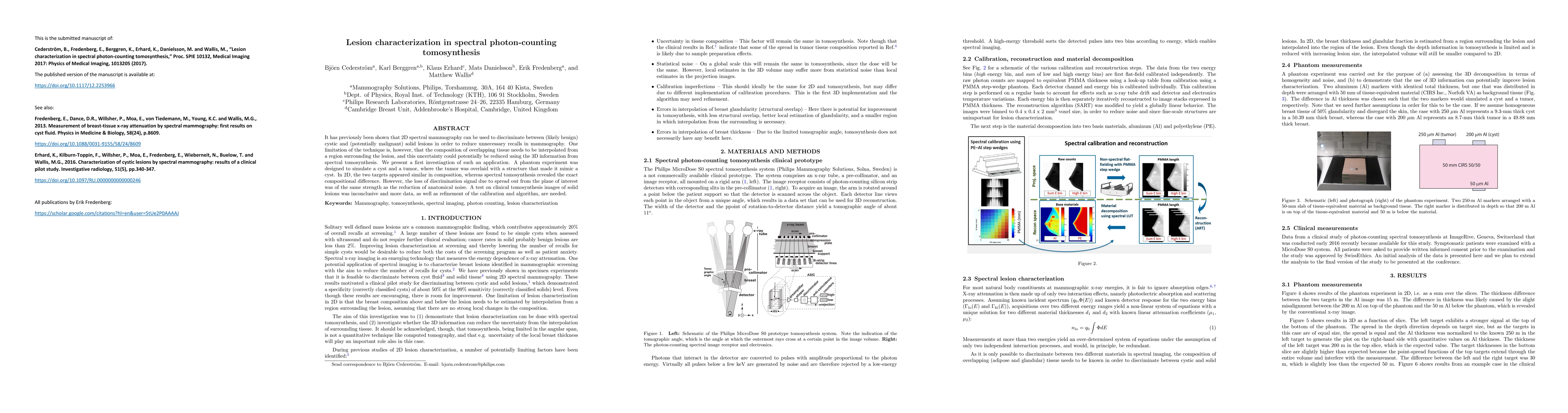
It has previously been shown that 2D spectral mammography can be used to discriminate between (likely benign) cystic and (potentially malignant) solid lesions in order to reduce unnecessary recalls ...
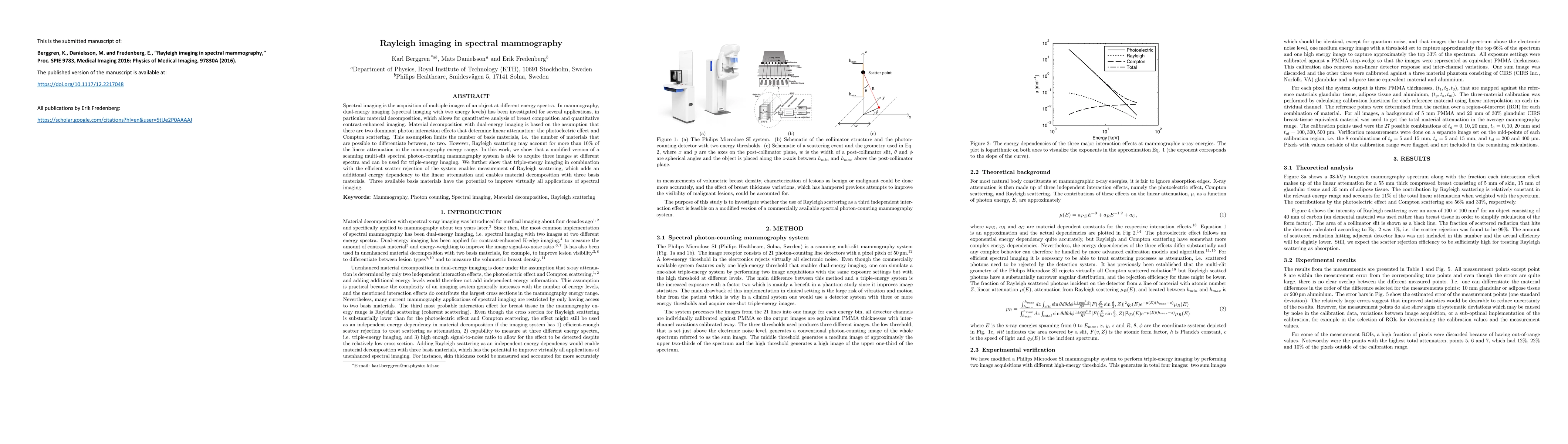
Spectral imaging is the acquisition of multiple images of an object at different energy spectra. In mammography, dual-energy imaging (spectral imaging with two energy levels) has been investigated f...
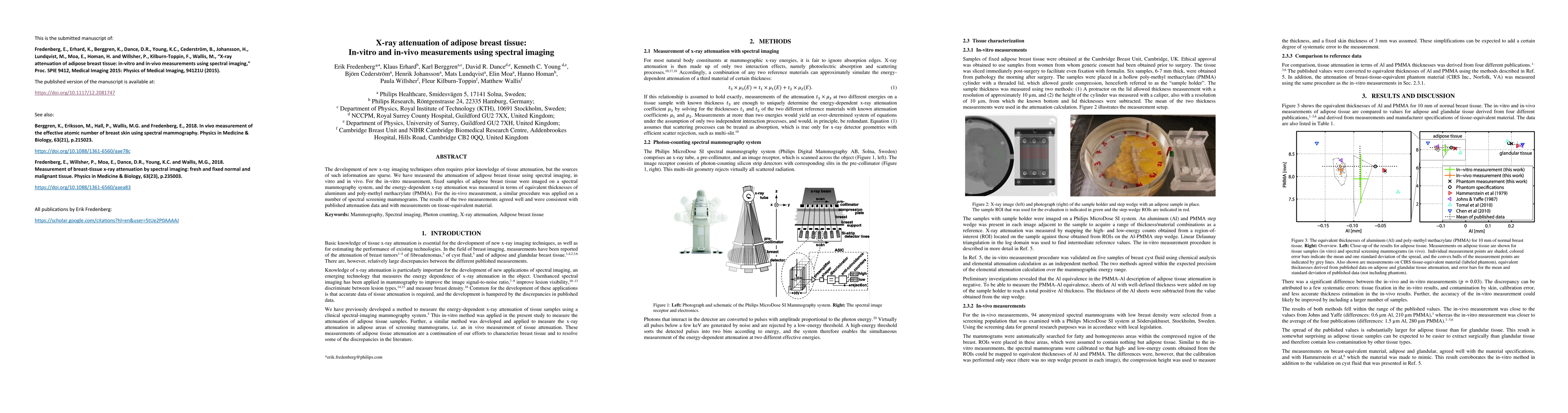
The development of new x-ray imaging techniques often requires prior knowledge of tissue attenuation, but the sources of such information are sparse. We have measured the attenuation of adipose brea...
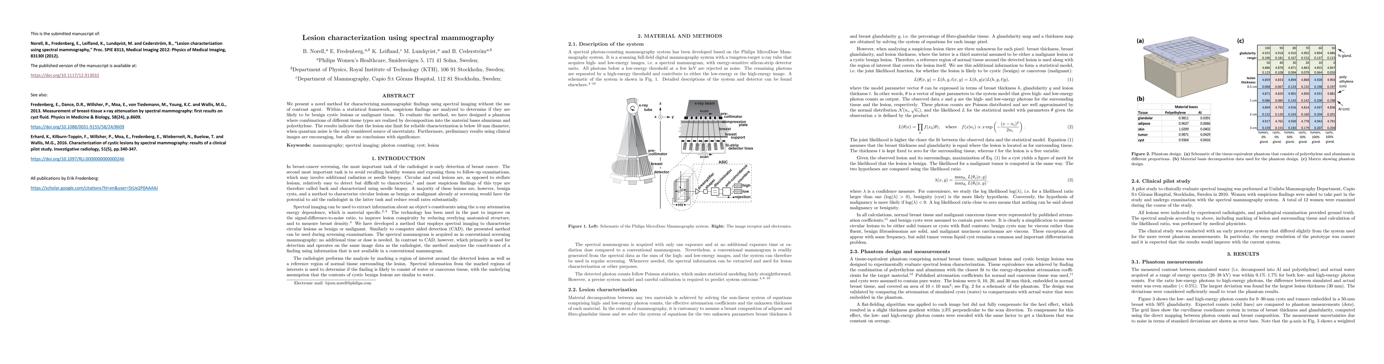
We present a novel method for characterizing mammographic findings using spectral imaging without the use of contrast agent. Within a statistical framework, suspicious findings are analyzed to deter...
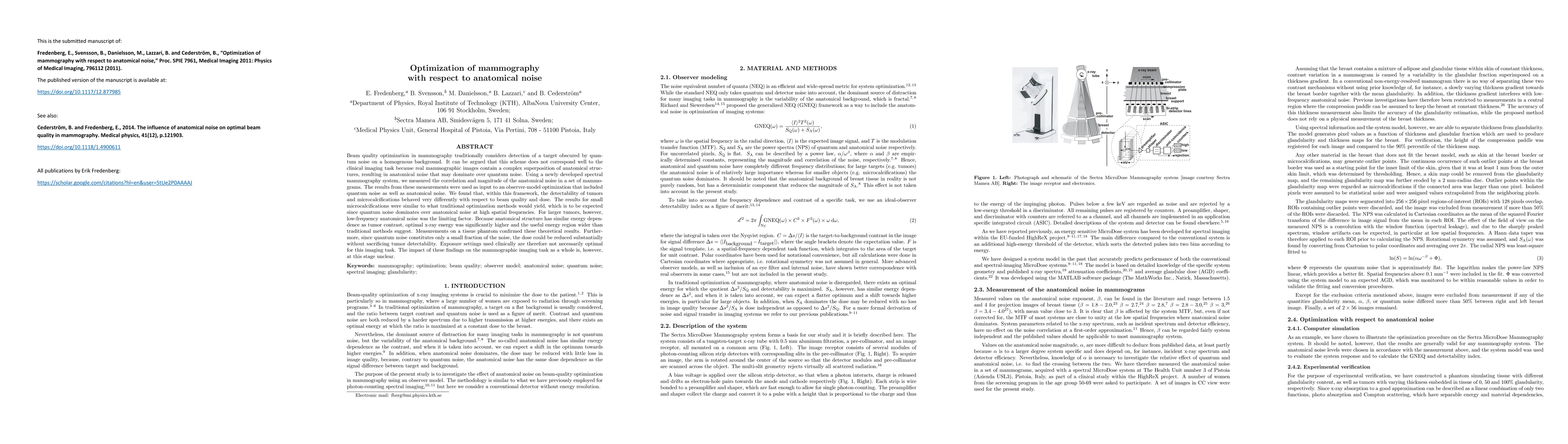
Beam quality optimization in mammography traditionally considers detection of a target obscured by quantum noise on a homogenous background. It can be argued that this scheme does not correspond wel...
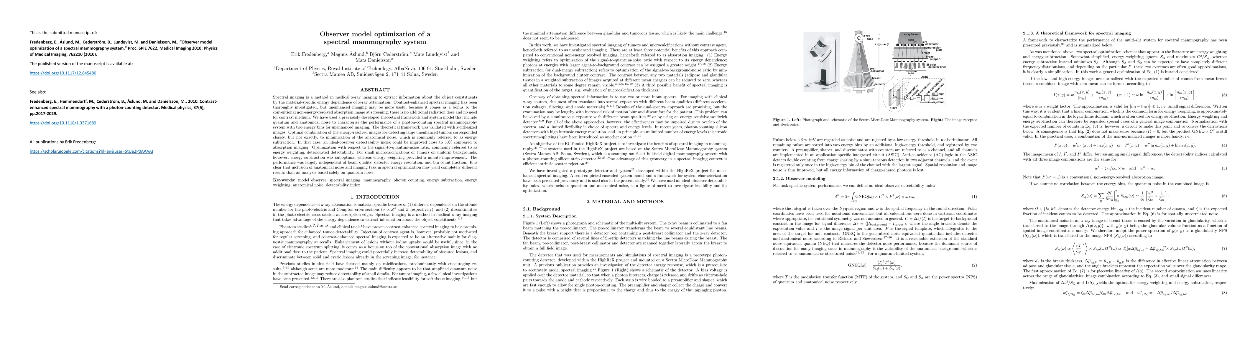
Spectral imaging is a method in medical x-ray imaging to extract information about the object constituents by the material-specific energy dependence of x-ray attenuation. Contrast-enhanced spectral...
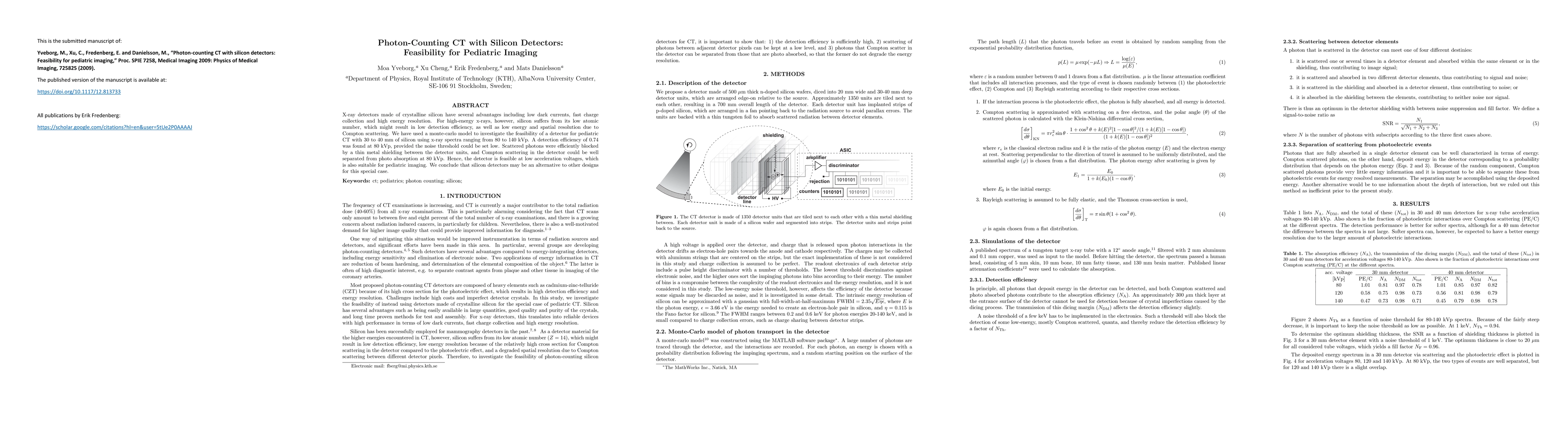
X-ray detectors made of crystalline silicon have several advantages including low dark currents, fast charge collection and high energy resolution. For high-energy x-rays, however, silicon suffers f...
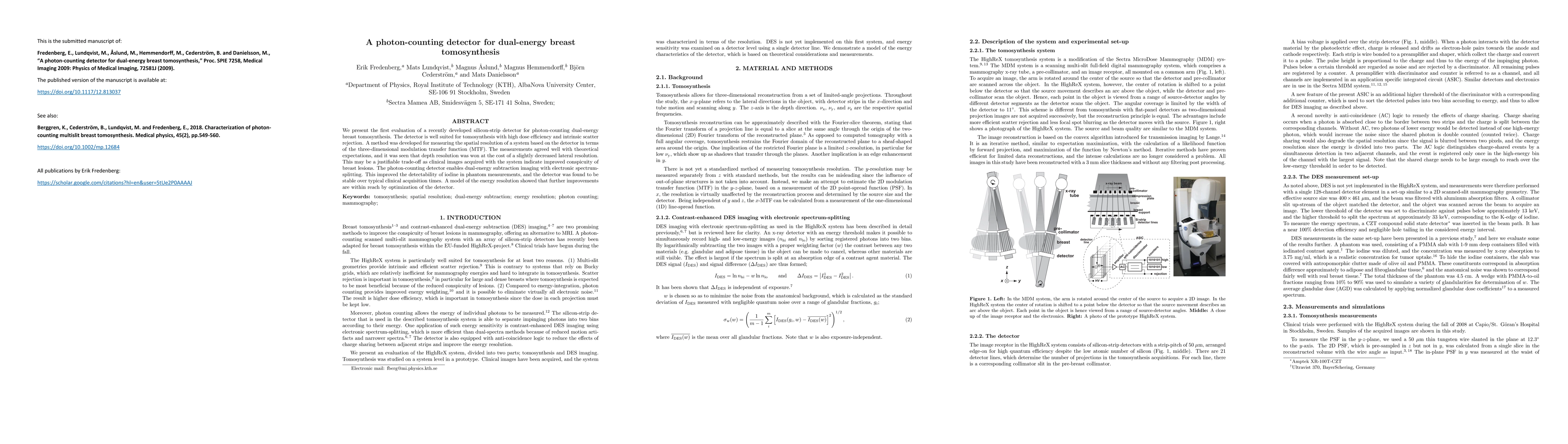
We present the first evaluation of a recently developed silicon-strip detector for photon-counting dual-energy breast tomosynthesis. The detector is well suited for tomosynthesis with high dose effi...
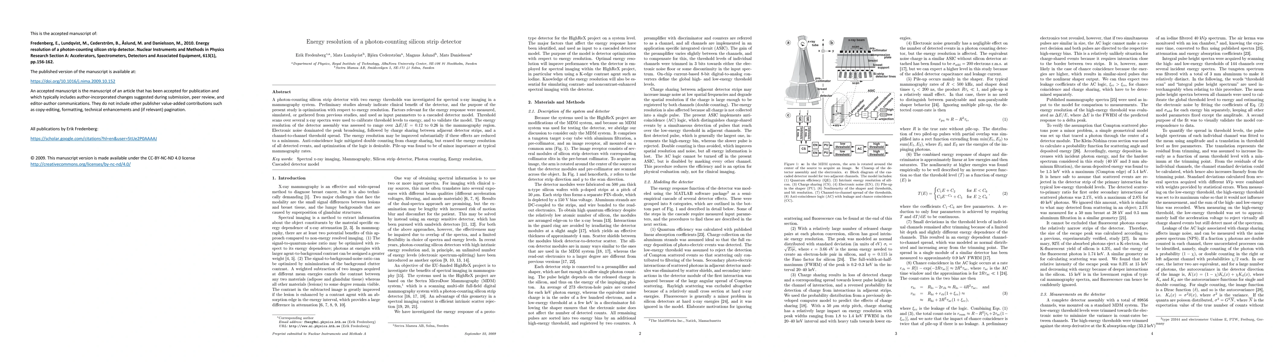
A photon-counting silicon strip detector with two energy thresholds was investigated for spectral X-ray imaging in a mammography system. Preliminary studies already indicate clinical benefit of the ...
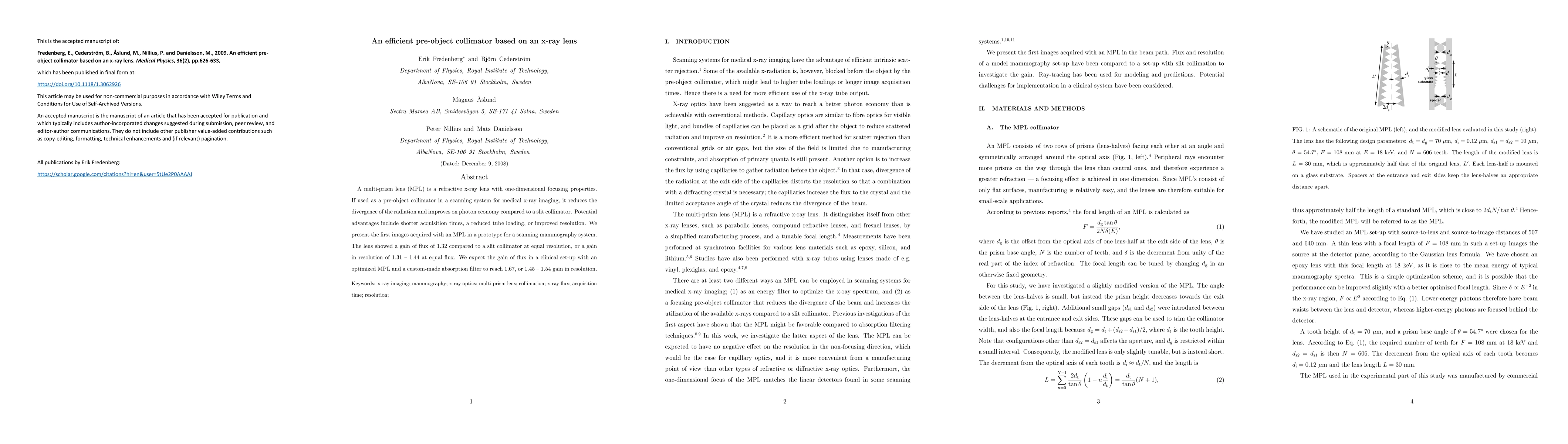
A multi-prism lens (MPL) is a refractive x-ray lens with one-dimensional focusing properties. If used as a pre-object collimator in a scanning system for medical x-ray imaging, it reduces the diverg...
Purpose: Spectral imaging is a method in medical x-ray imaging to extract information about the object constituents by the material-specific energy dependence of x-ray attenuation. The authors have ...
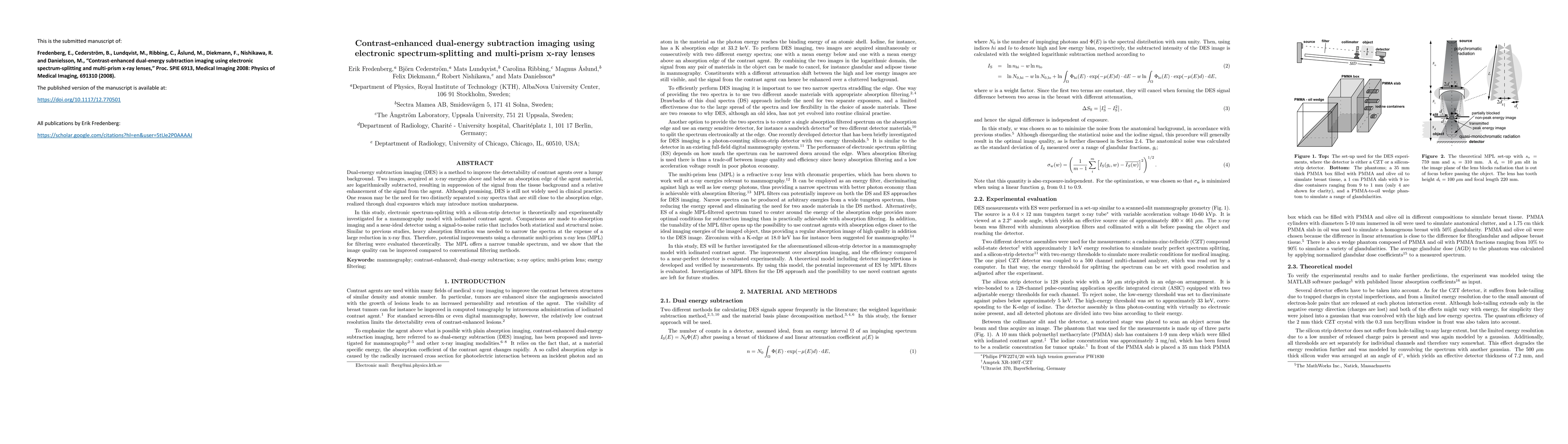
Dual-energy subtraction imaging (DES) is a method to improve the detectability of contrast agents over a lumpy background. Two images, acquired at x-ray energies above and below an absorption edge o...
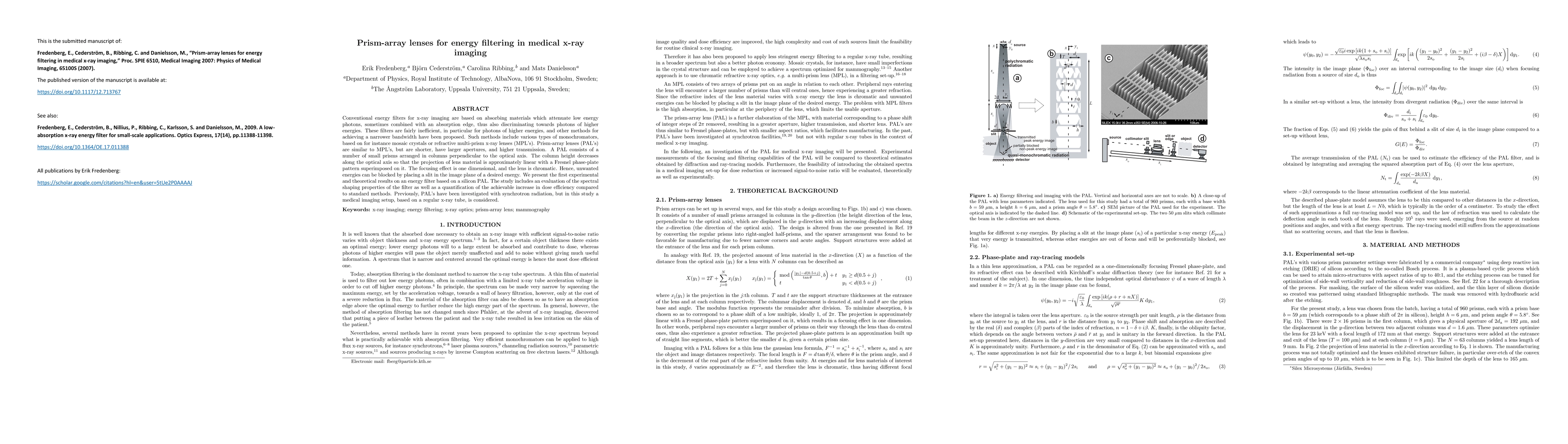
Conventional energy filters for x-ray imaging are based on absorbing materials which attenuate low energy photons, sometimes combined with an absorption edge, thus also discriminating towards photon...
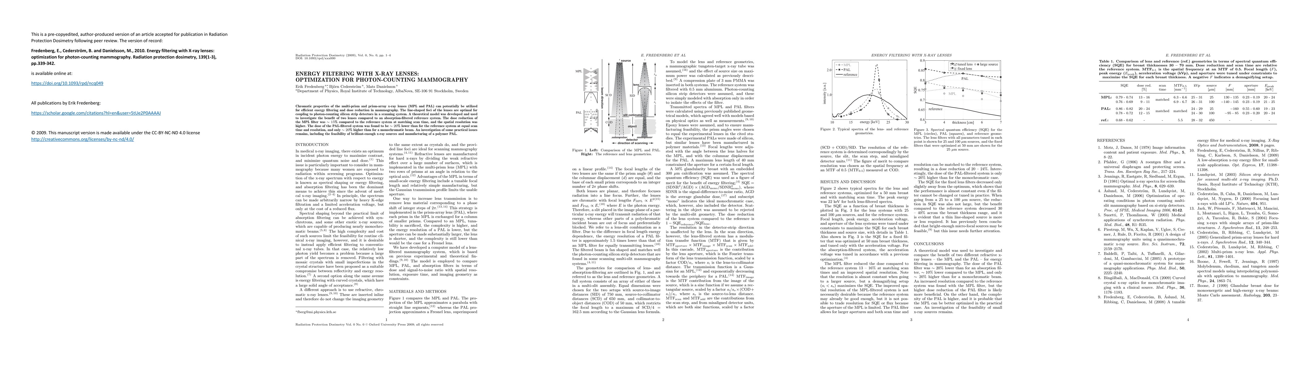
Chromatic properties of the multi-prism and prism-array X-ray lenses (MPL and PAL) can potentially be utilized for efficient energy filtering and dose reduction in mammography. The line-shaped foci ...
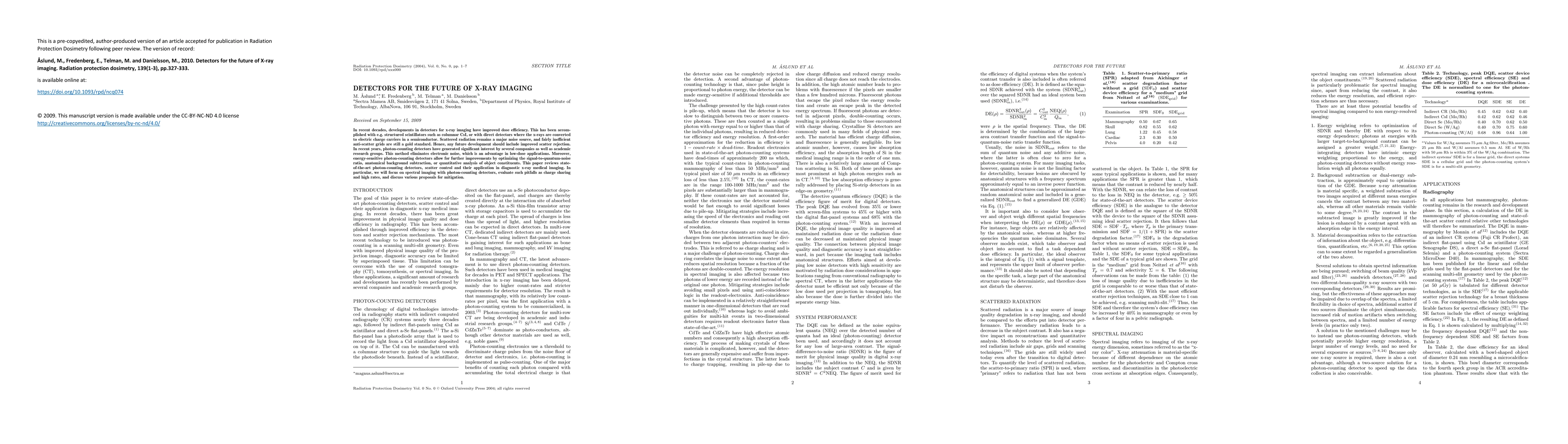
In recent decades, developments in detectors for X-ray imaging have improved dose efficiency. This has been accomplished with for example, structured scintillators such as columnar CsI, or with dire...
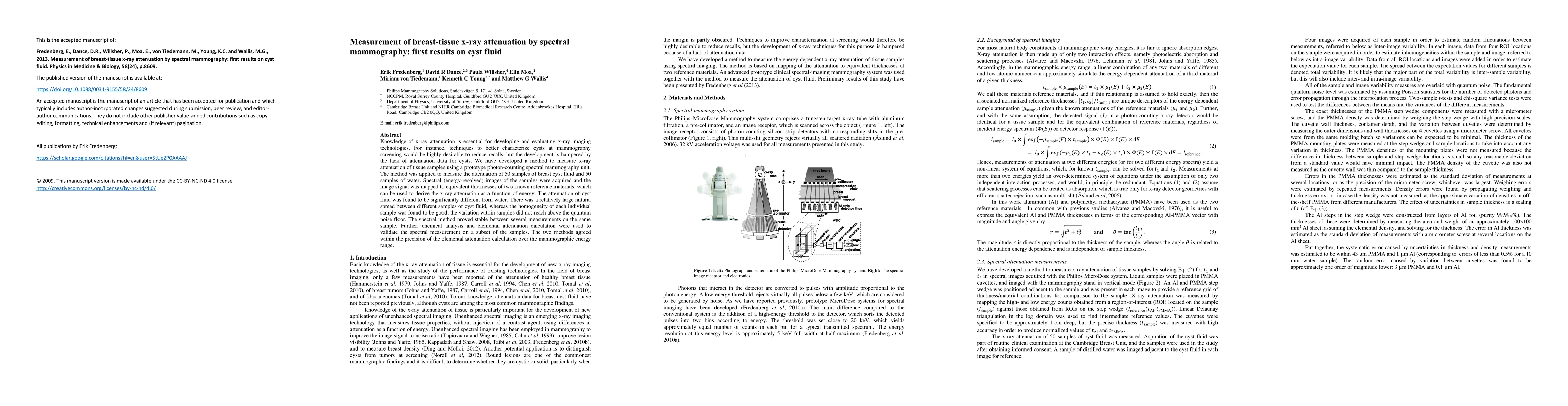
Knowledge of x-ray attenuation is essential for developing and evaluating x-ray imaging technologies. For instance, techniques to better characterize cysts at mammography screening would be highly d...
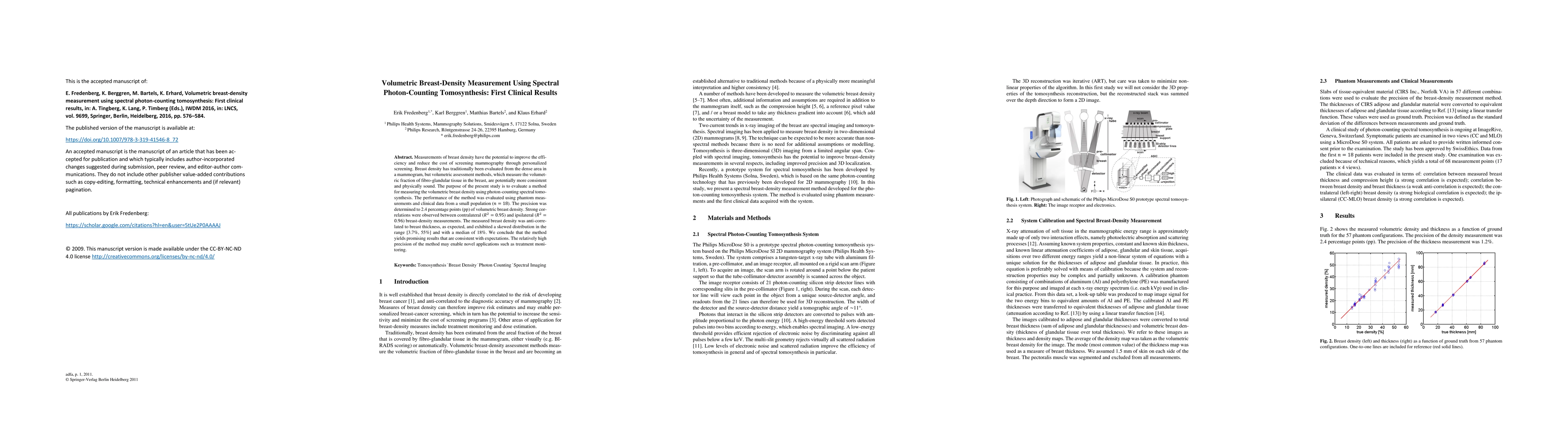
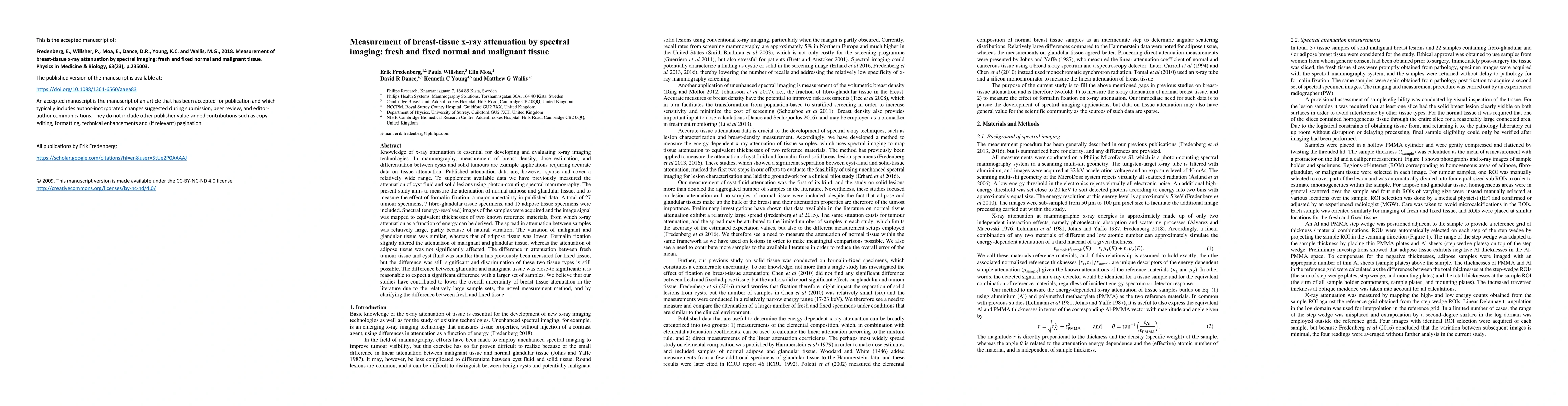
Measurements of breast density have the potential to improve the efficiency and reduce the cost of screening mammography through personalized screening. Breast density has traditionally been evaluat...
Knowledge of x-ray attenuation is essential for developing and evaluating x-ray imaging technologies. In mammography, measurement of breast density, dose estimation, and differentiation between cyst...
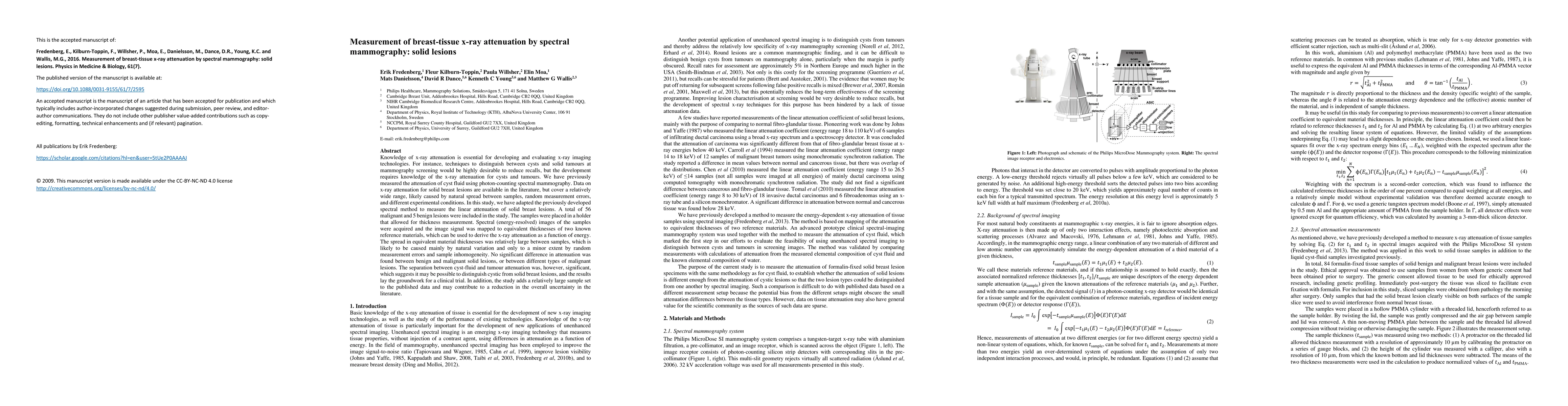
Knowledge of x-ray attenuation is essential for developing and evaluating x-ray imaging technologies. For instance, techniques to distinguish between cysts and solid tumours at mammography screening...
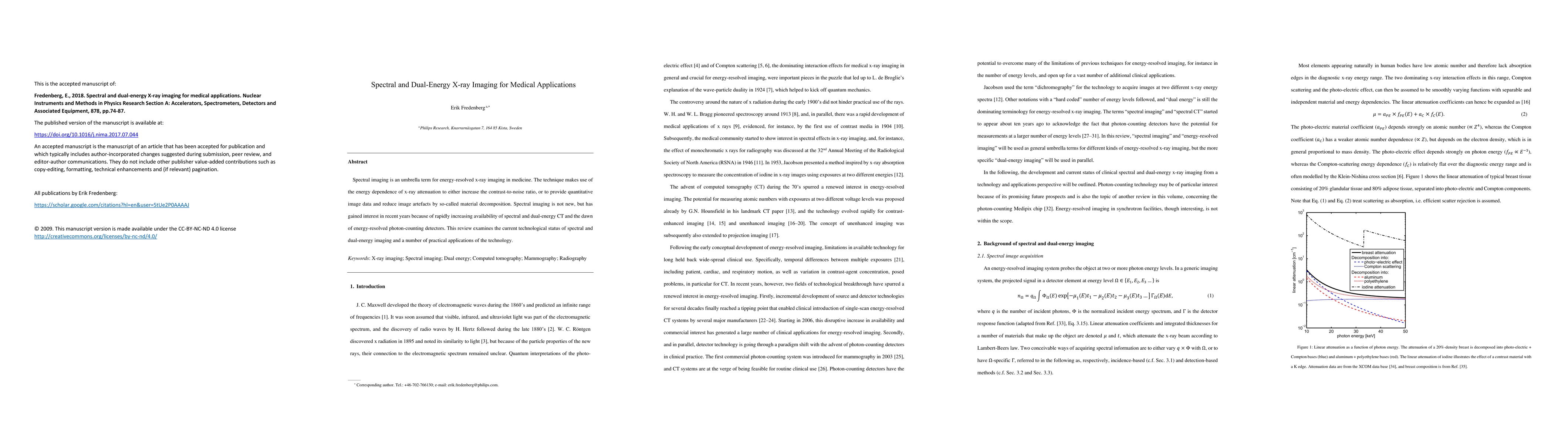
Spectral imaging is an umbrella term for energy-resolved x-ray imaging in medicine. The technique makes use of the energy dependence of x-ray attenuation to either increase the contrast-to-noise rat...