Academic Profile
Statistics
Similar Authors
Papers on arXiv
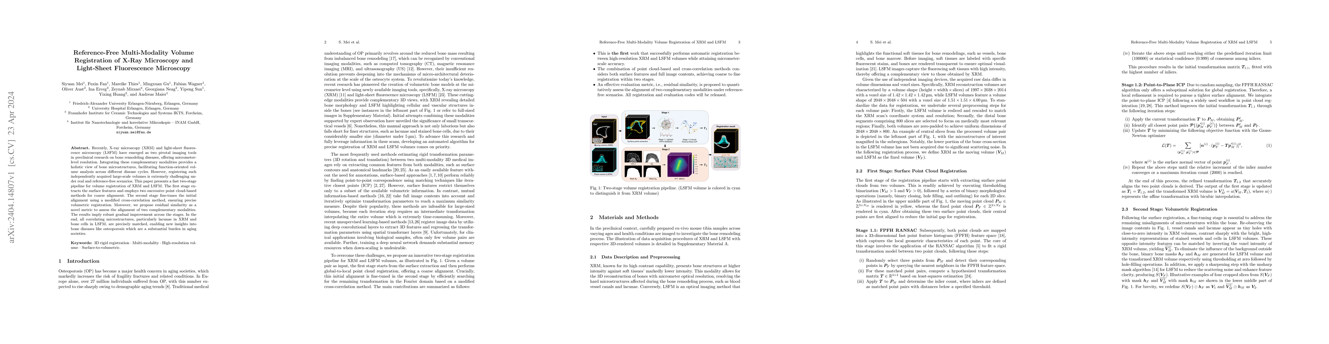
Recently, X-ray microscopy (XRM) and light-sheet fluorescence microscopy (LSFM) have emerged as two pivotal imaging tools in preclinical research on bone remodeling diseases, offering micrometer-lev...
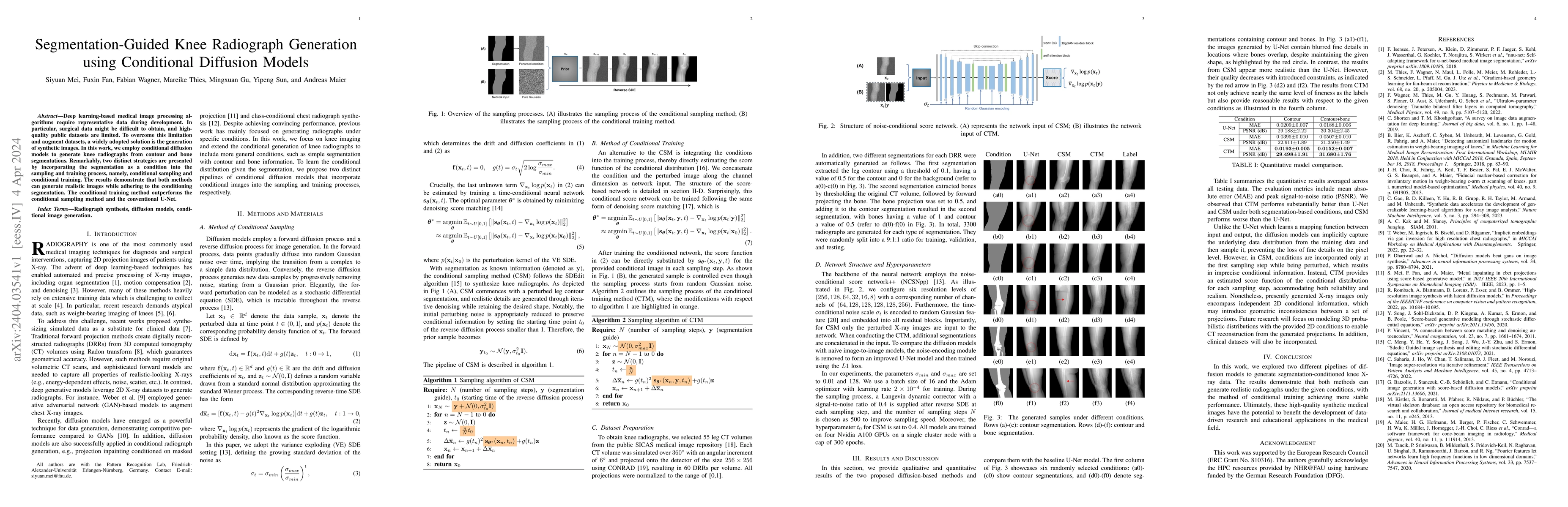
Deep learning-based medical image processing algorithms require representative data during development. In particular, surgical data might be difficult to obtain, and high-quality public datasets ar...
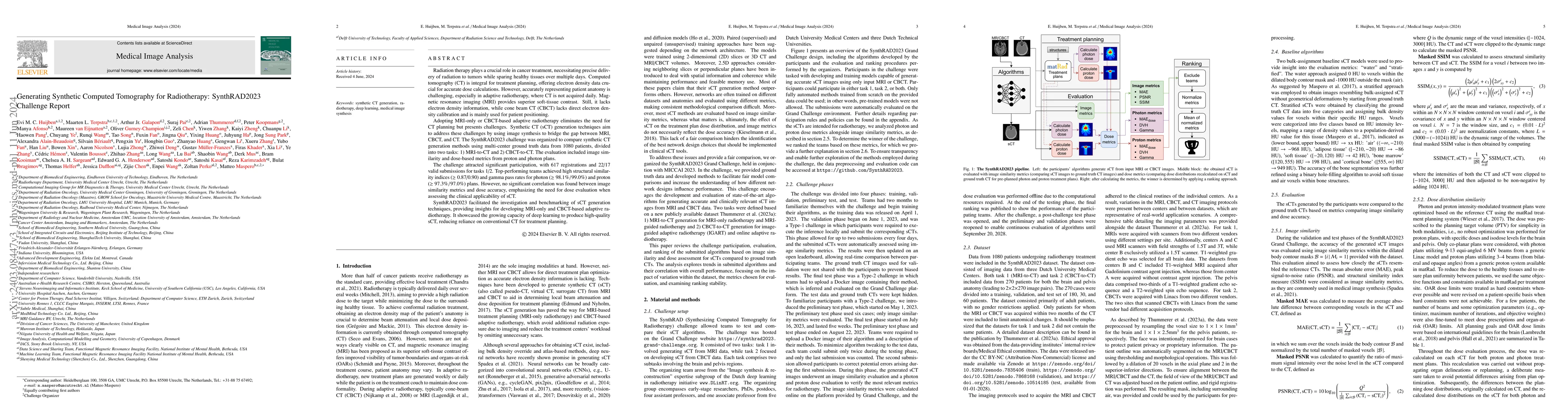
Radiation therapy plays a crucial role in cancer treatment, necessitating precise delivery of radiation to tumors while sparing healthy tissues over multiple days. Computed tomography (CT) is integr...
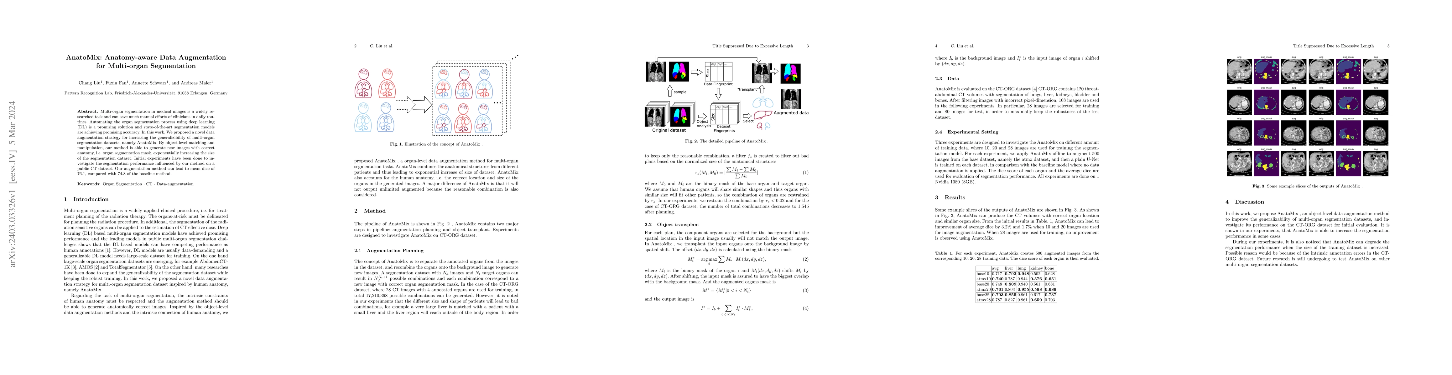
Multi-organ segmentation in medical images is a widely researched task and can save much manual efforts of clinicians in daily routines. Automating the organ segmentation process using deep learning...
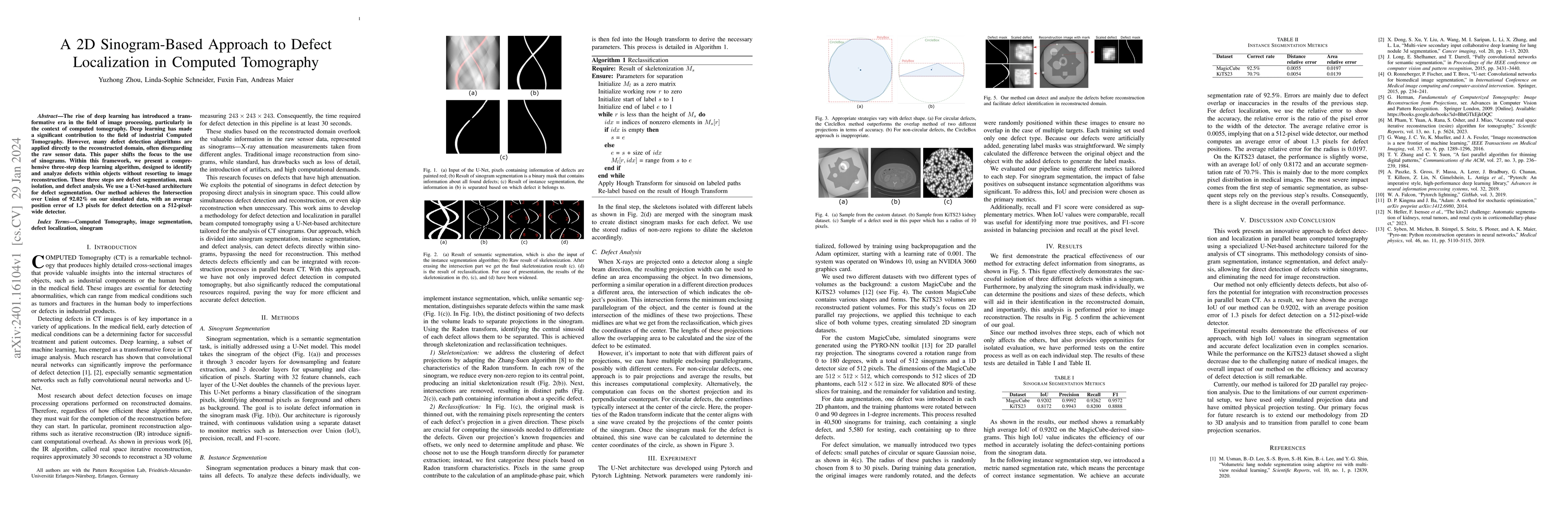
The rise of deep learning has introduced a transformative era in the field of image processing, particularly in the context of computed tomography. Deep learning has made a significant contribution ...
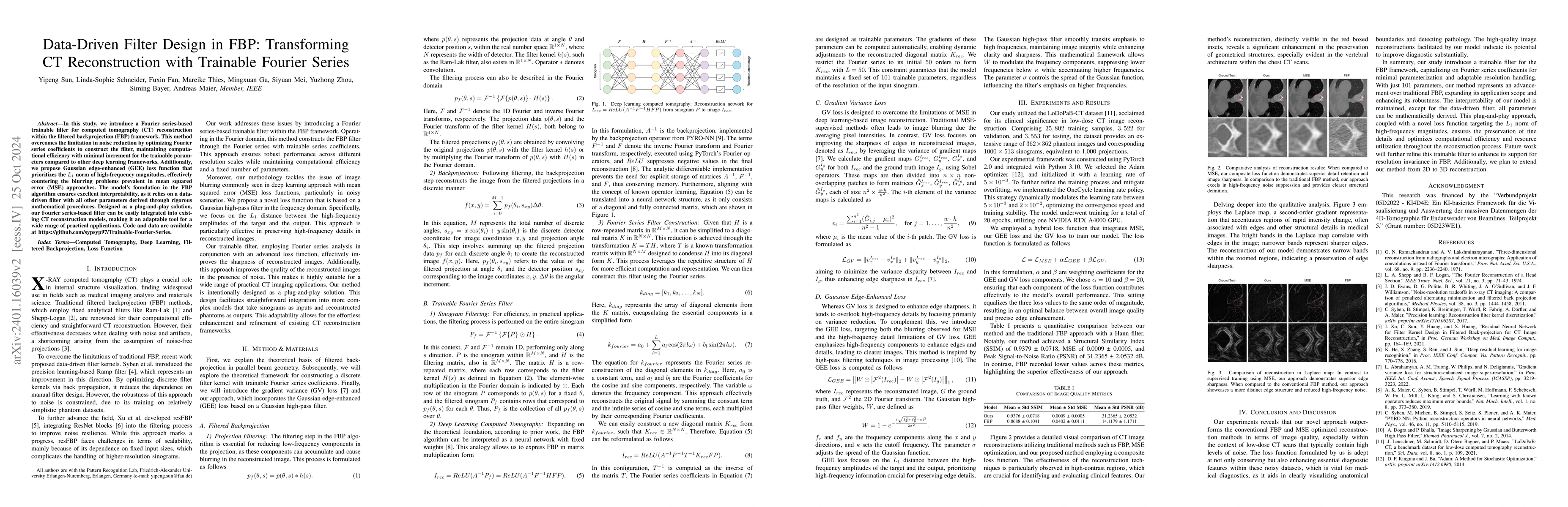
In this study, we introduce a Fourier series-based trainable filter for computed tomography (CT) reconstruction within the filtered backprojection (FBP) framework. This method overcomes the limitati...
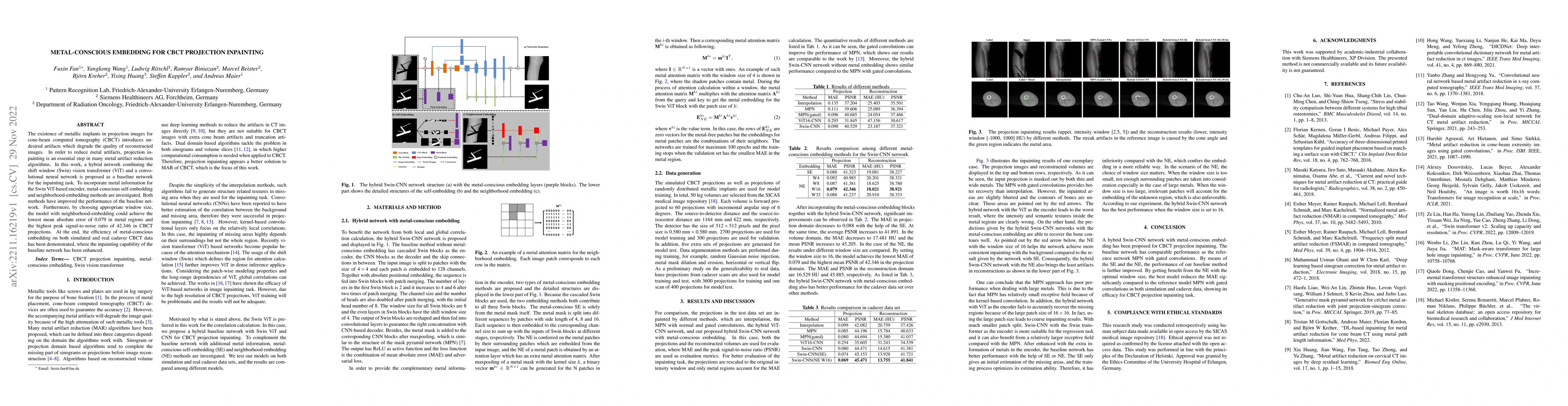
The existence of metallic implants in projection images for cone-beam computed tomography (CBCT) introduces undesired artifacts which degrade the quality of reconstructed images. In order to reduce ...
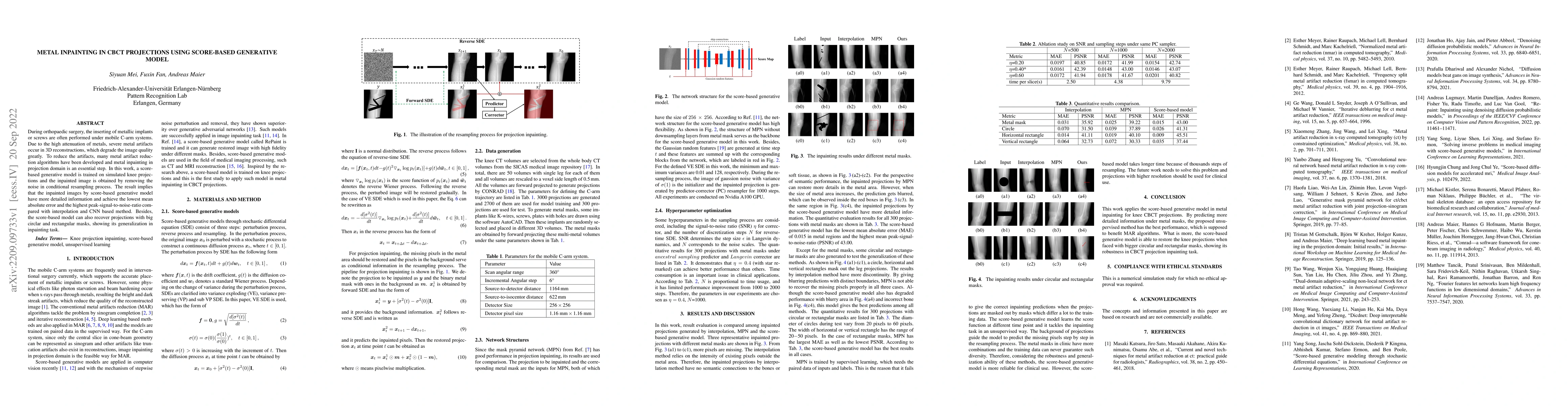
During orthopaedic surgery, the inserting of metallic implants or screws are often performed under mobile C-arm systems. Due to the high attenuation of metals, severe metal artifacts occur in 3D rec...
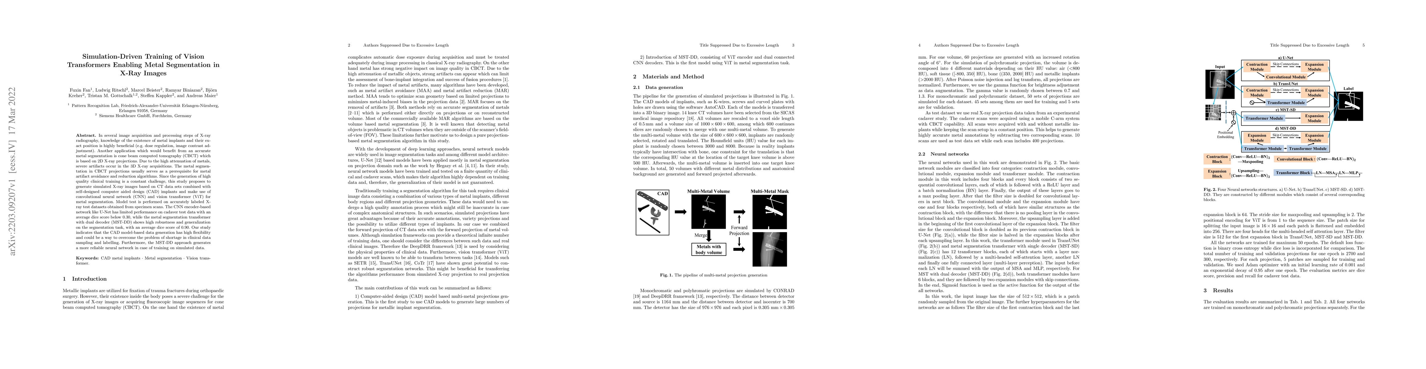
In several image acquisition and processing steps of X-ray radiography, knowledge of the existence of metal implants and their exact position is highly beneficial (e.g. dose regulation, image contra...
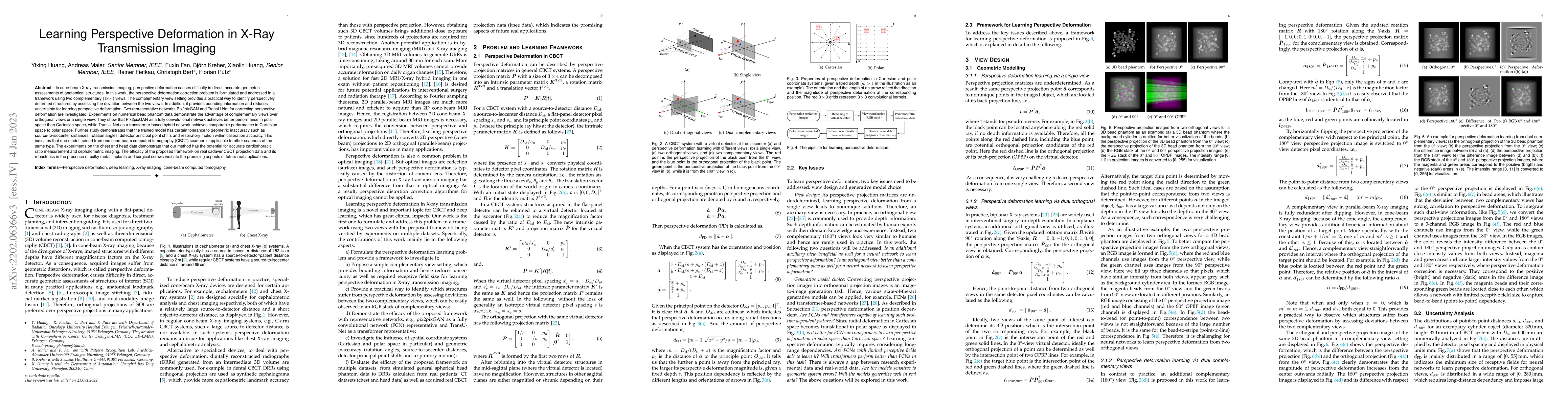
In cone-beam X-ray transmission imaging, perspective deformation causes difficulty in direct, accurate geometric assessments of anatomical structures. In this work, the perspective deformation corre...
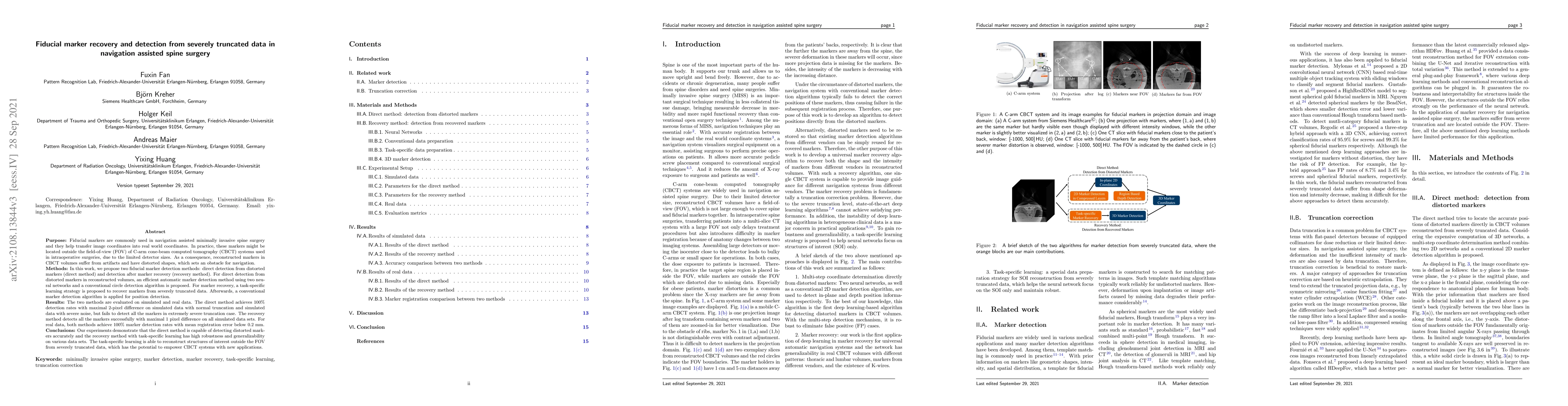
Fiducial markers are commonly used in navigation assisted minimally invasive spine surgery (MISS) and they help transfer image coordinates into real world coordinates. In practice, these markers mig...
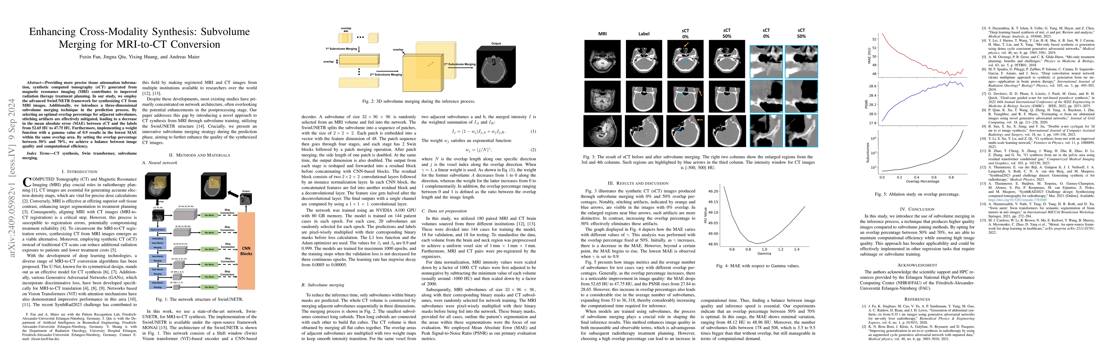
Providing more precise tissue attenuation information, synthetic computed tomography (sCT) generated from magnetic resonance imaging (MRI) contributes to improved radiation therapy treatment planning....

Cone-beam computed tomography (CBCT) is widely used in interventional surgeries and radiation oncology. Due to the limited size of flat-panel detectors, anatomical structures might be missing outside ...
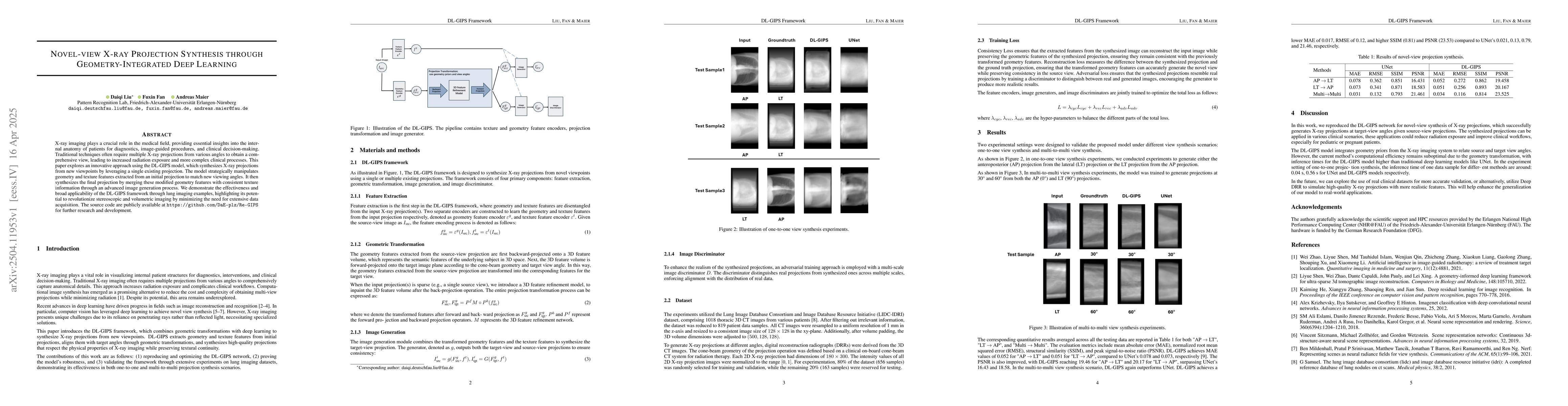
X-ray imaging plays a crucial role in the medical field, providing essential insights into the internal anatomy of patients for diagnostics, image-guided procedures, and clinical decision-making. Trad...
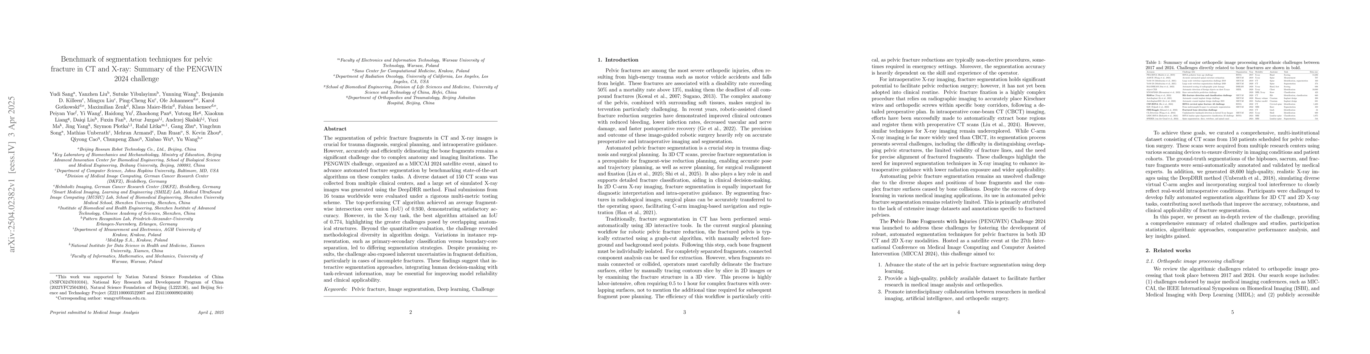
The segmentation of pelvic fracture fragments in CT and X-ray images is crucial for trauma diagnosis, surgical planning, and intraoperative guidance. However, accurately and efficiently delineating th...
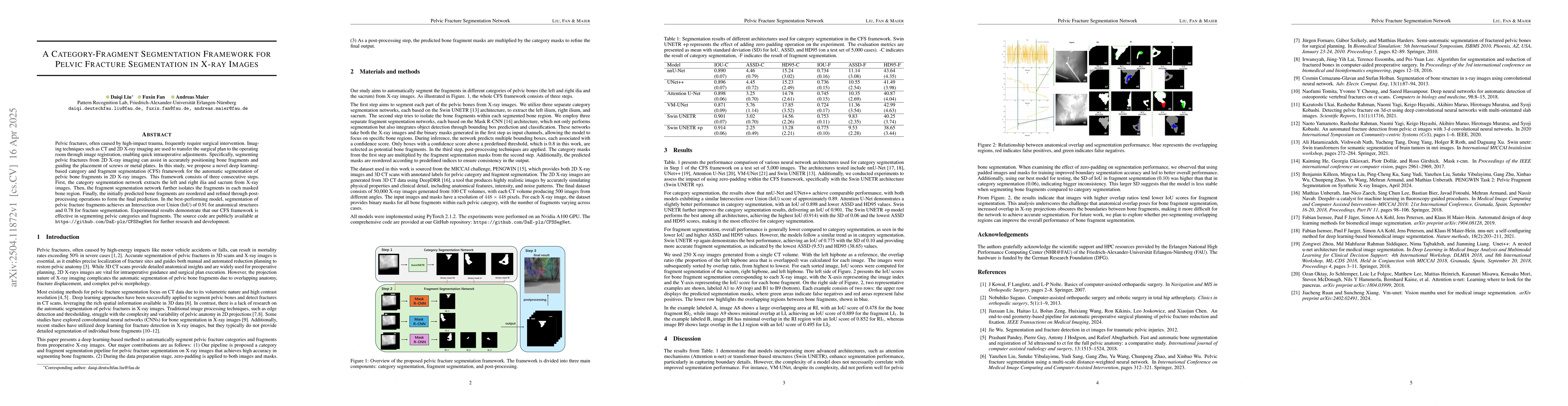
Pelvic fractures, often caused by high-impact trauma, frequently require surgical intervention. Imaging techniques such as CT and 2D X-ray imaging are used to transfer the surgical plan to the operati...
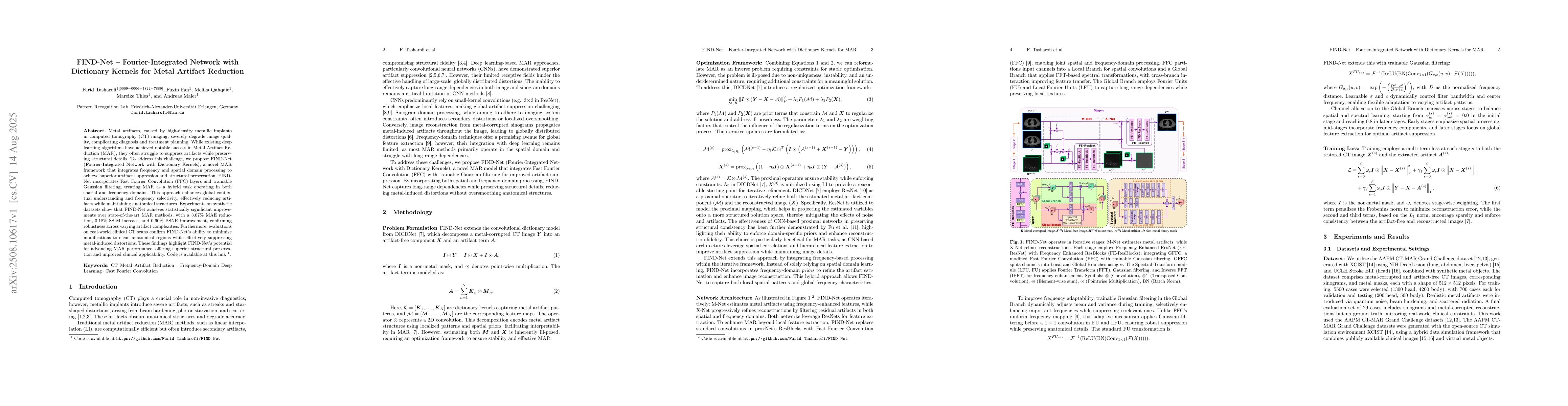
Metal artifacts, caused by high-density metallic implants in computed tomography (CT) imaging, severely degrade image quality, complicating diagnosis and treatment planning. While existing deep learni...
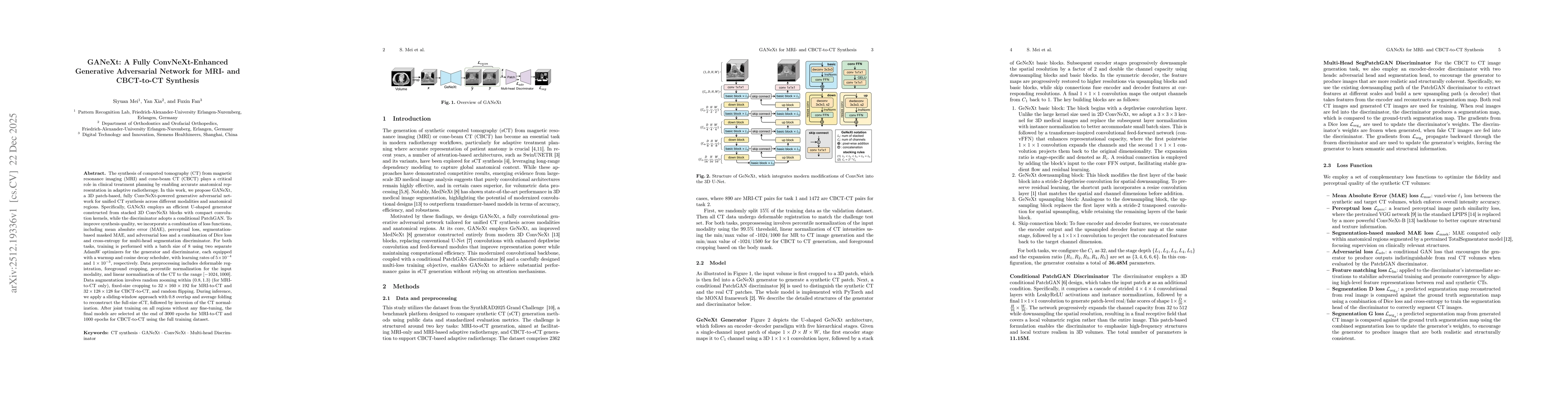
The synthesis of computed tomography (CT) from magnetic resonance imaging (MRI) and cone-beam CT (CBCT) plays a critical role in clinical treatment planning by enabling accurate anatomical representat...
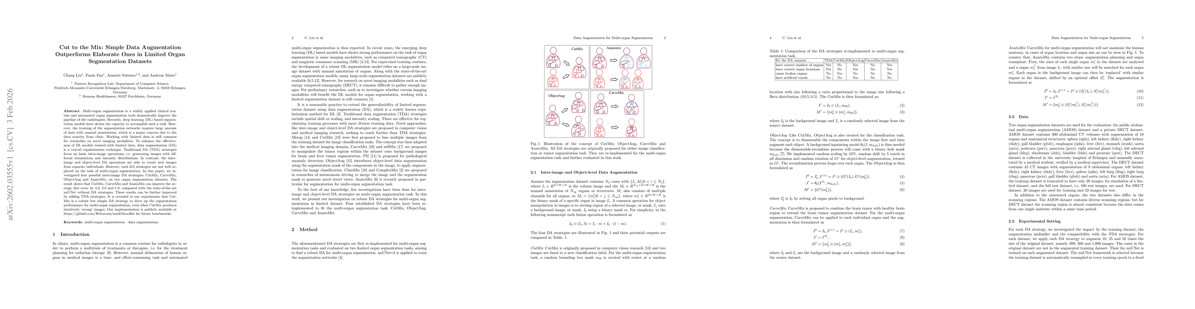
Multi-organ segmentation is a widely applied clinical routine and automated organ segmentation tools dramatically improve the pipeline of the radiologists. Recently, deep learning (DL) based segmentat...
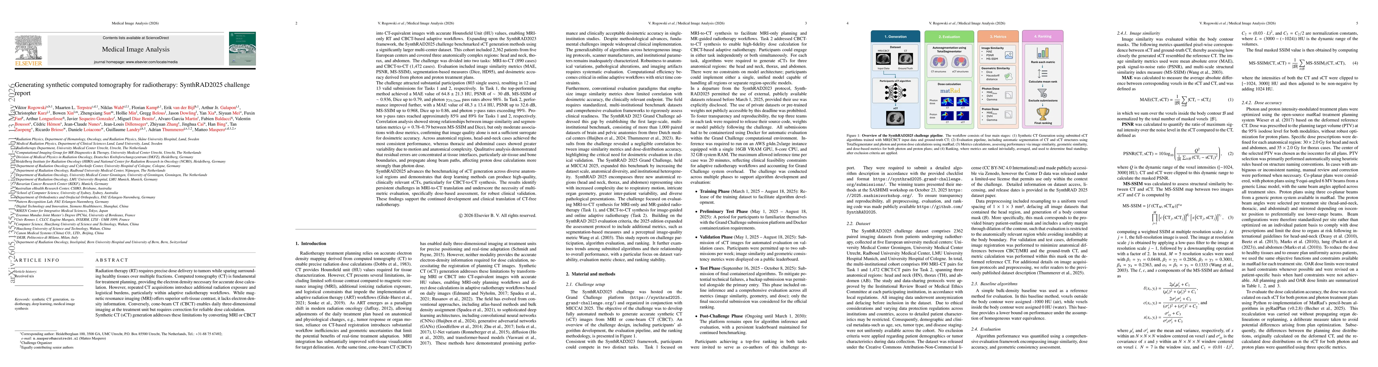
Radiation therapy (RT) requires precise dose delivery over multiple fractions, with CT fundamental for treatment planning due to its electron density information. Repeated CT acquisitions impose radia...
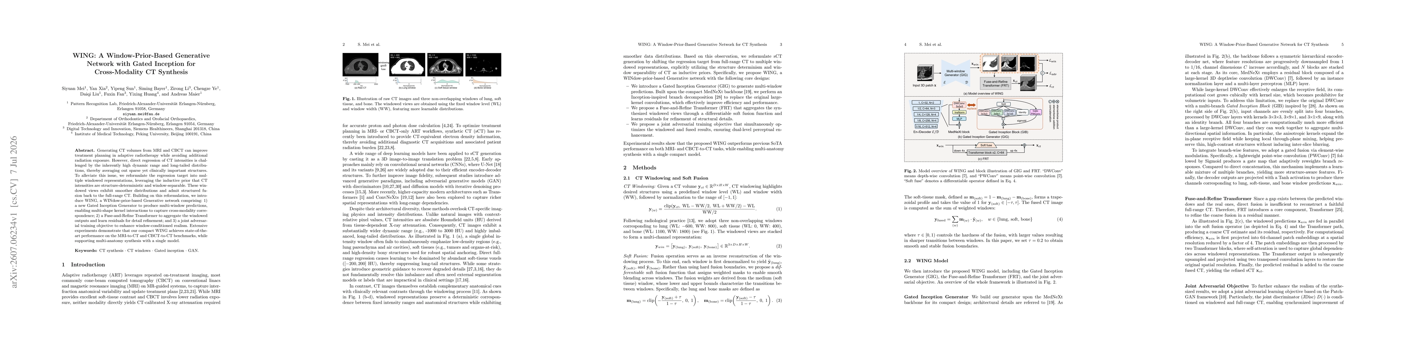
Generating CT volumes from MRI and CBCT can improve treatment planning in adaptive radiotherapy while avoiding additional radiation exposure. However, direct regression of CT intensities is challenged...