01
MethodologyHow they did it
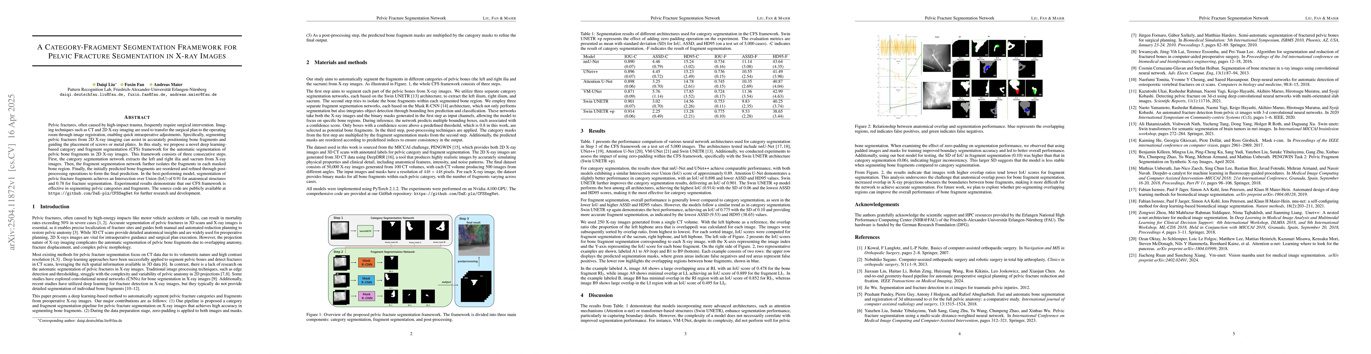
The research proposes a Category-Fragment Segmentation (CFS) framework for automatic segmentation of pelvic bone fragments in 2D X-ray images. It consists of three steps: category segmentation, fragment segmentation, and post-processing. The category segmentation uses SwinUNETR architecture, while fragment segmentation employs MaskR-CNN architecture. The dataset, sourced from the MICCAI challenge PENGWIN, contains 50,000 X-ray images generated from 100 CT volumes.
Discussion 0