Academic Profile
Statistics
Similar Authors
Papers on arXiv
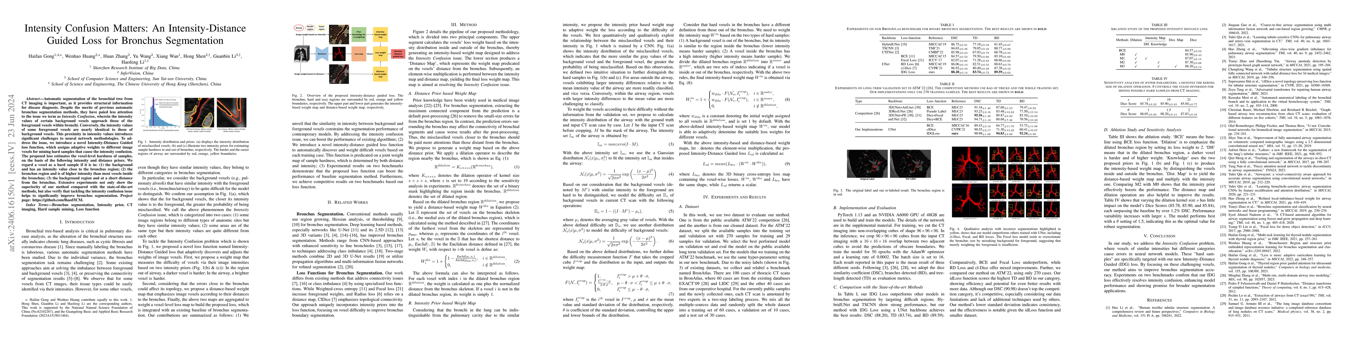
Automatic segmentation of the bronchial tree from CT imaging is important, as it provides structural information for disease diagnosis. Despite the merits of previous automatic bronchus segmentation m...
Recently, self-supervised learning (SSL) methods have been used in pre-training the segmentation models for 2D and 3D medical images. Most of these methods are based on reconstruction, contrastive l...
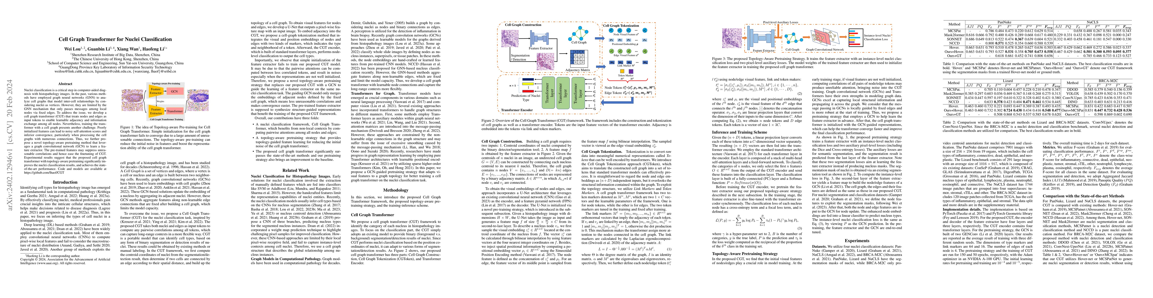
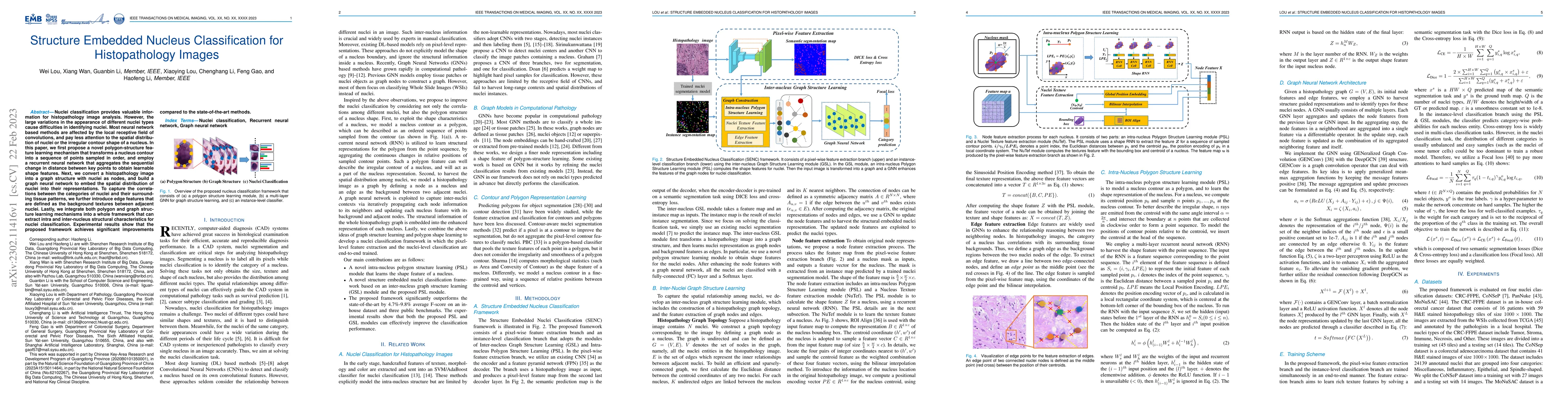
Nuclei classification is a critical step in computer-aided diagnosis with histopathology images. In the past, various methods have employed graph neural networks (GNN) to analyze cell graphs that mo...
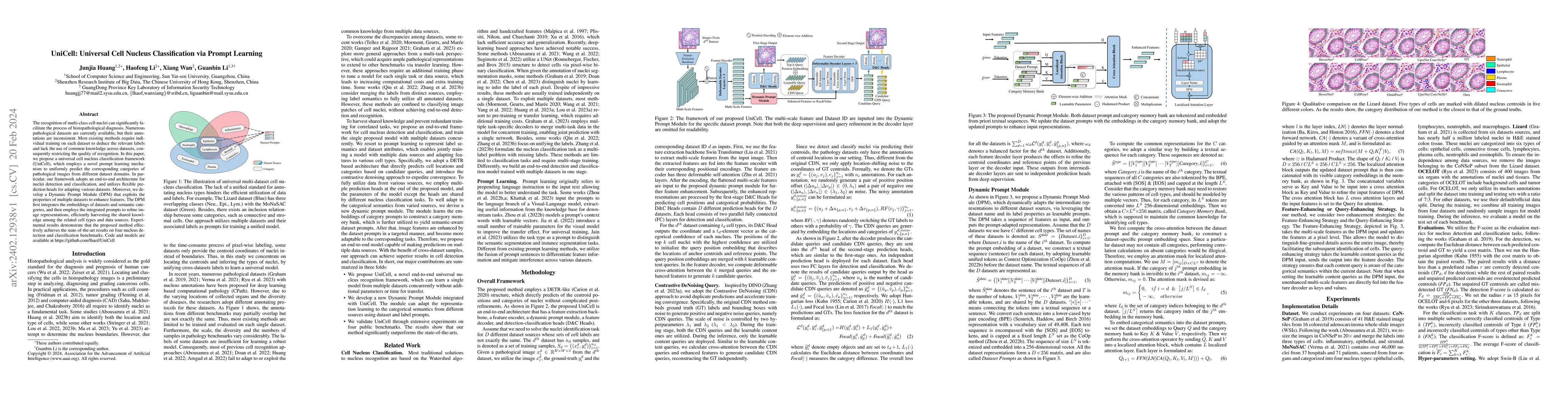
The recognition of multi-class cell nuclei can significantly facilitate the process of histopathological diagnosis. Numerous pathological datasets are currently available, but their annotations are ...
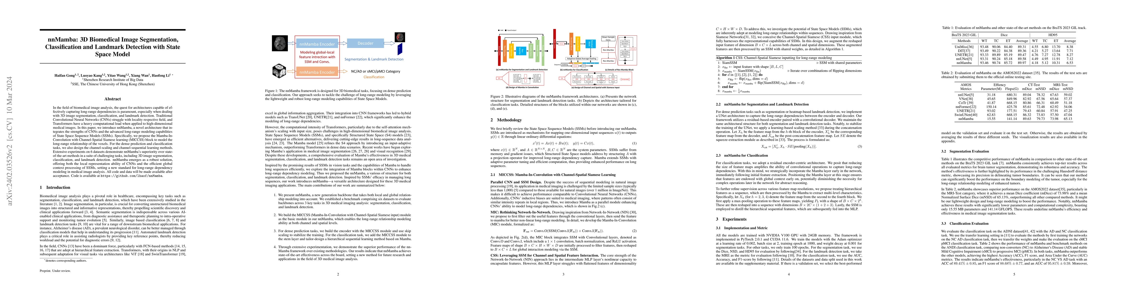
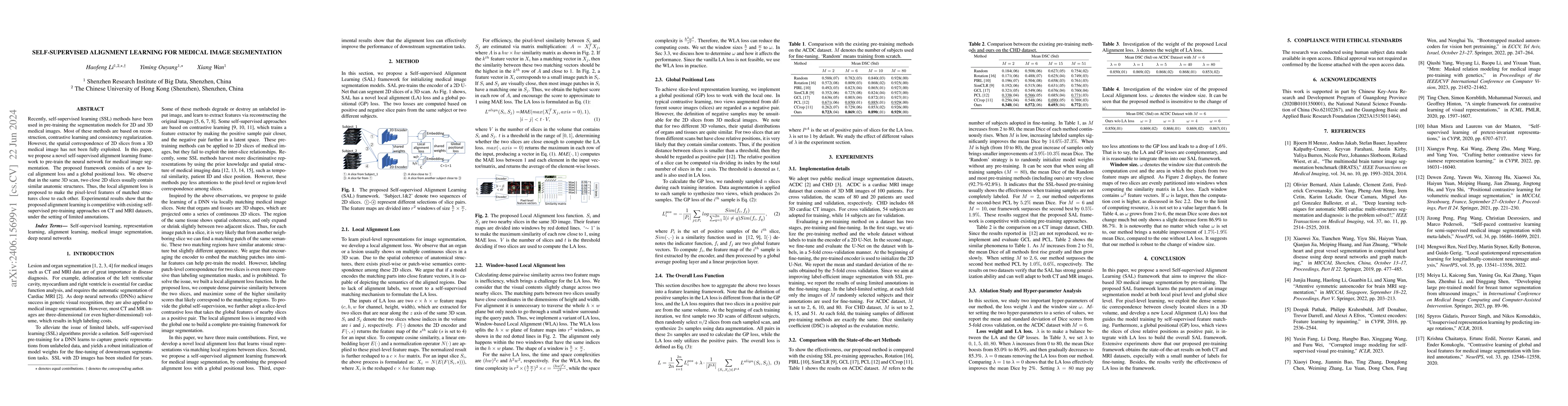
In the field of biomedical image analysis, the quest for architectures capable of effectively capturing long-range dependencies is paramount, especially when dealing with 3D image segmentation, clas...
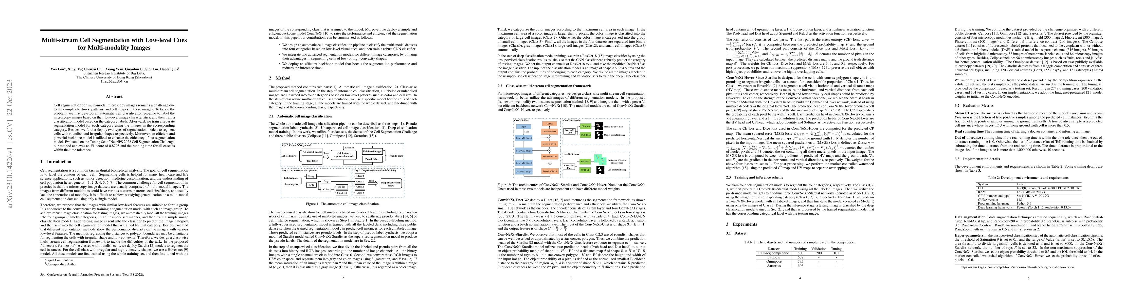
Cell segmentation for multi-modal microscopy images remains a challenge due to the complex textures, patterns, and cell shapes in these images. To tackle the problem, we first develop an automatic c...
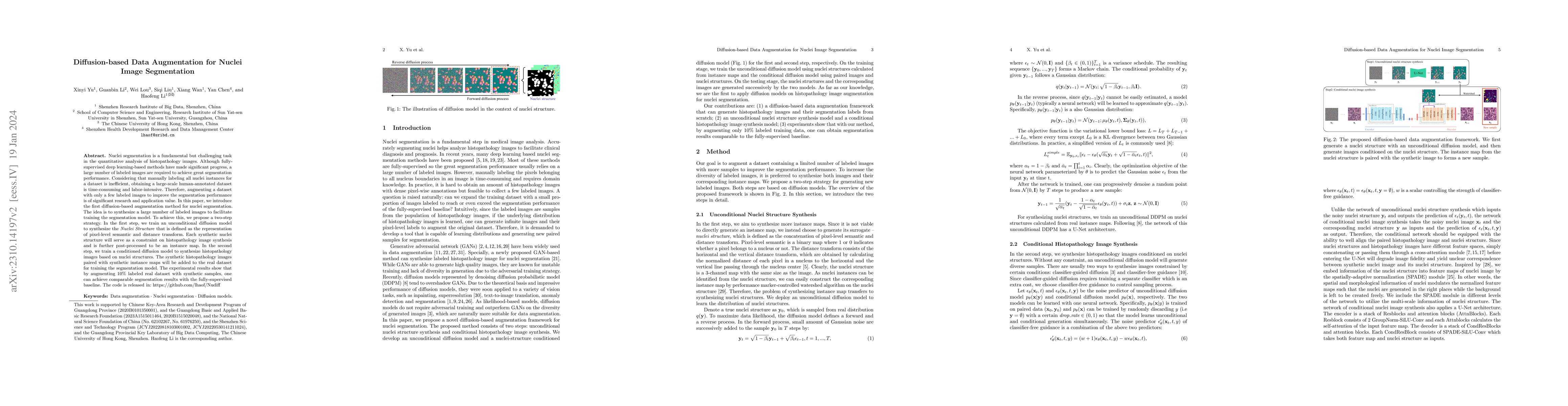
Nuclei segmentation is a fundamental but challenging task in the quantitative analysis of histopathology images. Although fully-supervised deep learning-based methods have made significant progress,...
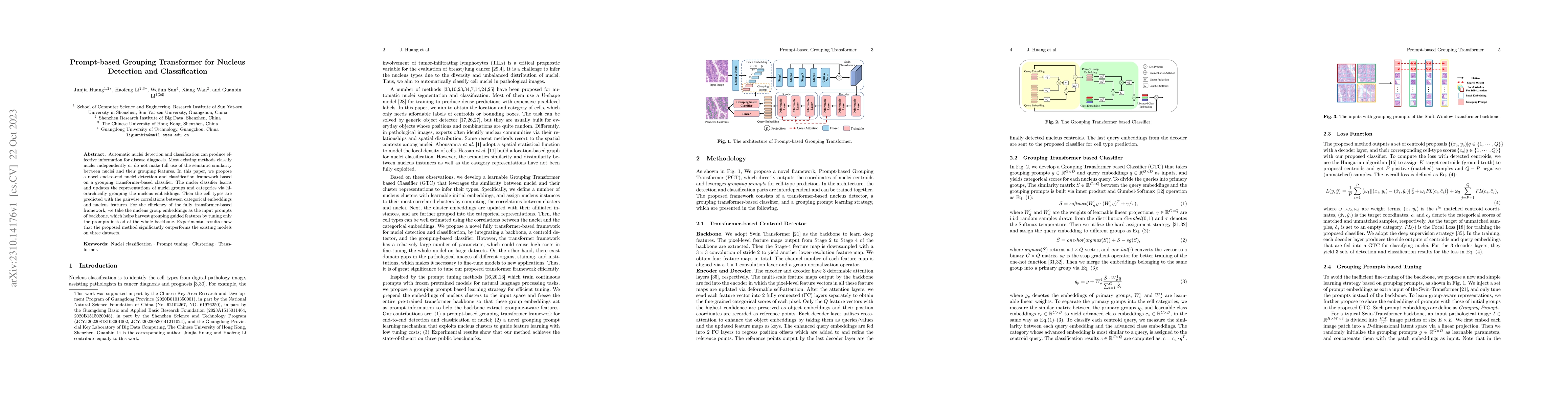
Automatic nuclei detection and classification can produce effective information for disease diagnosis. Most existing methods classify nuclei independently or do not make full use of the semantic sim...

Automatic tissue segmentation of fetal brain images is essential for the quantitative analysis of prenatal neurodevelopment. However, producing voxel-level annotations of fetal brain imaging is time...
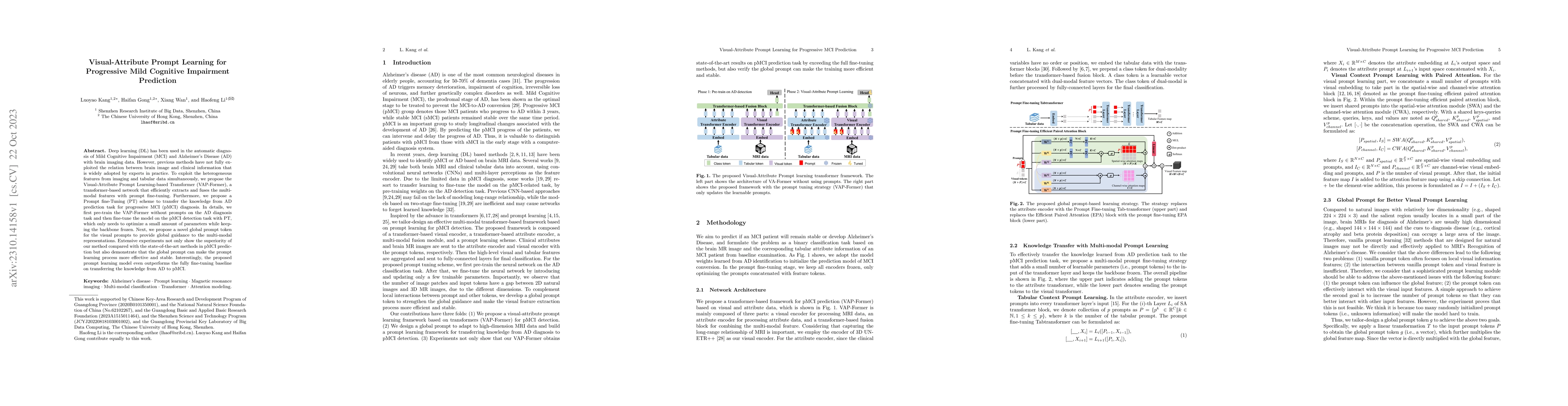
Deep learning (DL) has been used in the automatic diagnosis of Mild Cognitive Impairment (MCI) and Alzheimer's Disease (AD) with brain imaging data. However, previous methods have not fully exploite...
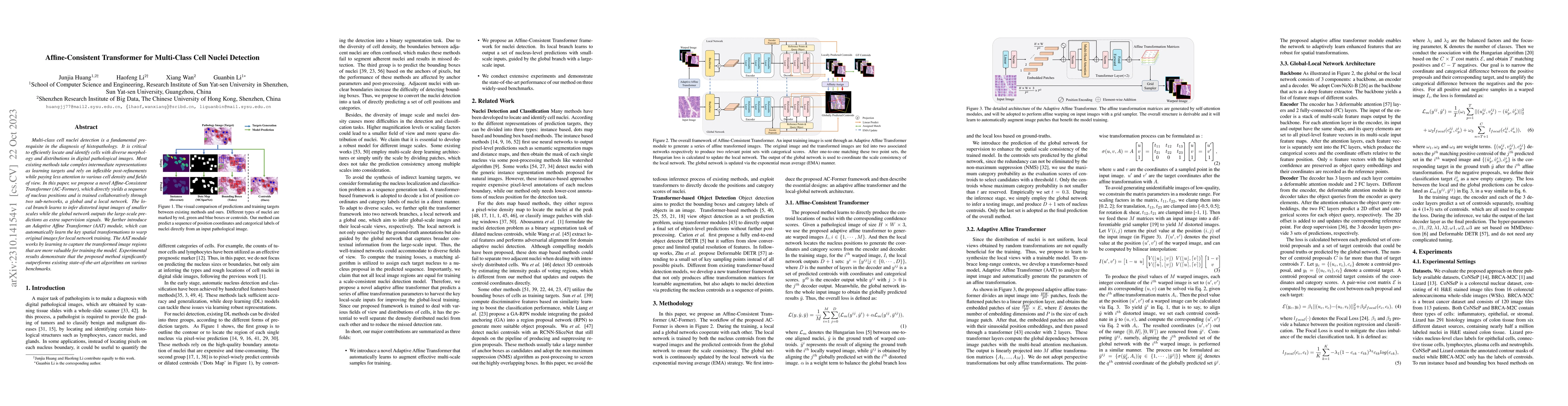
Multi-class cell nuclei detection is a fundamental prerequisite in the diagnosis of histopathology. It is critical to efficiently locate and identify cells with diverse morphology and distributions ...
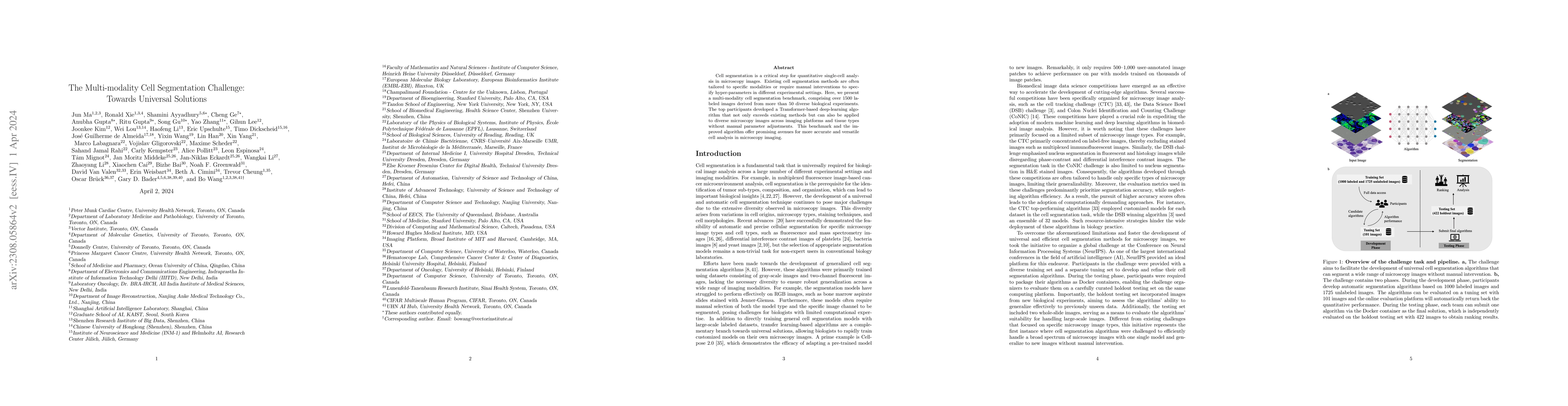
Cell segmentation is a critical step for quantitative single-cell analysis in microscopy images. Existing cell segmentation methods are often tailored to specific modalities or require manual interv...
Nuclei classification provides valuable information for histopathology image analysis. However, the large variations in the appearance of different nuclei types cause difficulties in identifying nuc...
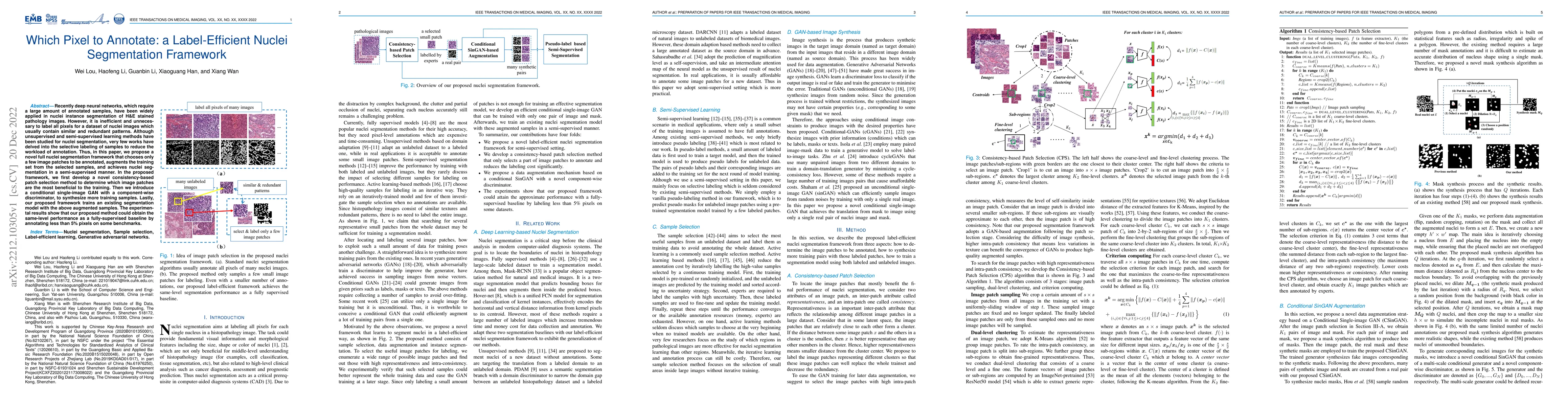
Recently deep neural networks, which require a large amount of annotated samples, have been widely applied in nuclei instance segmentation of H\&E stained pathology images. However, it is inefficien...
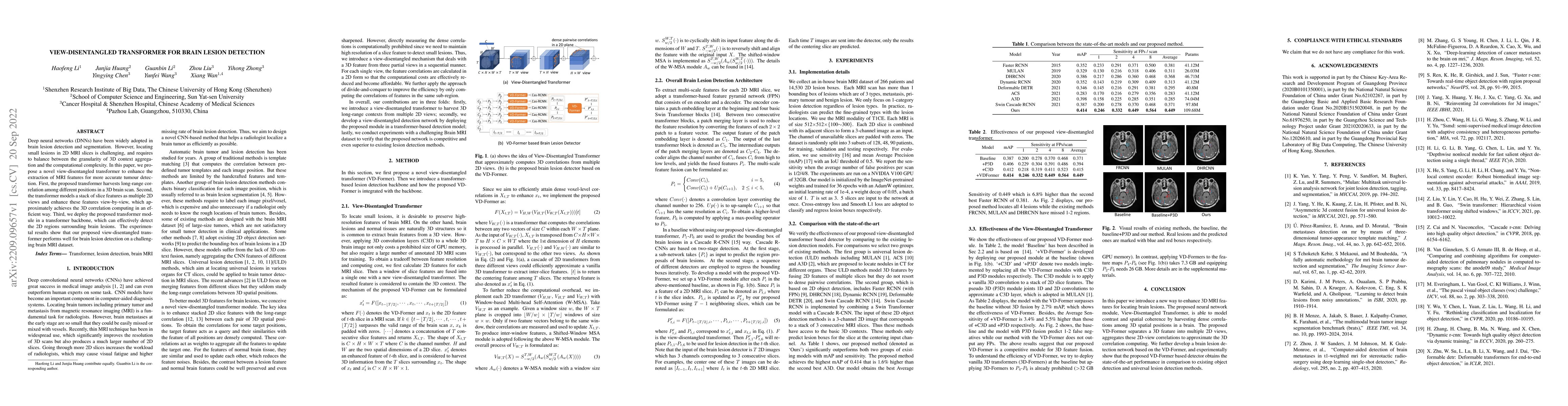
Deep neural networks (DNNs) have been widely adopted in brain lesion detection and segmentation. However, locating small lesions in 2D MRI slices is challenging, and requires to balance between the ...
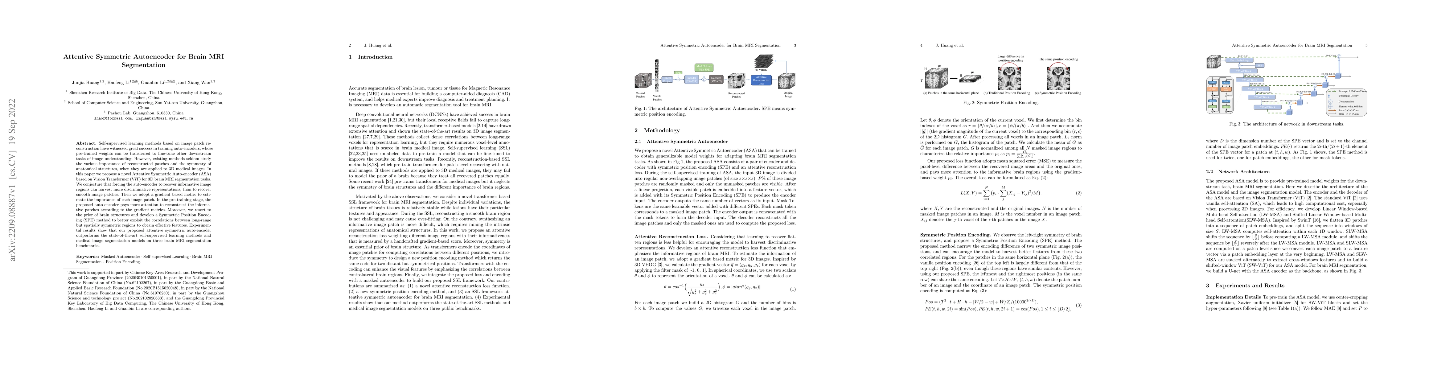
Self-supervised learning methods based on image patch reconstruction have witnessed great success in training auto-encoders, whose pre-trained weights can be transferred to fine-tune other downstrea...
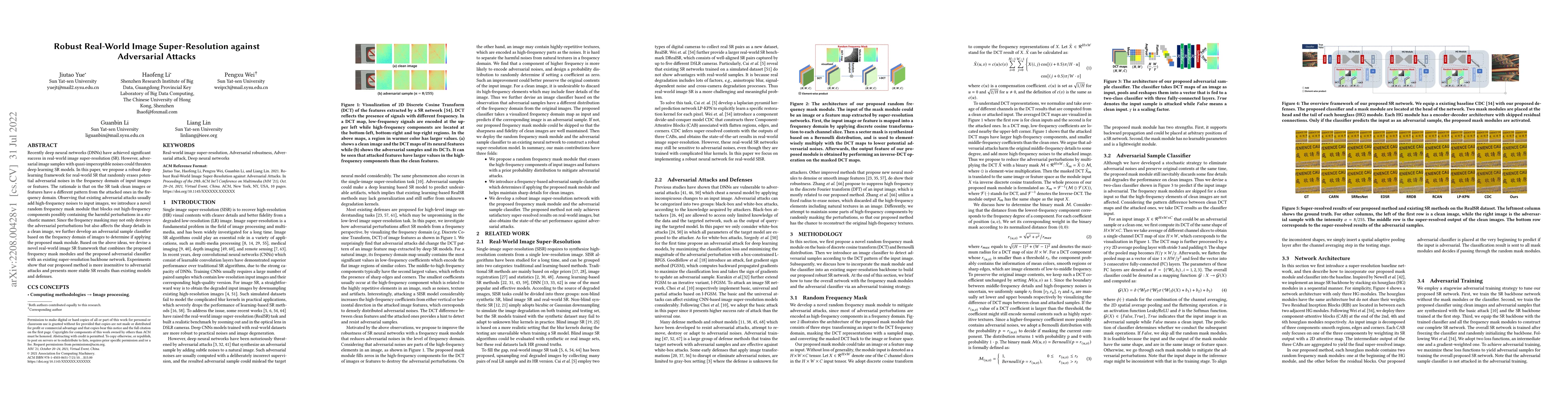
Recently deep neural networks (DNNs) have achieved significant success in real-world image super-resolution (SR). However, adversarial image samples with quasi-imperceptible noises could threaten de...
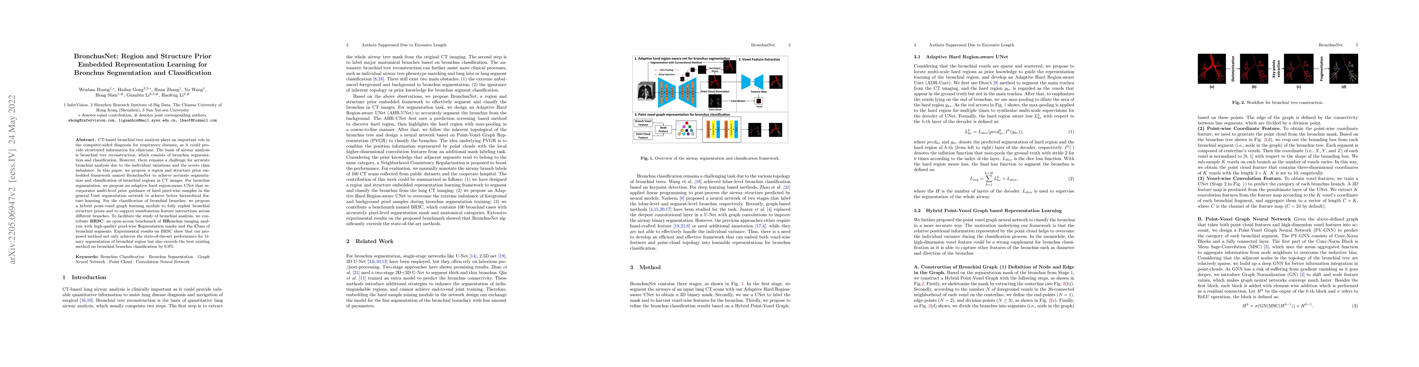
CT-based bronchial tree analysis plays an important role in the computer-aided diagnosis for respiratory diseases, as it could provide structured information for clinicians. The basis of airway anal...
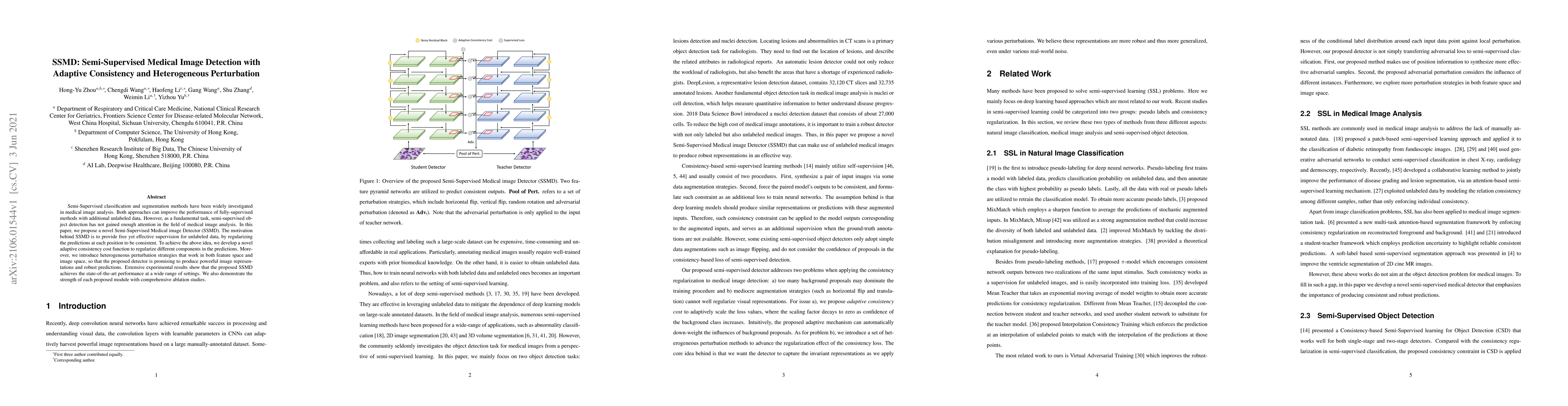
Semi-Supervised classification and segmentation methods have been widely investigated in medical image analysis. Both approaches can improve the performance of fully-supervised methods with addition...
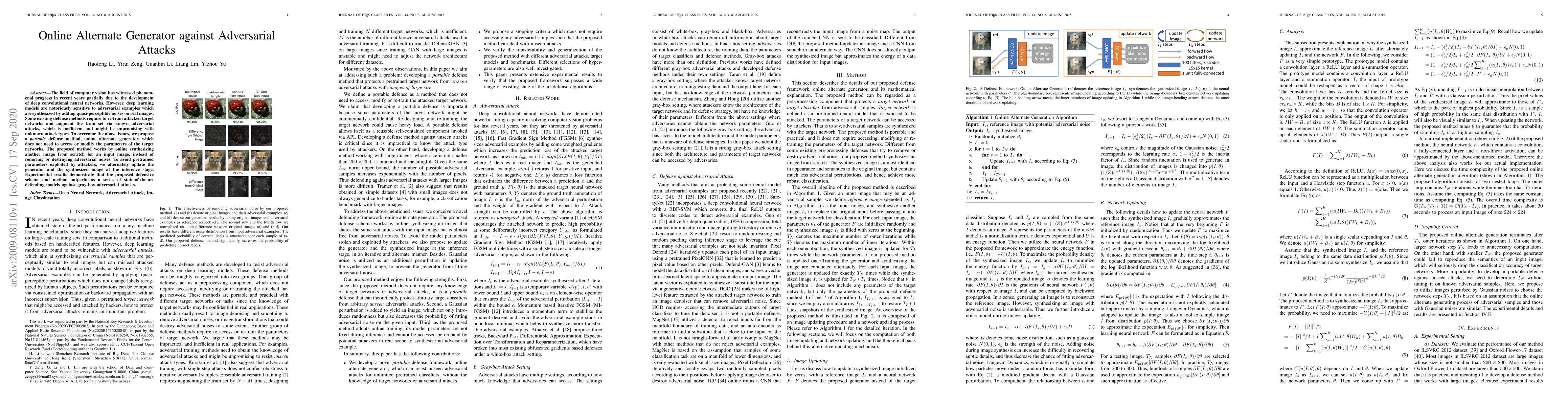
The field of computer vision has witnessed phenomenal progress in recent years partially due to the development of deep convolutional neural networks. However, deep learning models are notoriously s...
Recently deep convolutional neural networks have achieved significant success in salient object detection. However, existing state-of-the-art methods require high-end GPUs to achieve real-time perfo...
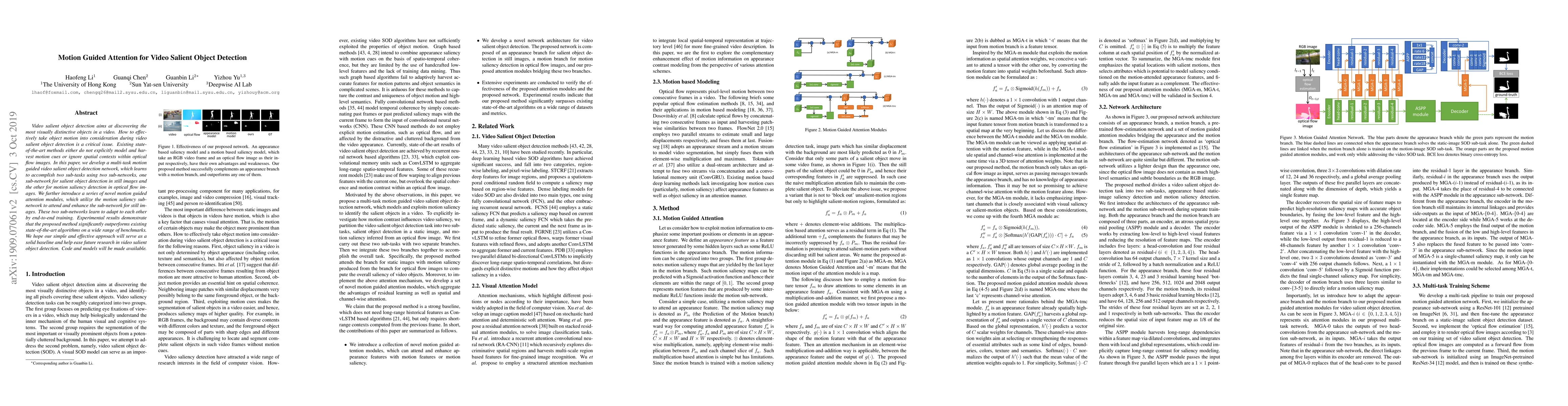
Video salient object detection aims at discovering the most visually distinctive objects in a video. How to effectively take object motion into consideration during video salient object detection is...
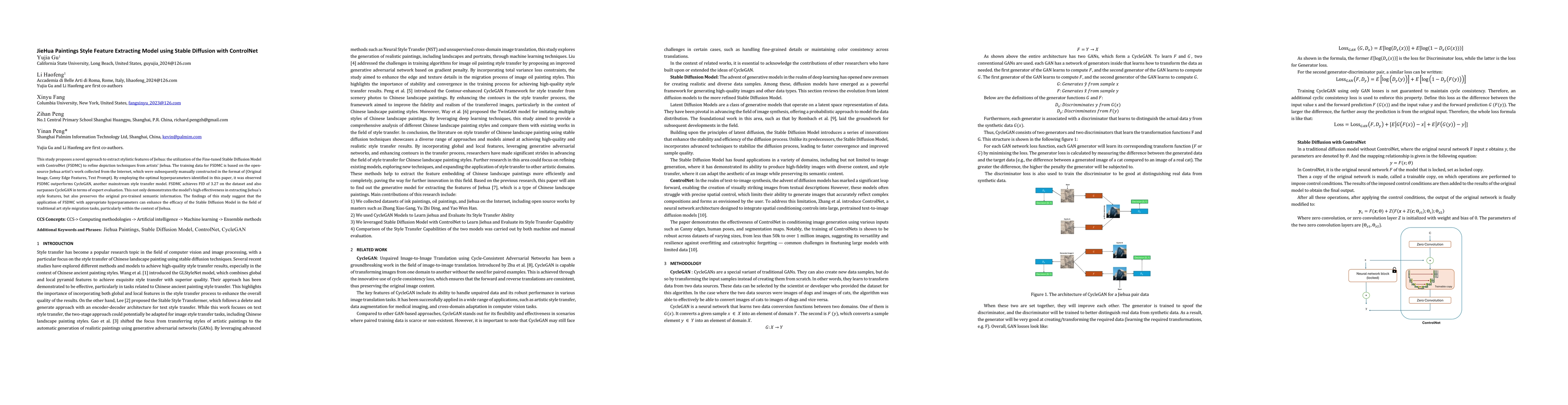
This study proposes a novel approach to extract stylistic features of Jiehua: the utilization of the Fine-tuned Stable Diffusion Model with ControlNet (FSDMC) to refine depiction techniques from artis...
The scarcity and complexity of voxel-level annotations in 3D medical imaging present significant challenges, particularly due to the domain gap between labeled datasets from well-resourced centers and...
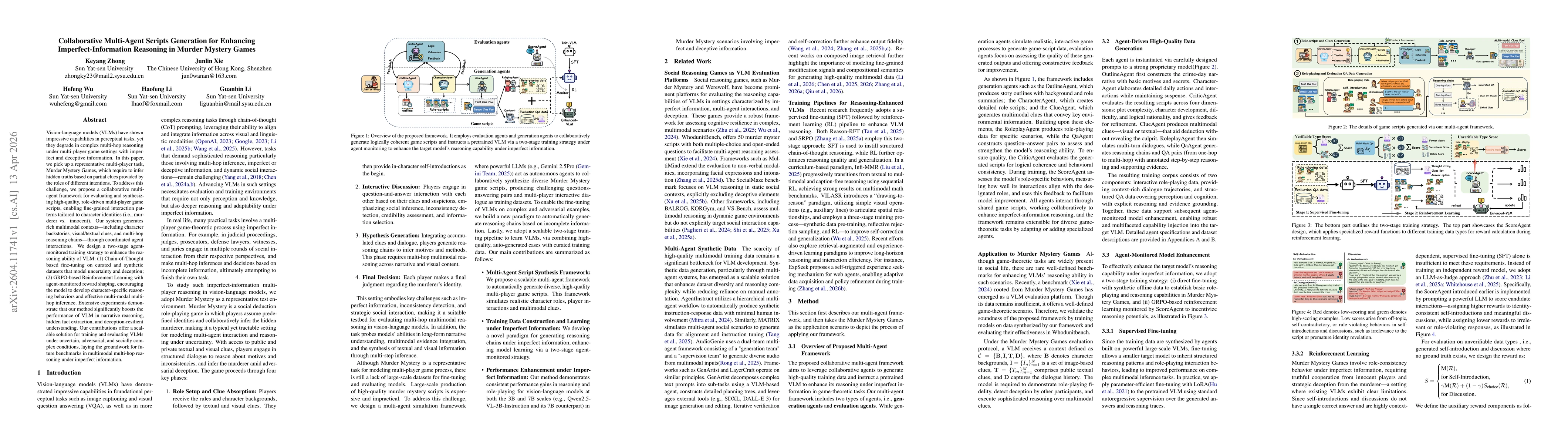
Vision-language models (VLMs) have shown impressive capabilities in perceptual tasks, yet they degrade in complex multi-hop reasoning under multiplayer game settings with imperfect and deceptive infor...
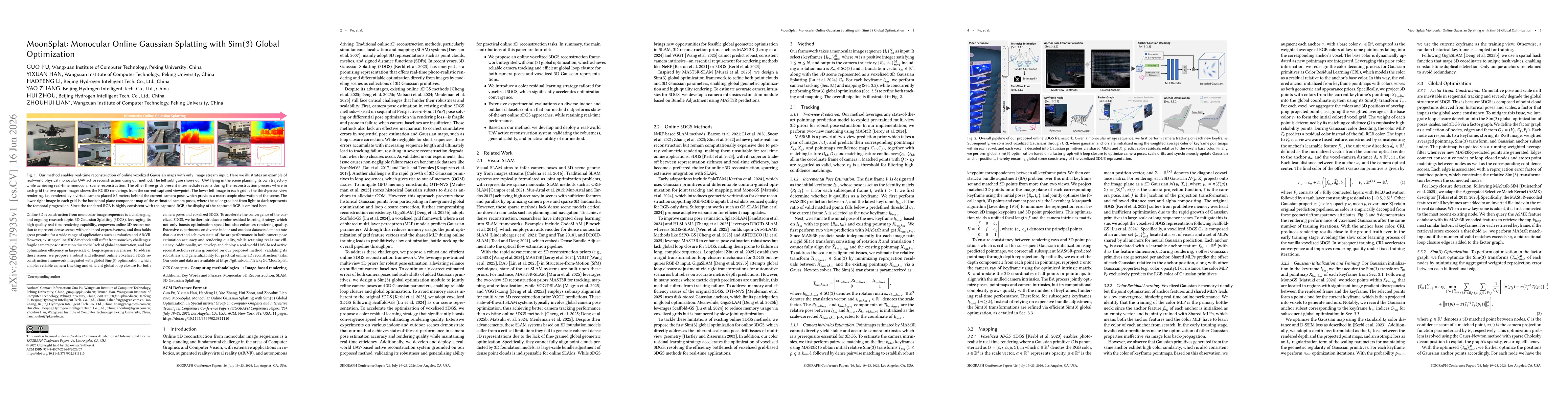
Online 3D reconstruction from monocular image sequences is a challenging and ongoing research topic. 3D Gaussian Splatting (3DGS), leveraging its high-quality real-time rendering capability, empowers ...
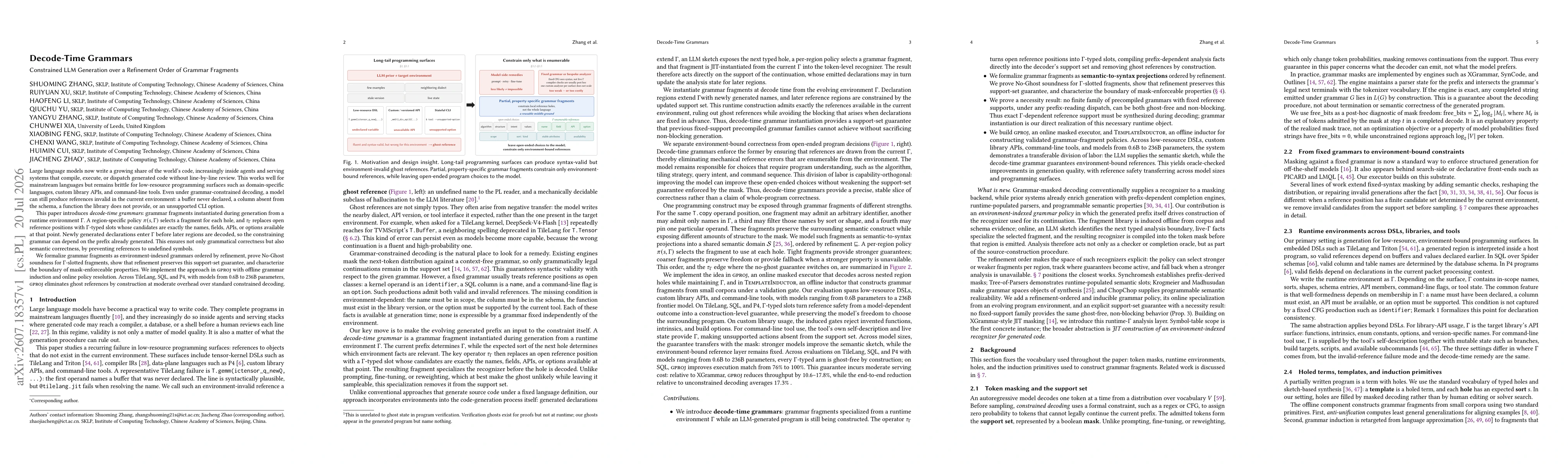
Large language models now write a growing share of the world's code, increasingly inside agents and serving systems that compile, execute, or dispatch generated code without line-by-line review. This ...