Quick Summary:

This paper presents a multi-stream cell segmentation method for multi-modality microscopy images, utilizing low-level image features for automatic classification and segmentation. The method achieves an F1-score of 0.8795 on the NeurIPS 2022 Cell Segmentation Challenge Tuning Set, demonstrating efficient and accurate segmentation of both roundish and irregular cell shapes.

Summary

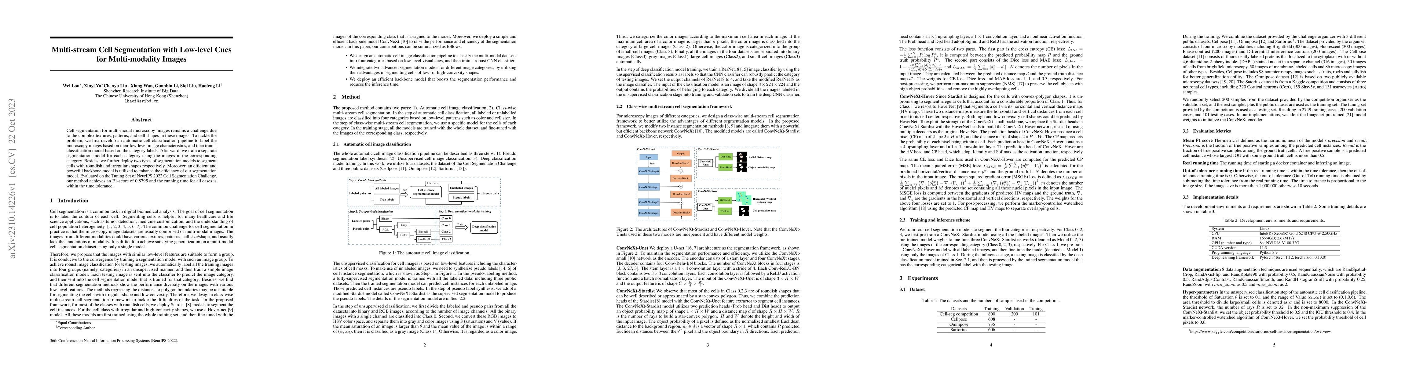
Cell segmentation for multi-modal microscopy images remains a challenge due to the complex textures, patterns, and cell shapes in these images. To tackle the problem, we first develop an automatic cell classification pipeline to label the microscopy images based on their low-level image characteristics, and then train a classification model based on the category labels. Afterward, we train a separate segmentation model for each category using the images in the corresponding category. Besides, we further deploy two types of segmentation models to segment cells with roundish and irregular shapes respectively. Moreover, an efficient and powerful backbone model is utilized to enhance the efficiency of our segmentation model. Evaluated on the Tuning Set of NeurIPS 2022 Cell Segmentation Challenge, our method achieves an F1-score of 0.8795 and the running time for all cases is within the time tolerance.

AI Key Findings

Get AI-generated insights about this paper's methodology, results, and significance.

Paper Details

PDF Preview

Download PDF

Key Terms

segmentation (0.323)
segmentation (0.306)
cell (0.275)
cell (0.264)
category (0.255)
category (0.245)
images (0.226)
microscopy (0.224)
shapes (0.219)
images (0.218)
microscopy (0.211)
shapes (0.210)
train (0.198)
train (0.190)
challenge (0.175)
challenge (0.171)
classification (0.163)
classification (0.162)
multi modal (0.153)
tolerance (0.149)

Citation Network

Current paper (gray), citations (green), references (blue)

Current Paper
Citations
References
Click to view
Title Authors Year Actions
Unsupervised Deep Learning Method for Cell Segmentation of Confocal Microscopy Images
Ognjen Pavić, Jorge Barrasa-Fano, Lazar Dasic, Tijana Geroski, A. Shapeti, H. Oosterwyck, Vesna Ranković, Nenad Filipović
2024
View Paper
Cell Graph Transformer for Nuclei Classification
Wei Lou, Guanbin Li, Xiang Wan, Haofeng Li
2024
View Paper
Diffusion-Based Data Augmentation for Nuclei Image Segmentation
Xinyi Yu, Guanbin Li, Wei Lou, Siqi Liu, Xiang Wan, Yan Chen, Haofeng Li
2023
View Paper
Prompt-Based Grouping Transformer for Nucleus Detection and Classification
Junjia Huang, Haofeng Li, Weijun Sun, Xiang Wan, Guanbin Li
2023
View Paper
Affine-Consistent Transformer for Multi-Class Cell Nuclei Detection
Junjia Huang, Haofeng Li, Xiang Wan, Guanbin Li
2023
View Paper
The Multi-modality Cell Segmentation Challenge: Towards Universal Solutions
Jun Ma, Ronald Xie, Shamini Ayyadhury, Cheng Ge, Anubha Gupta, Ritu Gupta, Song Gu, Yao Zhang, Gihun Lee, Joonkee Kim, Wei Lou, Haofeng Li, Eric Upschulte, Timo Dickscheid, José Guilherme de Almeida, Yixin Wang, Li-Jun Han, Xin Yang, Marco Labagnara, S. Rahi, Carly Kempster, A. Pollitt, L. Espinosa, T. Mignot, J. Middeke, Jan-Niklas Eckardt, Wangkai Li, Zhaoyang Li, Xiao-Wei Cai, Bizhe Bai, N. Greenwald, D. V. Valen, Erin Weisbart, B. Cimini, Zhuoshi Li, Chao Zuo, Oscar Bruck, Gary D. Bader, Bohui Wang
2023
View Paper
Structure Embedded Nucleus Classification for Histopathology Images
Wei Lou, Xiang Wan, Guanbin Li, X. Lou, Chenghang Li, Feng Gao, Haofeng Li
2023
View Paper

Comments (0)

This website uses cookies to ensure you get the best experience. Learn more