Academic Profile
Statistics
Similar Authors
Papers on arXiv
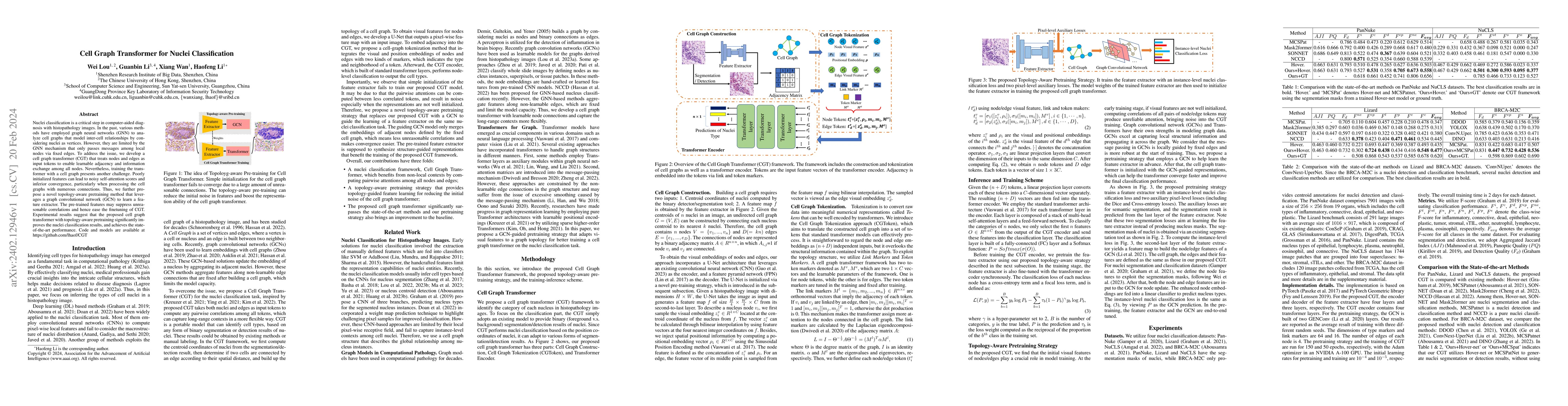
Nuclei classification is a critical step in computer-aided diagnosis with histopathology images. In the past, various methods have employed graph neural networks (GNN) to analyze cell graphs that mo...
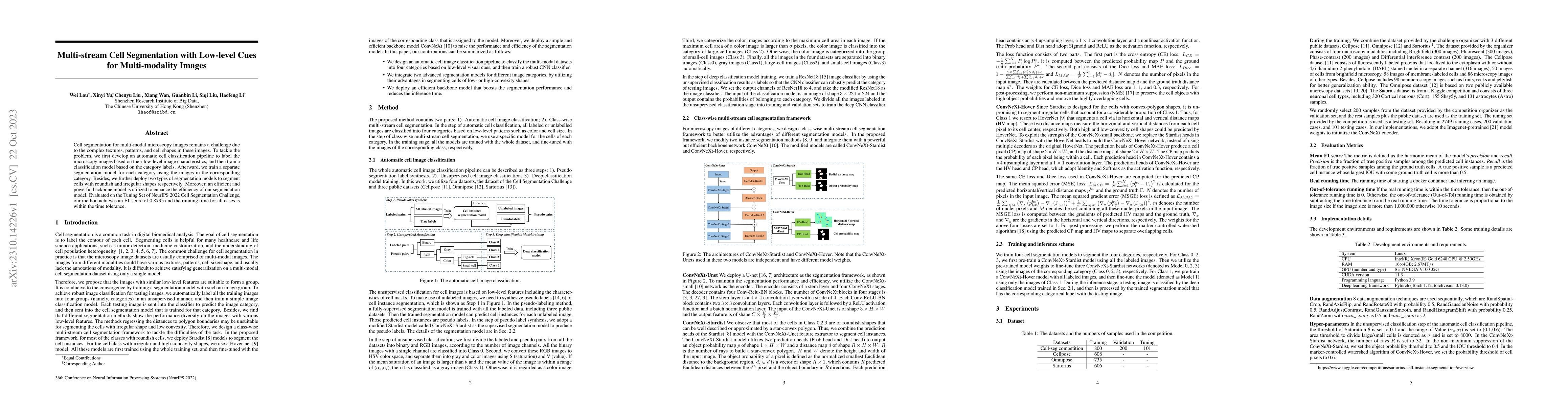
Cell segmentation for multi-modal microscopy images remains a challenge due to the complex textures, patterns, and cell shapes in these images. To tackle the problem, we first develop an automatic c...
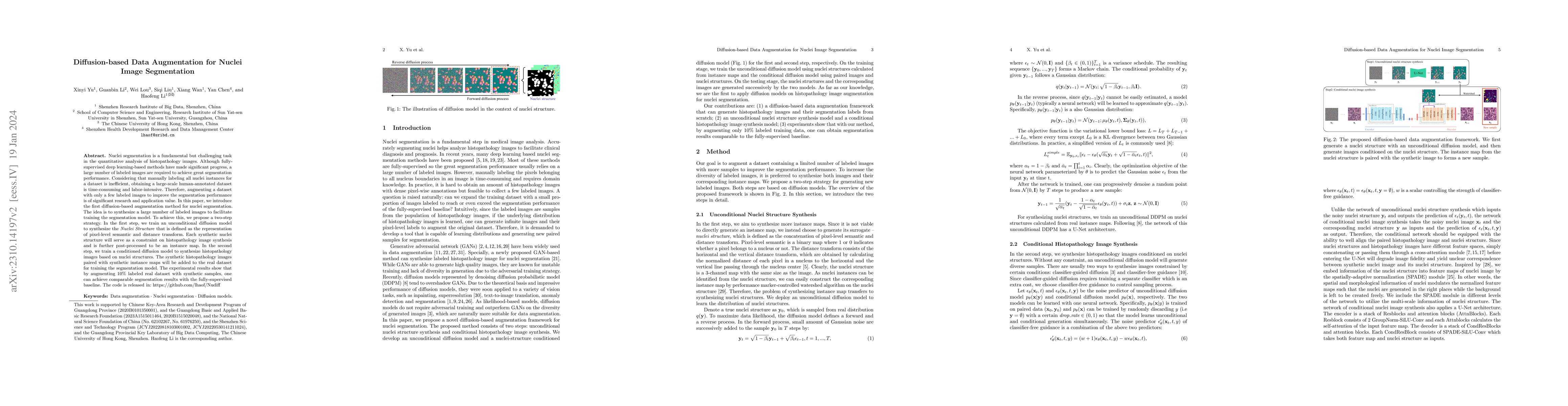
Nuclei segmentation is a fundamental but challenging task in the quantitative analysis of histopathology images. Although fully-supervised deep learning-based methods have made significant progress,...
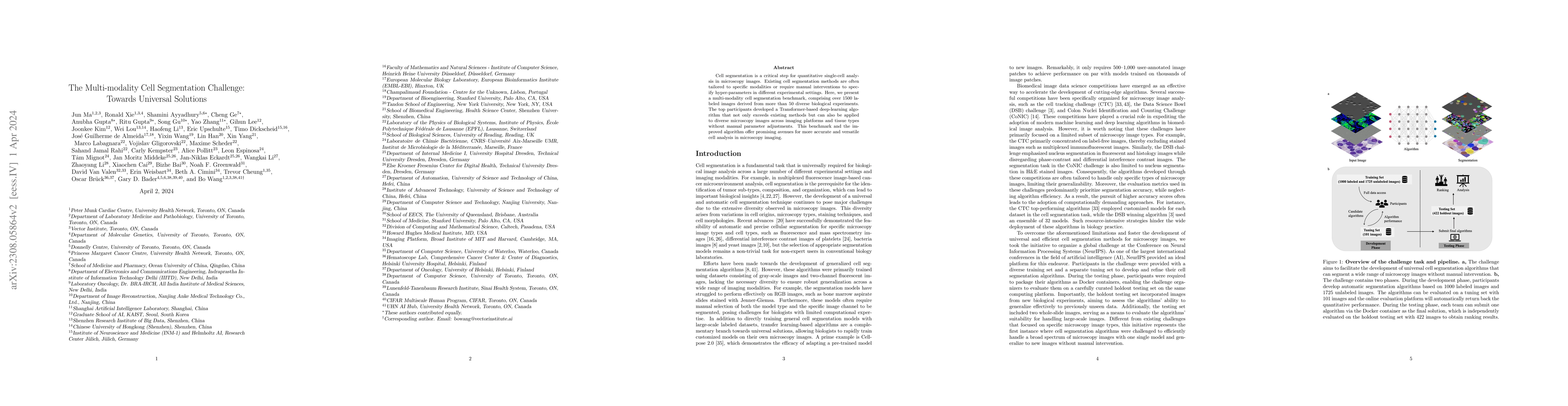
Cell segmentation is a critical step for quantitative single-cell analysis in microscopy images. Existing cell segmentation methods are often tailored to specific modalities or require manual interv...
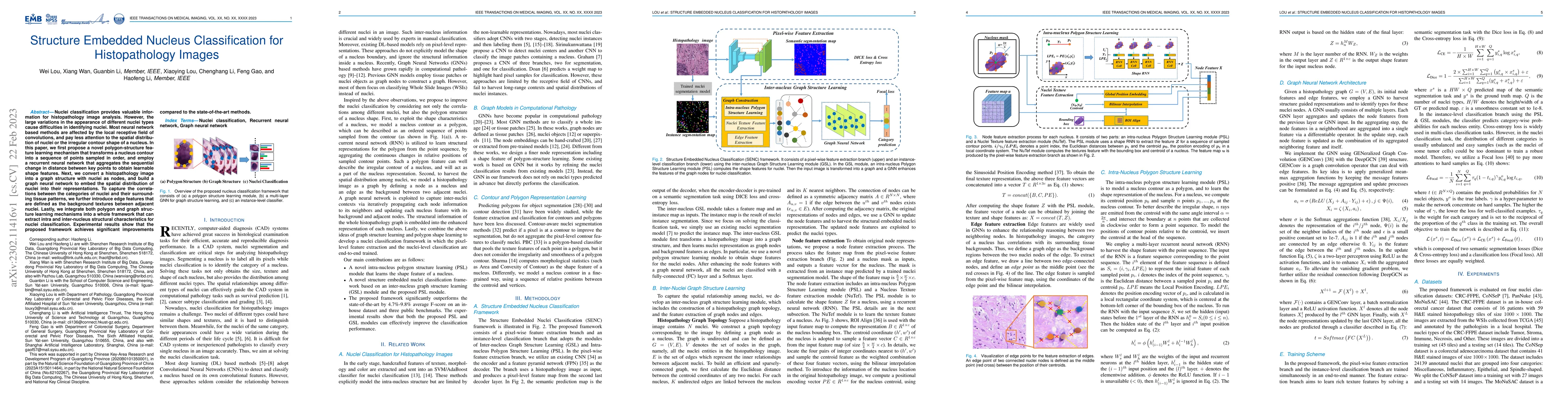
Nuclei classification provides valuable information for histopathology image analysis. However, the large variations in the appearance of different nuclei types cause difficulties in identifying nuc...
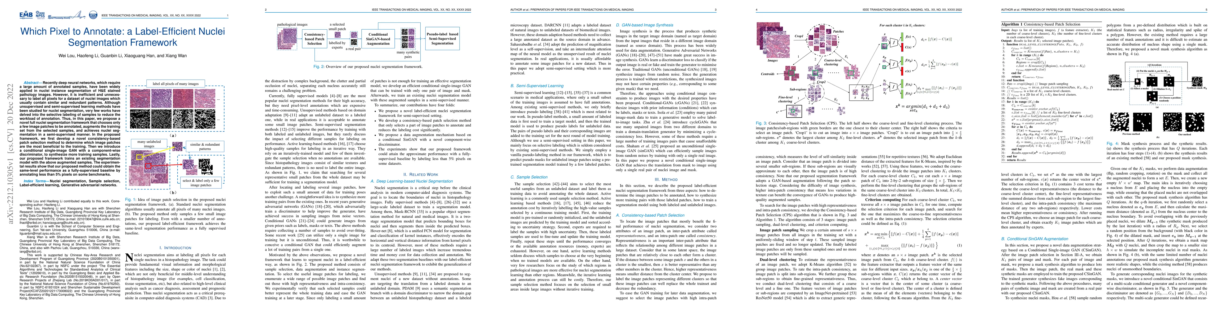
Recently deep neural networks, which require a large amount of annotated samples, have been widely applied in nuclei instance segmentation of H\&E stained pathology images. However, it is inefficien...
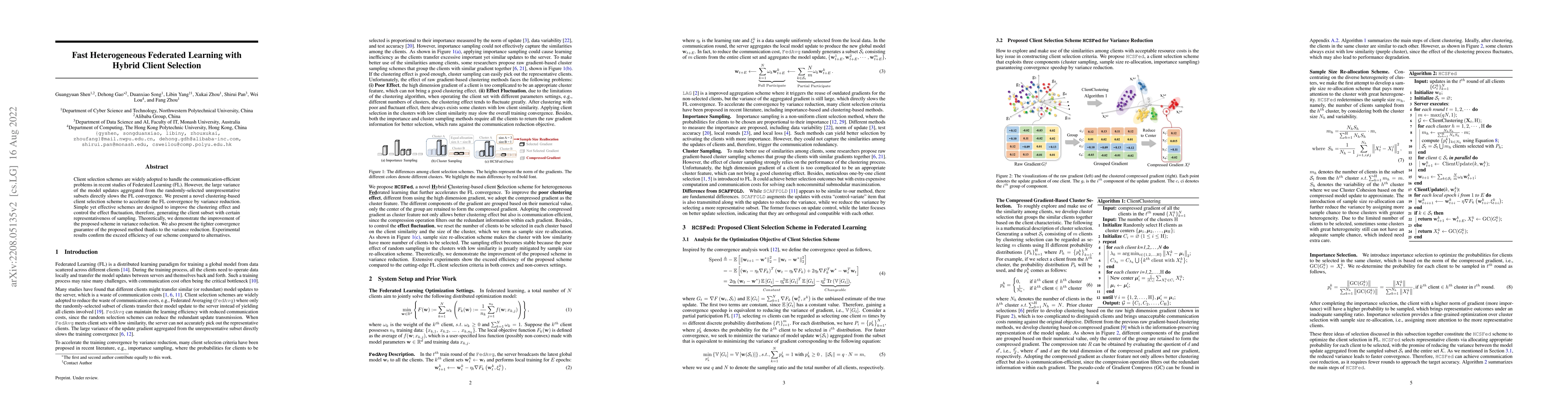
Client selection schemes are widely adopted to handle the communication-efficient problems in recent studies of Federated Learning (FL). However, the large variance of the model updates aggregated f...

Client selection strategies are widely adopted to handle the communication-efficient problem in recent studies of Federated Learning (FL). However, due to the large variance of the selected subset's...