Academic Profile
Statistics
Similar Authors
Papers on arXiv
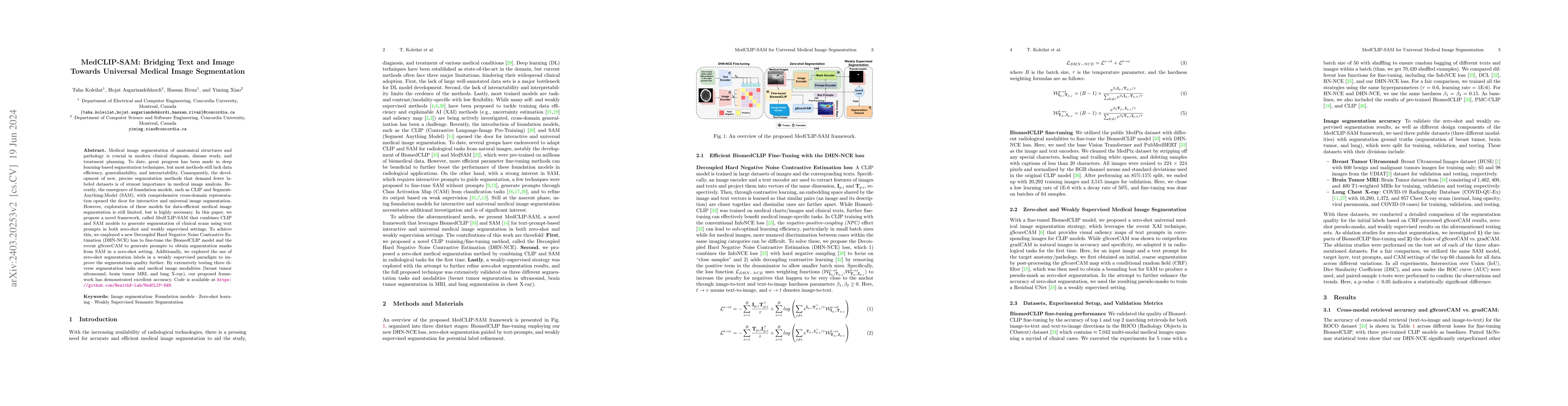
Medical image segmentation of anatomical structures and pathology is crucial in modern clinical diagnosis, disease study, and treatment planning. To date, great progress has been made in deep learning...
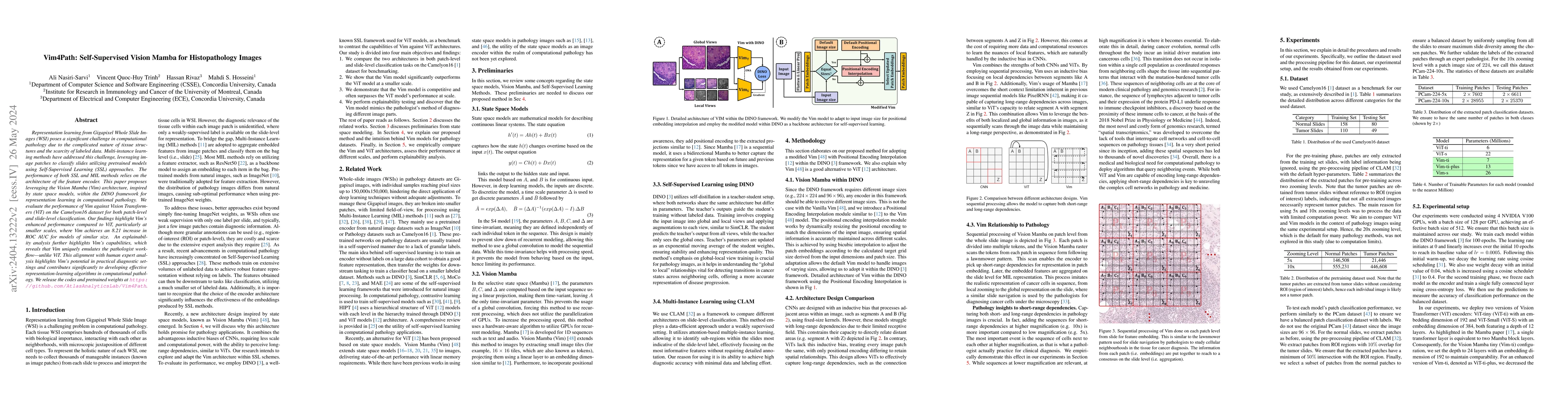
Representation learning from Gigapixel Whole Slide Images (WSI) poses a significant challenge in computational pathology due to the complicated nature of tissue structures and the scarcity of labele...
As deep learning has become the state-of-the-art for computer-assisted diagnosis, interpretability of the automatic decisions is crucial for clinical deployment. While various methods were proposed ...
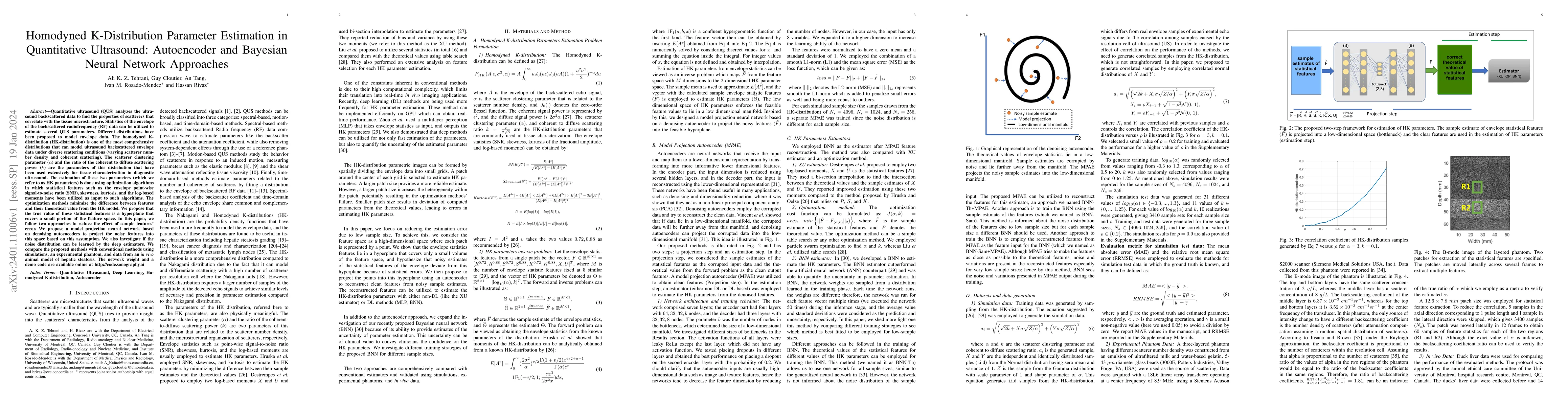
Quantitative ultrasound (QUS) analyzes the ultrasound backscattered data to find the properties of scatterers that correlate with the tissue microstructure. Statistics of the envelope of the backsca...
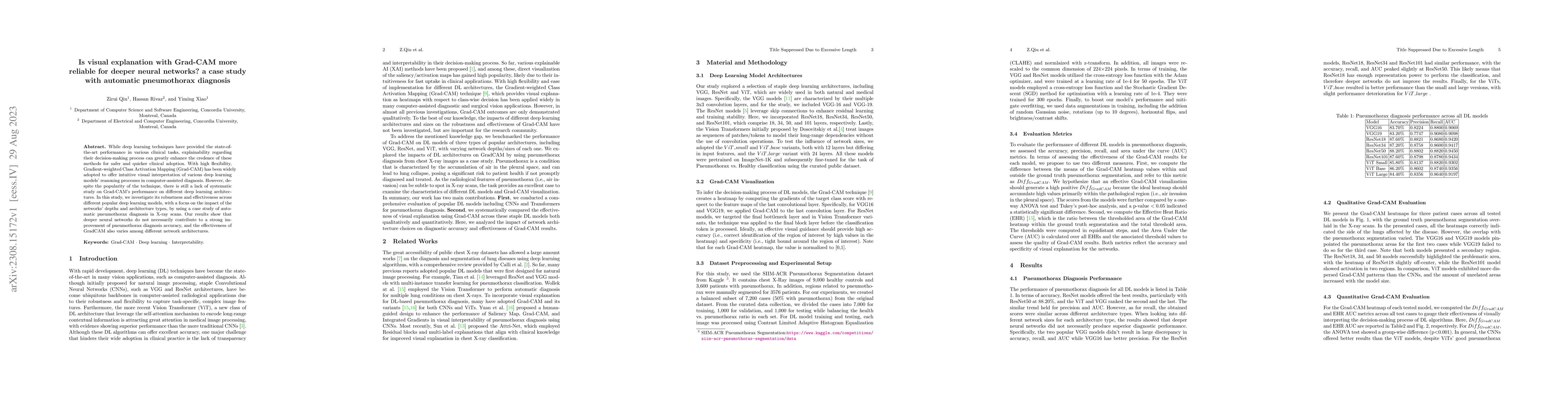
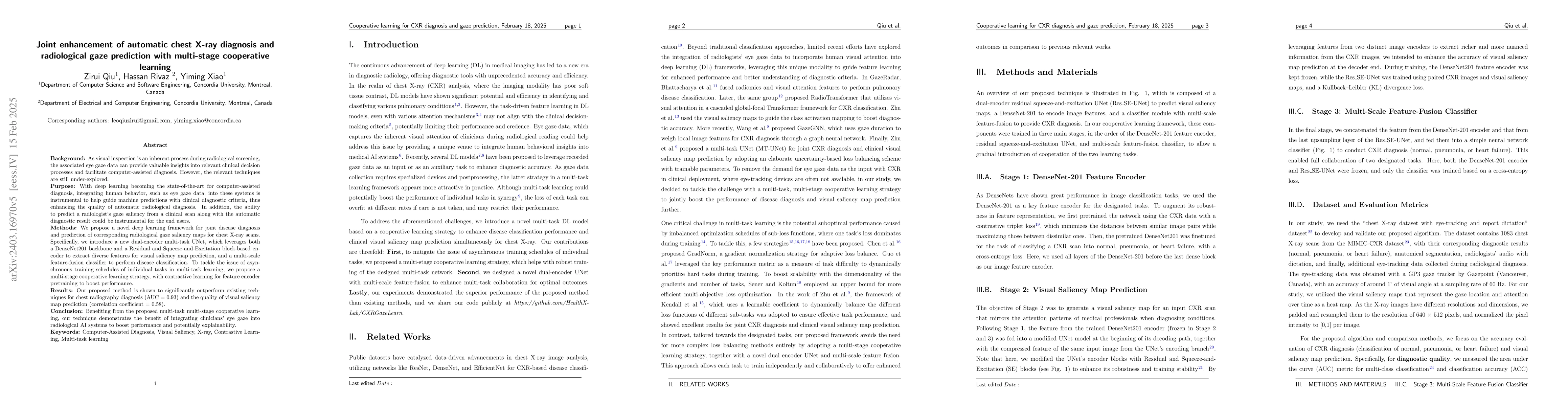
While deep learning techniques have provided the state-of-the-art performance in various clinical tasks, explainability regarding their decision-making process can greatly enhance the credence of th...
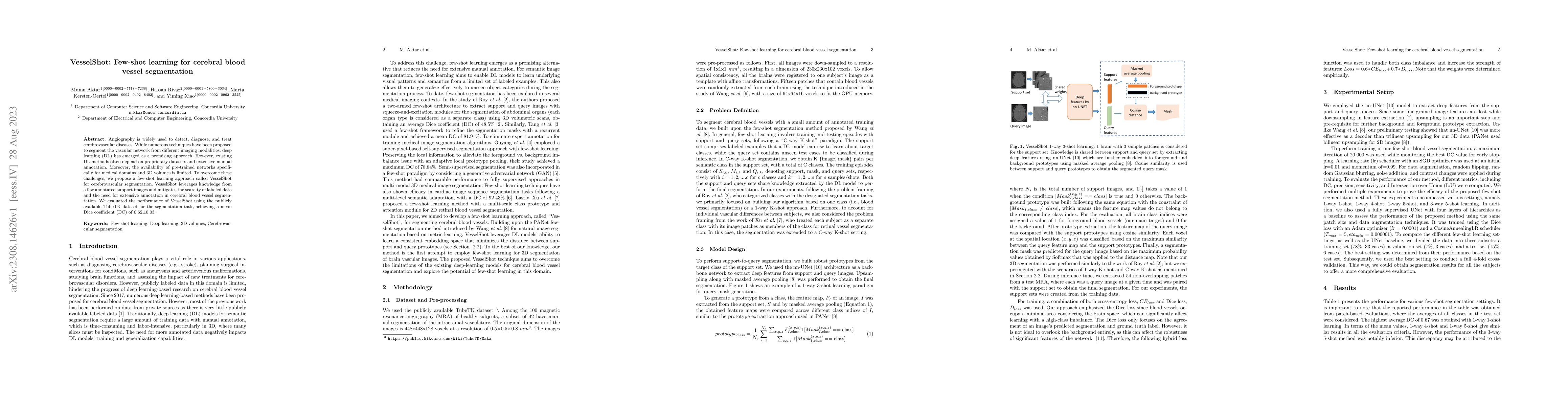
Angiography is widely used to detect, diagnose, and treat cerebrovascular diseases. While numerous techniques have been proposed to segment the vascular network from different imaging modalities, de...
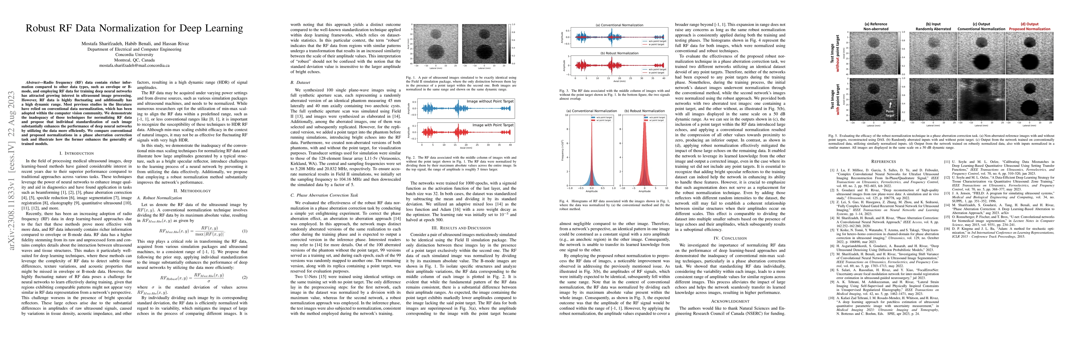
Radio frequency (RF) data contain richer information compared to other data types, such as envelope or B-mode, and employing RF data for training deep neural networks has attracted growing interest ...
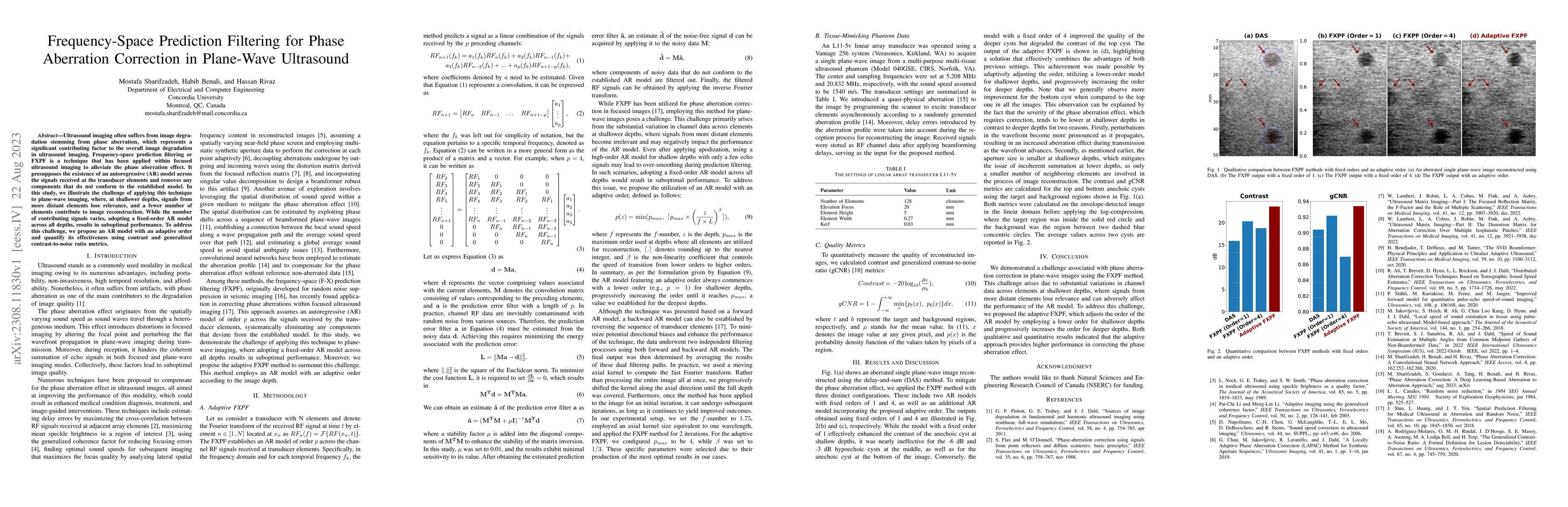
Ultrasound imaging often suffers from image degradation stemming from phase aberration, which represents a significant contributing factor to the overall image degradation in ultrasound imaging. Fre...
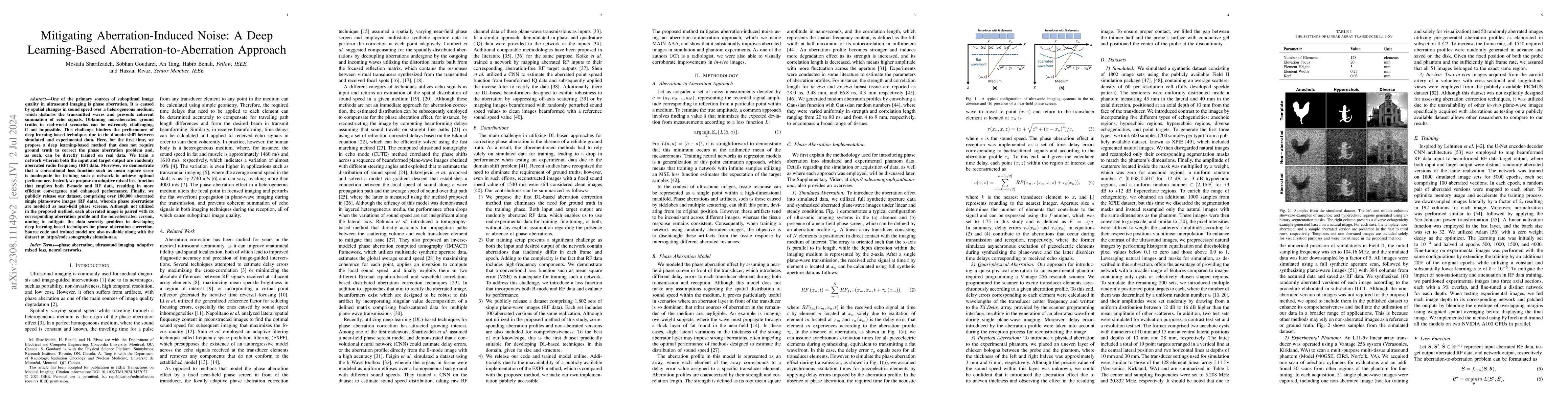
One of the primary sources of suboptimal image quality in ultrasound imaging is phase aberration. It is caused by spatial changes in sound speed over a heterogeneous medium, which disturbs the trans...
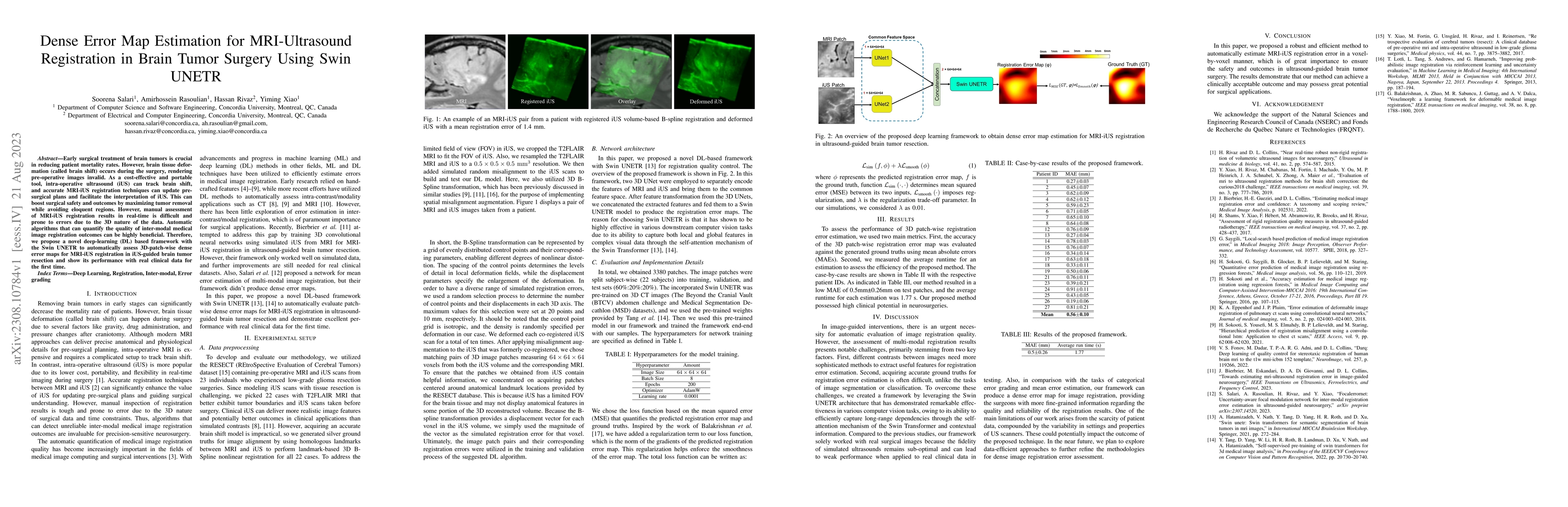
Early surgical treatment of brain tumors is crucial in reducing patient mortality rates. However, brain tissue deformation (called brain shift) occurs during the surgery, rendering pre-operative ima...
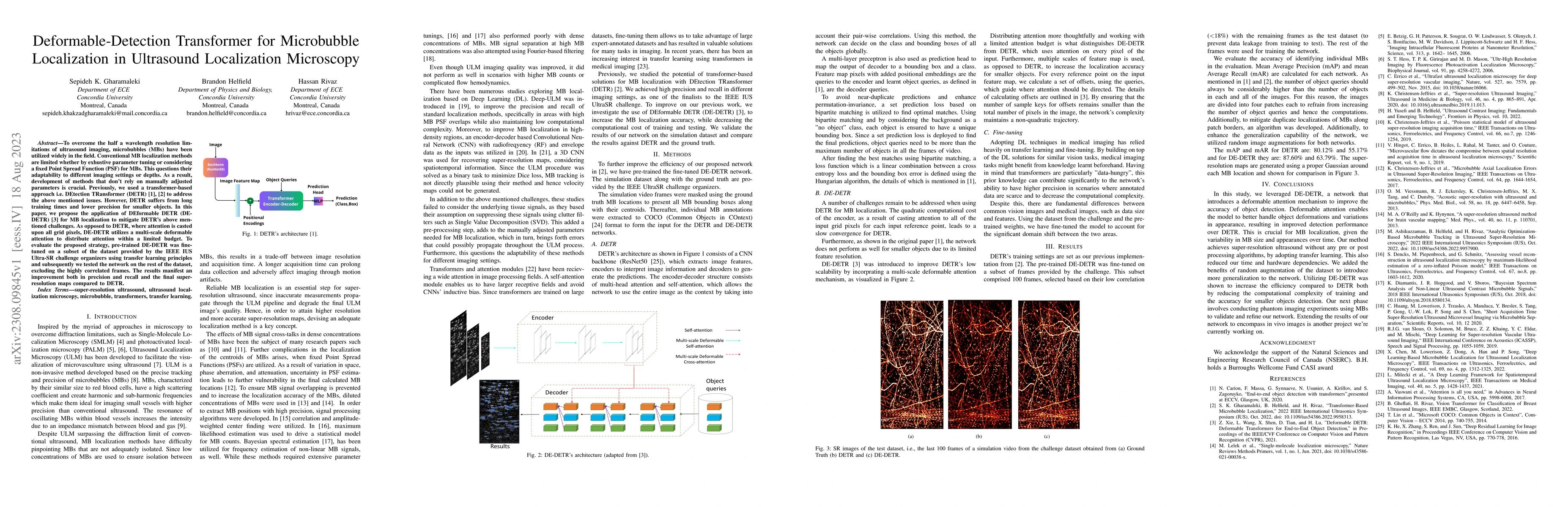
To overcome the half a wavelength resolution limitations of ultrasound imaging, microbubbles (MBs) have been utilized widely in the field. Conventional MB localization methods are limited whether by...
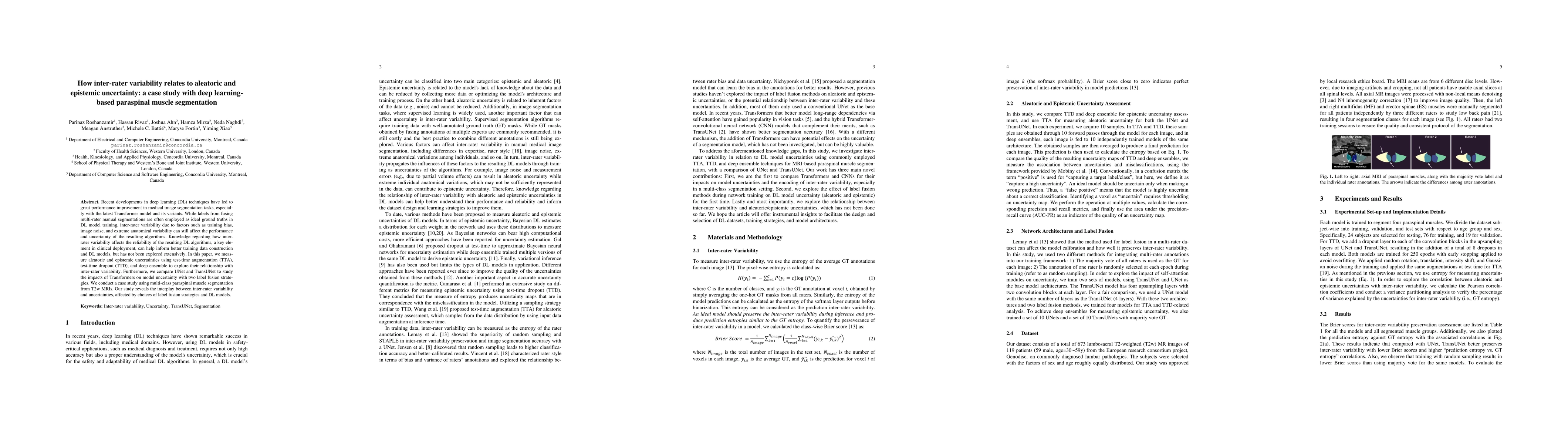
Recent developments in deep learning (DL) techniques have led to great performance improvement in medical image segmentation tasks, especially with the latest Transformer model and its variants. Whi...
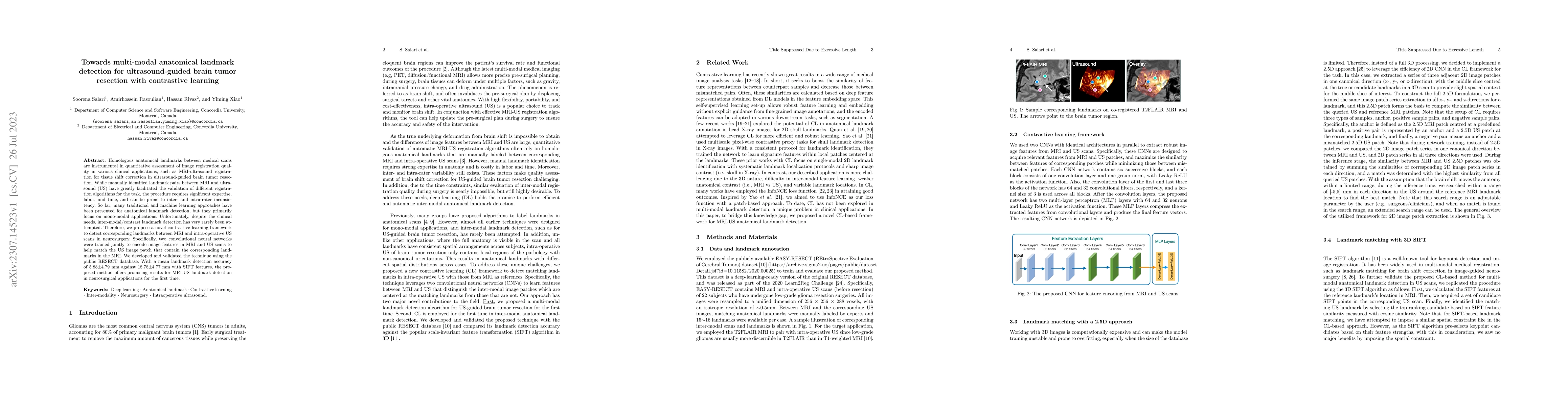
Homologous anatomical landmarks between medical scans are instrumental in quantitative assessment of image registration quality in various clinical applications, such as MRI-ultrasound registration ...
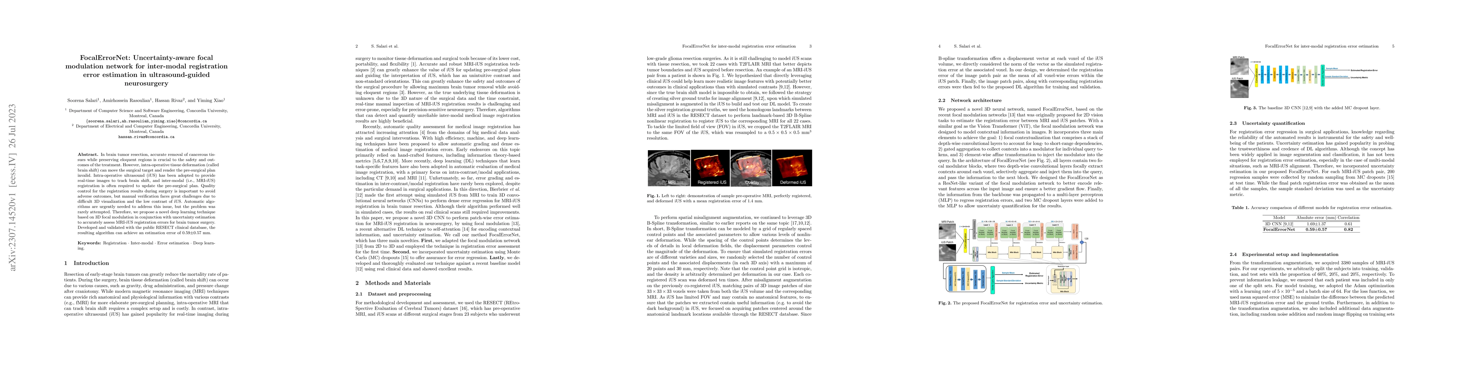
In brain tumor resection, accurate removal of cancerous tissues while preserving eloquent regions is crucial to the safety and outcomes of the treatment. However, intra-operative tissue deformation ...
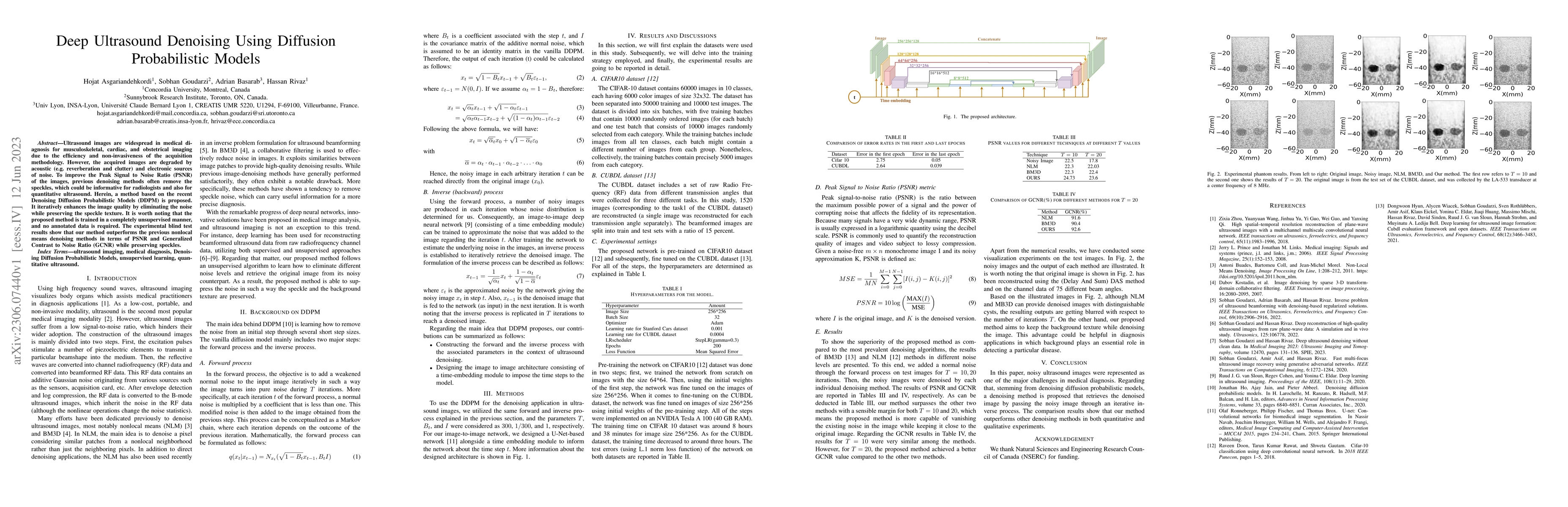
Ultrasound images are widespread in medical diagnosis for musculoskeletal, cardiac, and obstetrical imaging due to the efficiency and non-invasiveness of the acquisition methodology. However, the ac...
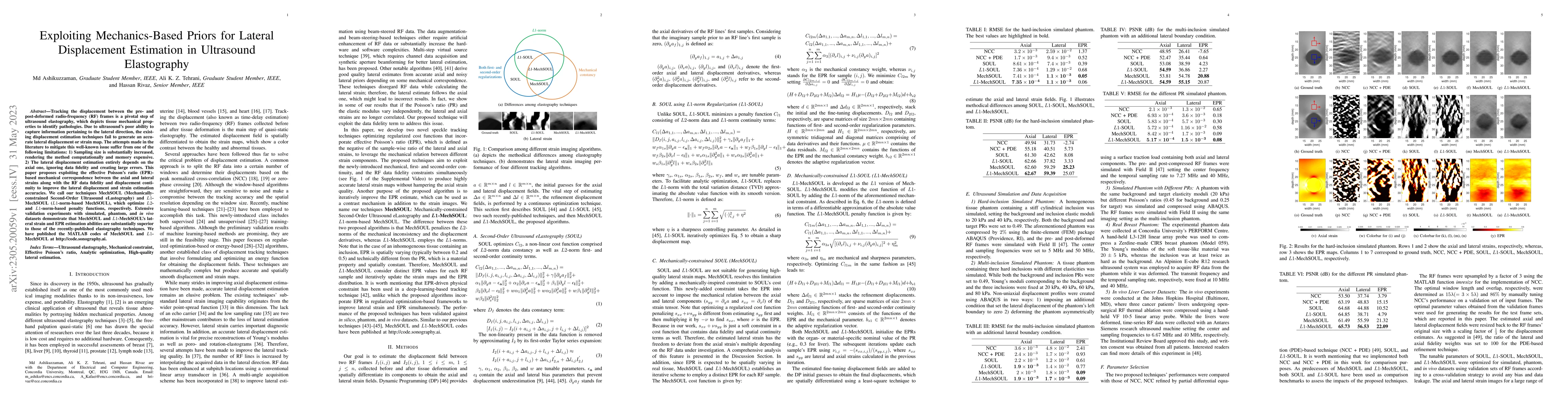
Tracking the displacement between the pre- and post-deformed radio-frequency (RF) frames is a pivotal step of ultrasound elastography, which depicts tissue mechanical properties to identify patholog...
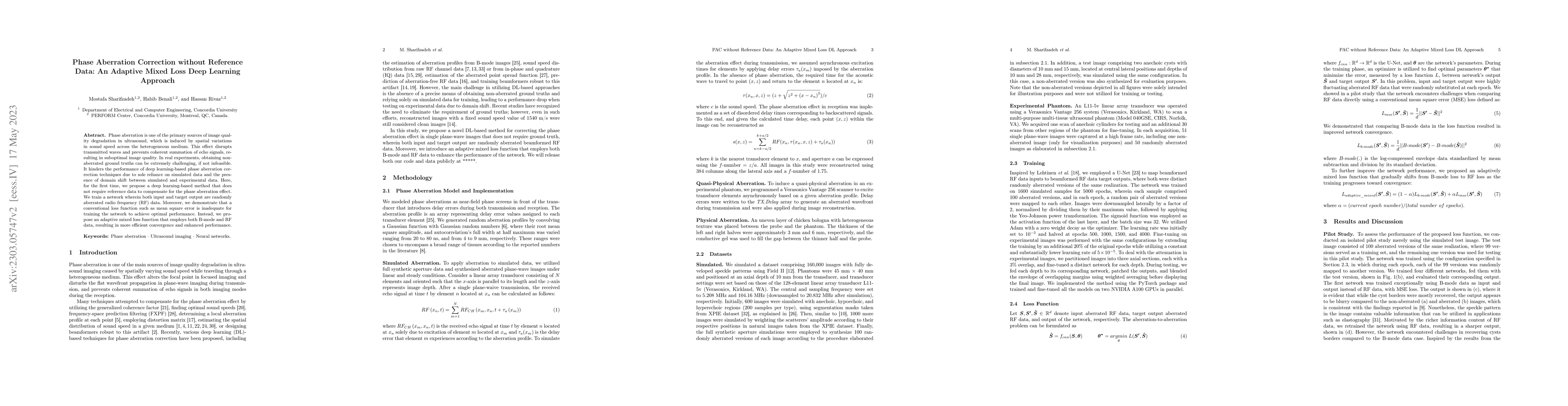
Phase aberration is one of the primary sources of image quality degradation in ultrasound, which is induced by spatial variations in sound speed across the heterogeneous medium. This effect disrupts...
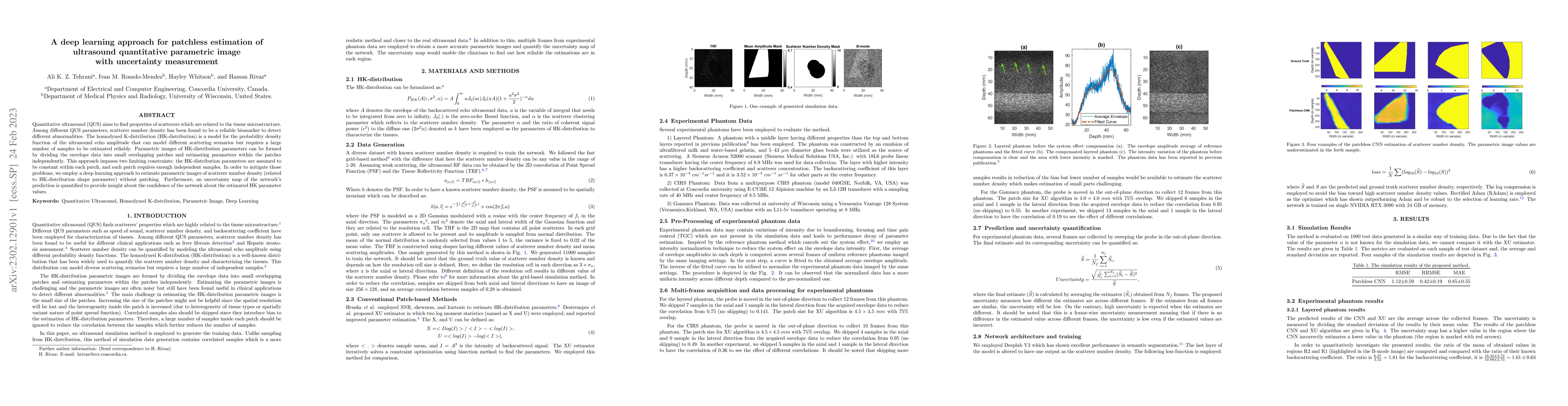
Quantitative ultrasound (QUS) aims to find properties of scatterers which are related to the tissue microstructure. Among different QUS parameters, scatterer number density has been found to be a re...
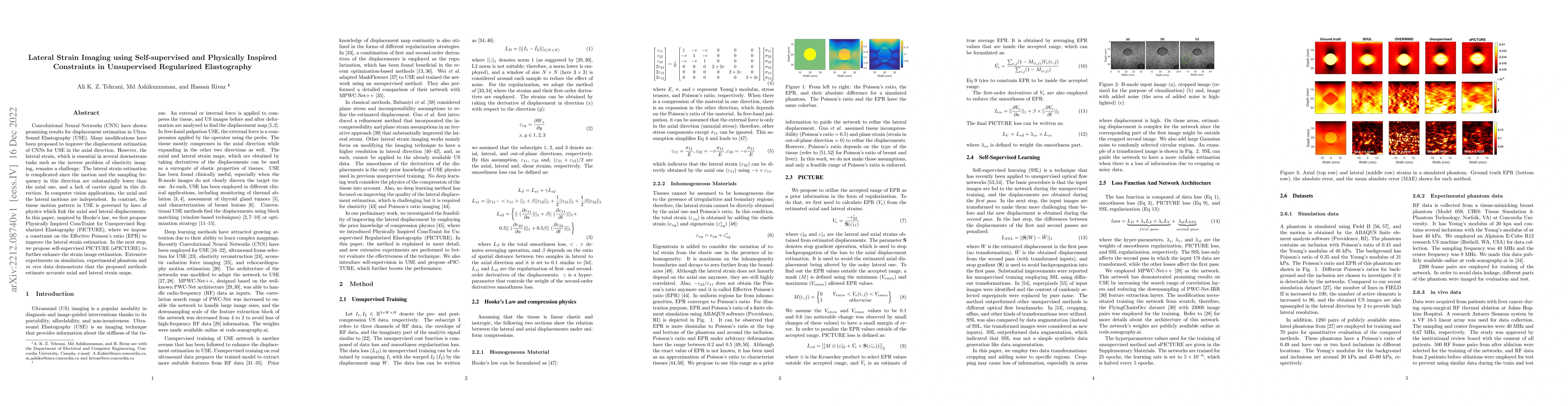
Convolutional Neural Networks (CNN) have shown promising results for displacement estimation in UltraSound Elastography (USE). Many modifications have been proposed to improve the displacement estim...
Quantitative ultrasound (QUS) allows estimating the intrinsic tissue properties. Speckle statistics are the QUS parameters that describe the first order statistics of ultrasound (US) envelope data. ...
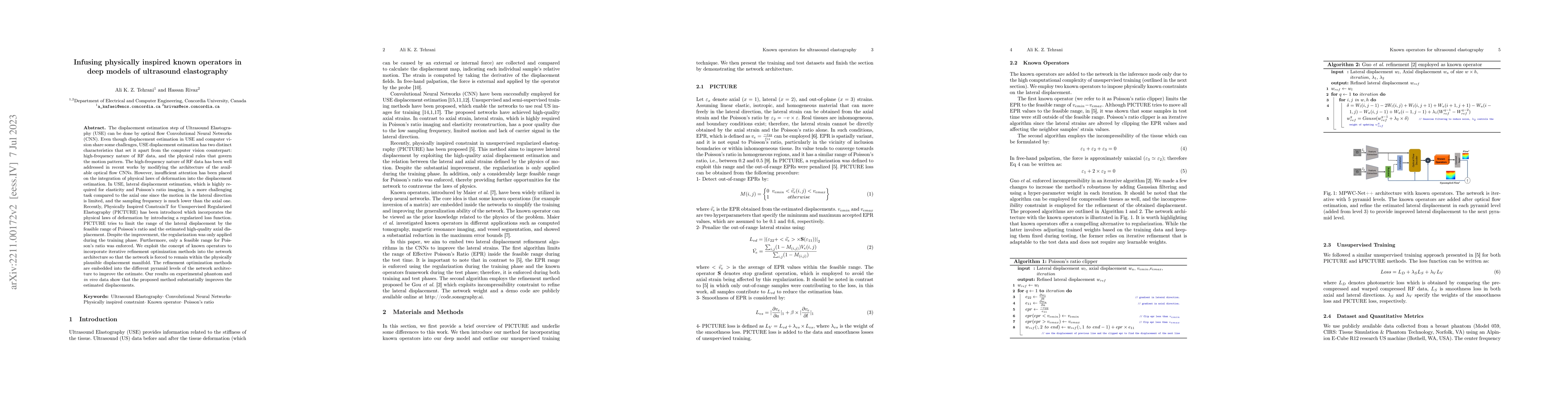
Convolutional Neural Networks (CNN) have been employed for displacement estimation in ultrasound elastography (USE). High-quality axial strains (derivative of the axial displacement in the axial dir...
Ultrasound Localization Microscopy (ULM) is an emerging technique that employs the localization of echogenic microbubbles (MBs) to finely sample and image the microcirculation beyond the diffraction...
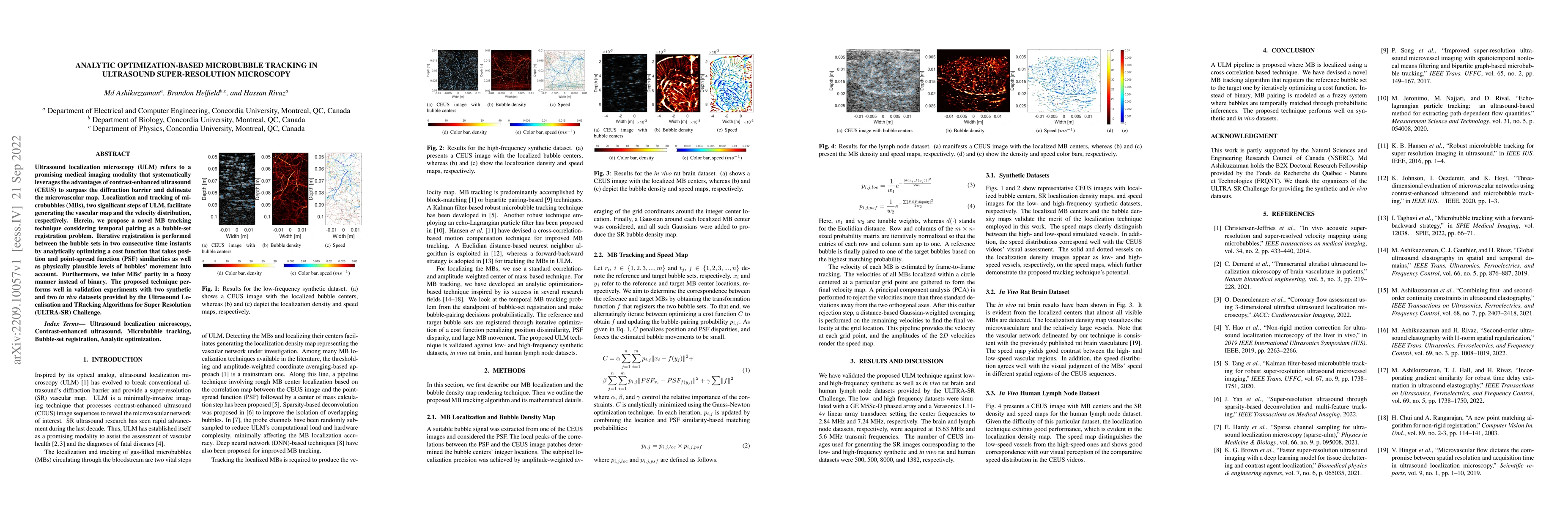
Ultrasound localization microscopy (ULM) refers to a promising medical imaging modality that systematically leverages the advantages of contrast-enhanced ultrasound (CEUS) to surpass the diffraction...
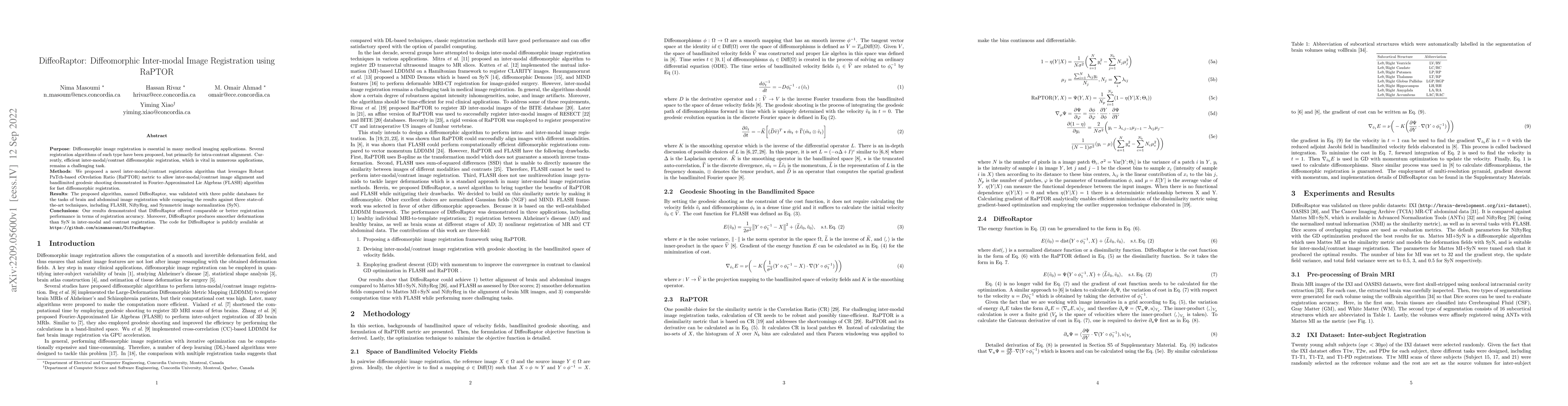
Purpose: Diffeomorphic image registration is essential in many medical imaging applications. Several registration algorithms of such type have been proposed, but primarily for intra-contrast alignme...
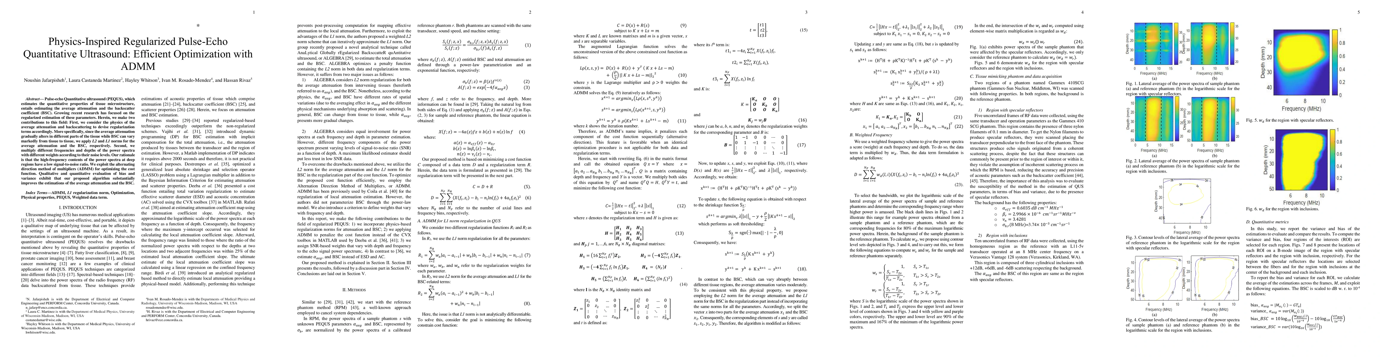
Pulse-echo Quantitative ultrasound (PEQUS), which estimates the quantitative properties of tissue microstructure, entails estimating the average attenuation and the backscatter coefficient (BSC). Gr...
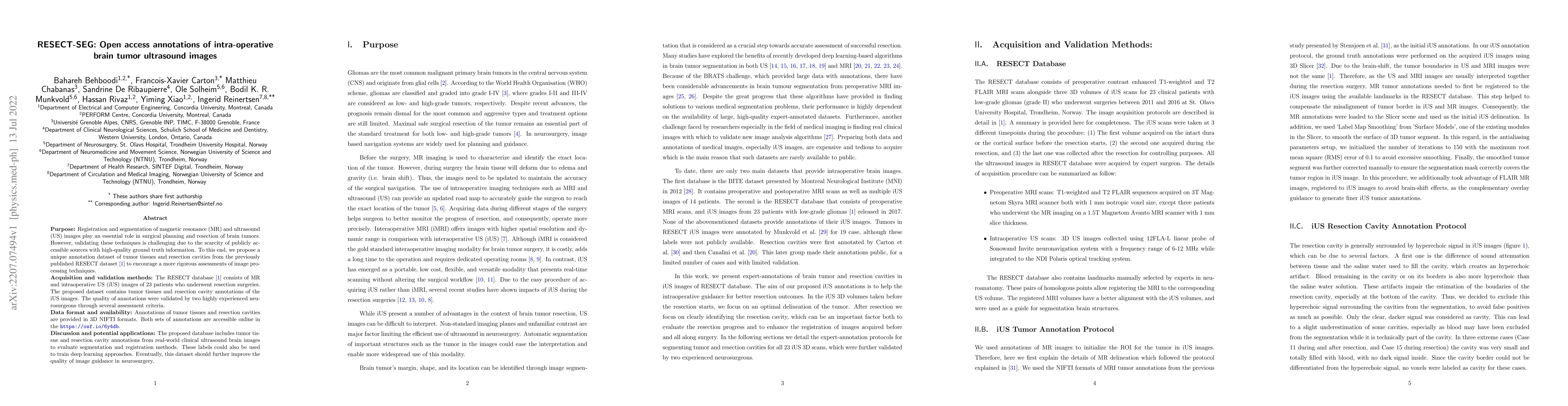
Purpose: Registration and segmentation of magnetic resonance (MR) and ultrasound (US) images play an essential role in surgical planning and resection of brain tumors. However, validating these tech...
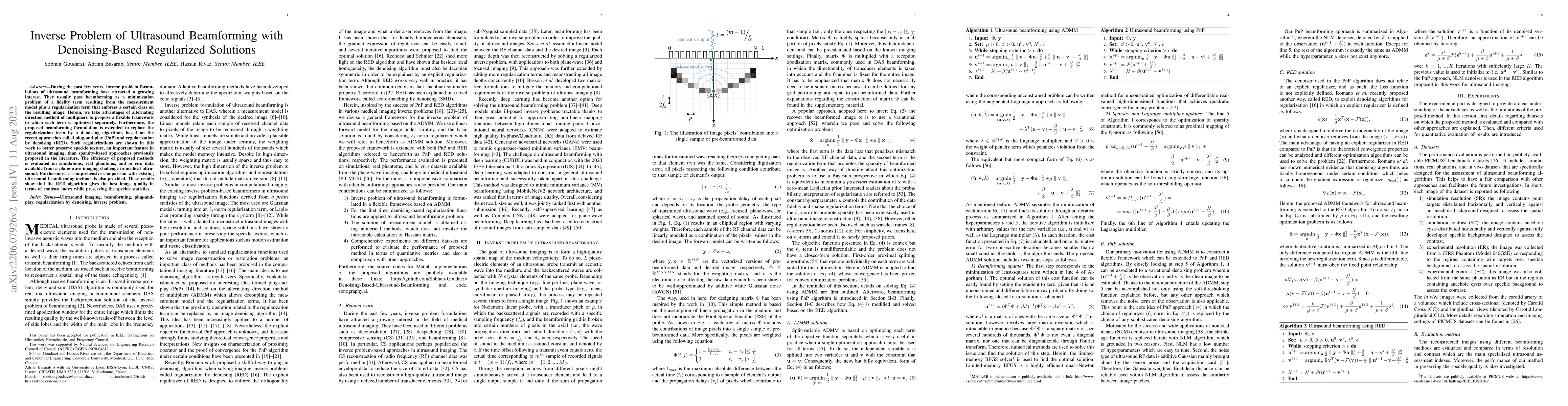
During the past few years, inverse problem formulations of ultrasound beamforming have attracted a growing interest. They usually pose beamforming as a minimization problem of a fidelity term result...
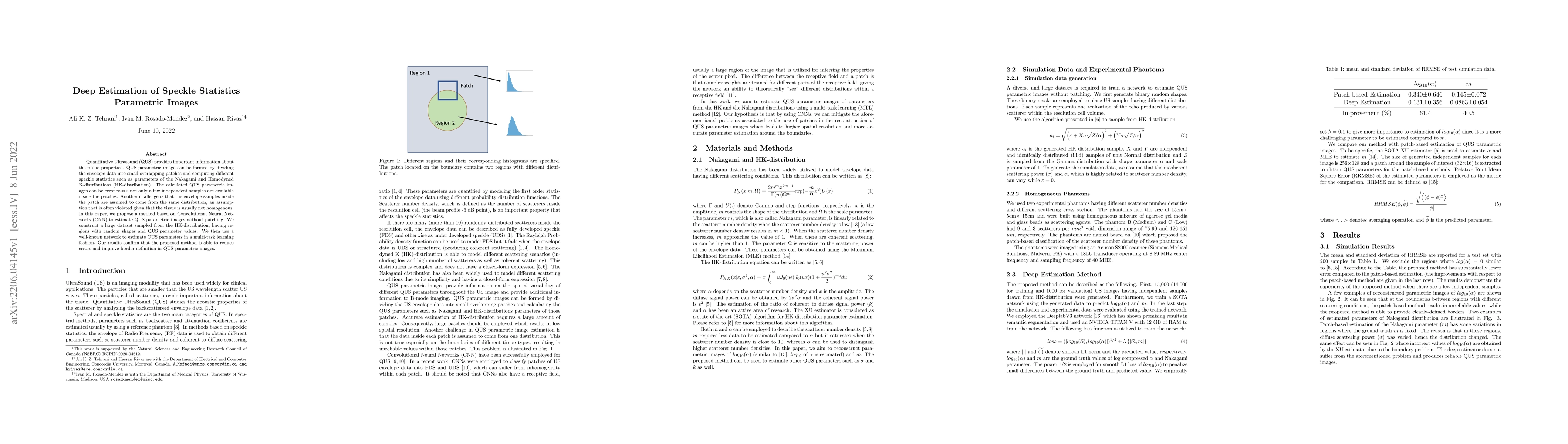
Quantitative Ultrasound (QUS) provides important information about the tissue properties. QUS parametric image can be formed by dividing the envelope data into small overlapping patches and computin...
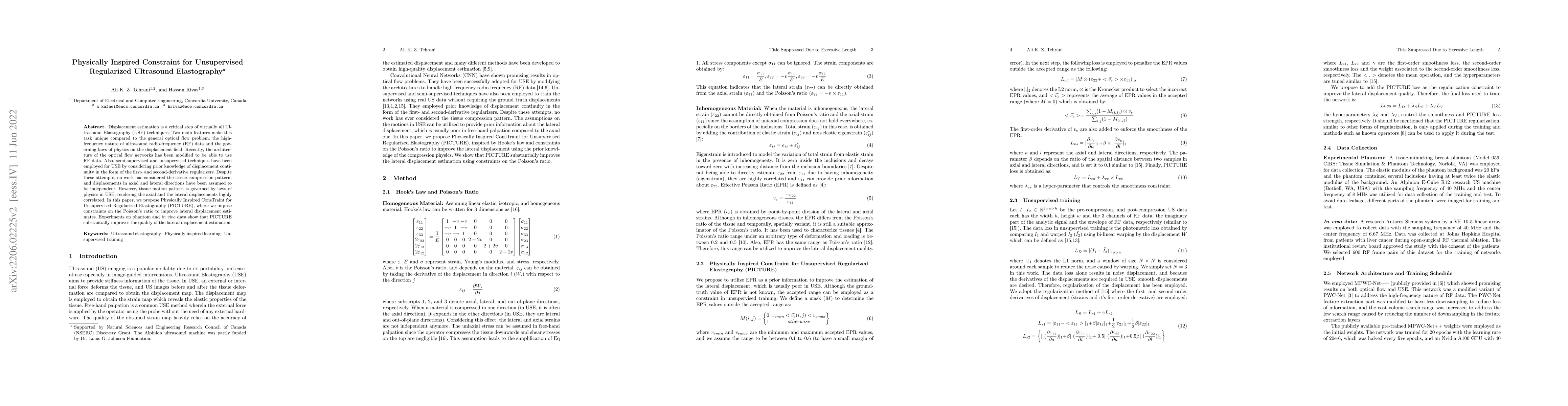
Displacement estimation is a critical step of virtually all Ultrasound Elastography (USE) techniques. Two main features make this task unique compared to the general optical flow problem: the high-f...
Energy-based ultrasound elastography techniques minimize a regularized cost function consisting of data and continuity terms to obtain local displacement estimates based on the local time-delay esti...
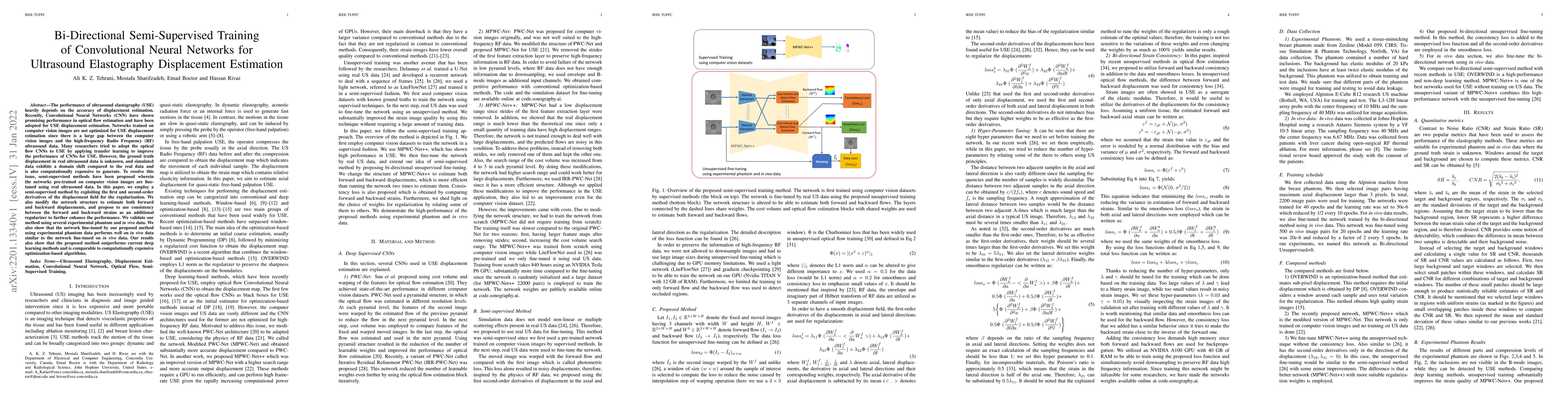
The performance of ultrasound elastography (USE) heavily depends on the accuracy of displacement estimation. Recently, Convolutional Neural Networks (CNN) have shown promising performance in optical...
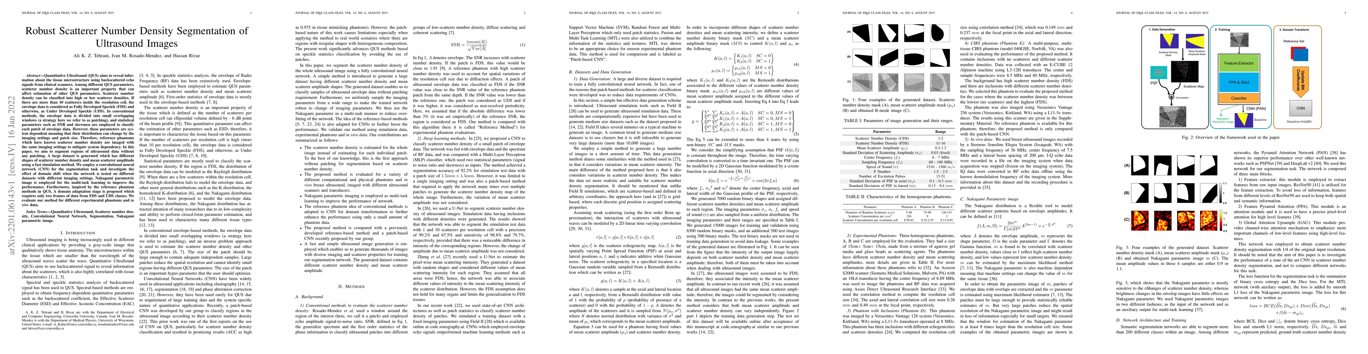
Quantitative UltraSound (QUS) aims to reveal information about the tissue microstructure using backscattered echo signals from clinical scanners. Among different QUS parameters, scatterer number den...
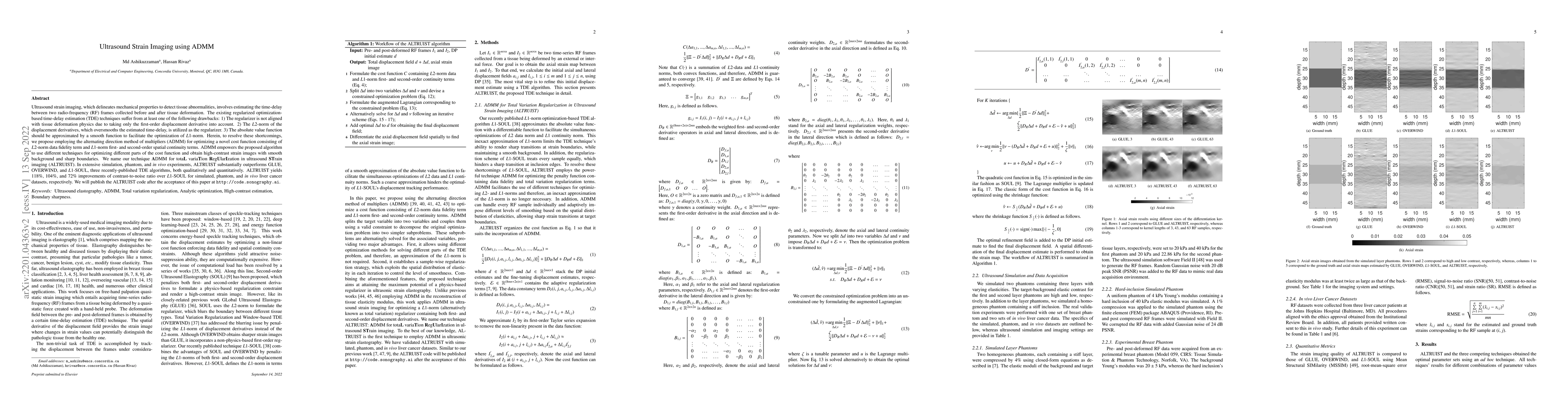
Ultrasound strain imaging, which delineates mechanical properties to detect tissue abnormalities, involves estimating the time-delay between two radio-frequency (RF) frames collected before and afte...
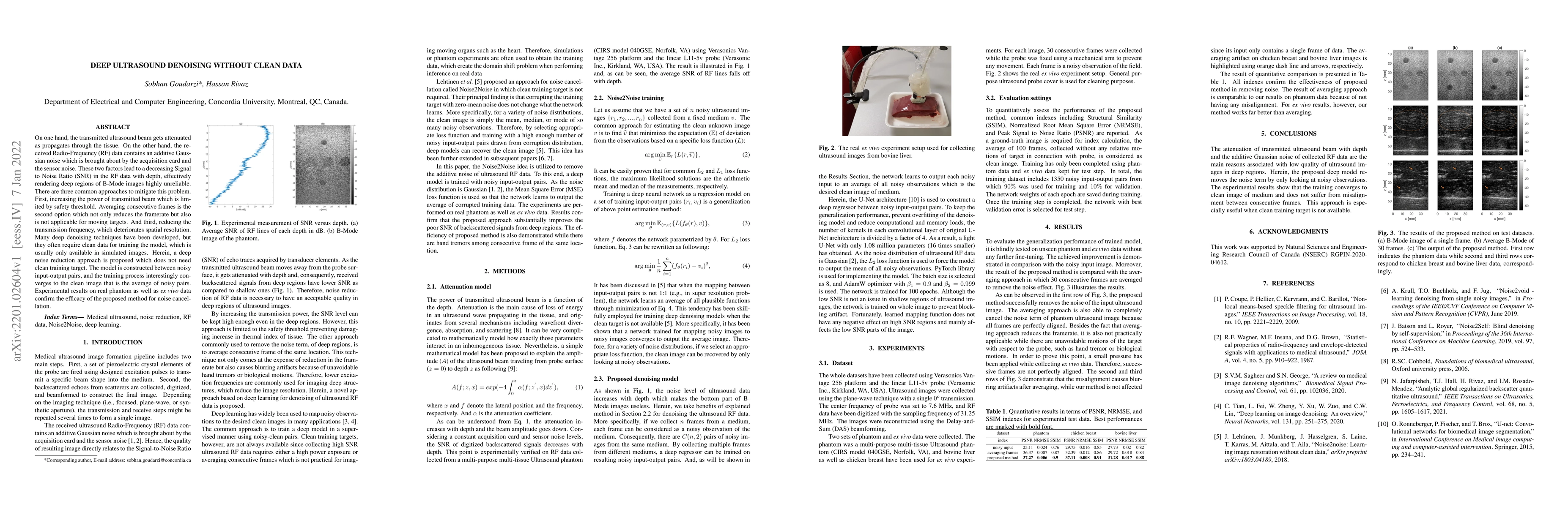
On one hand, the transmitted ultrasound beam gets attenuated as propagates through the tissue. On the other hand, the received Radio-Frequency (RF) data contains an additive Gaussian noise which is ...
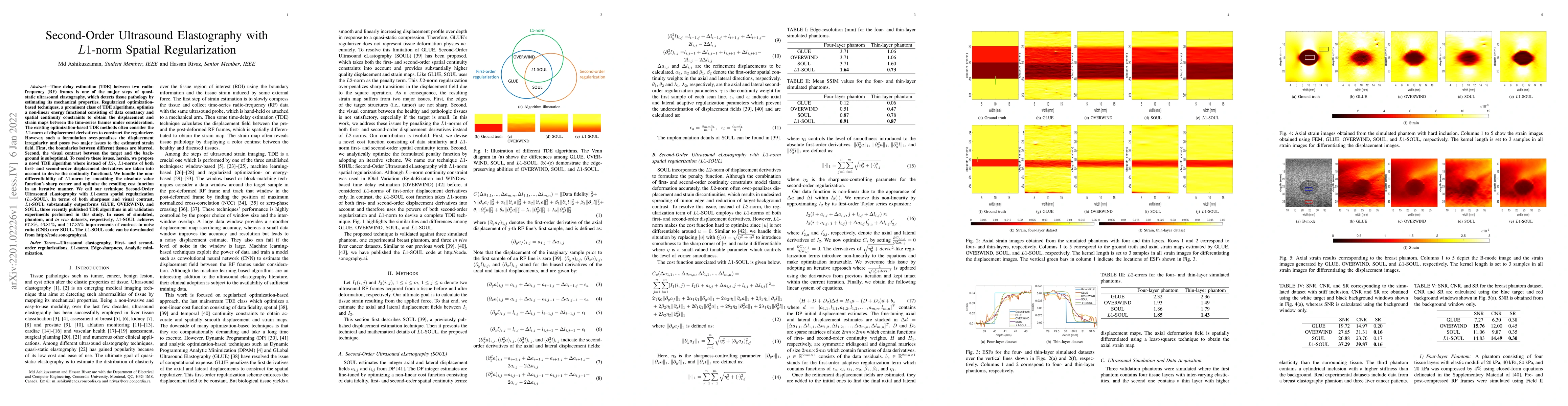
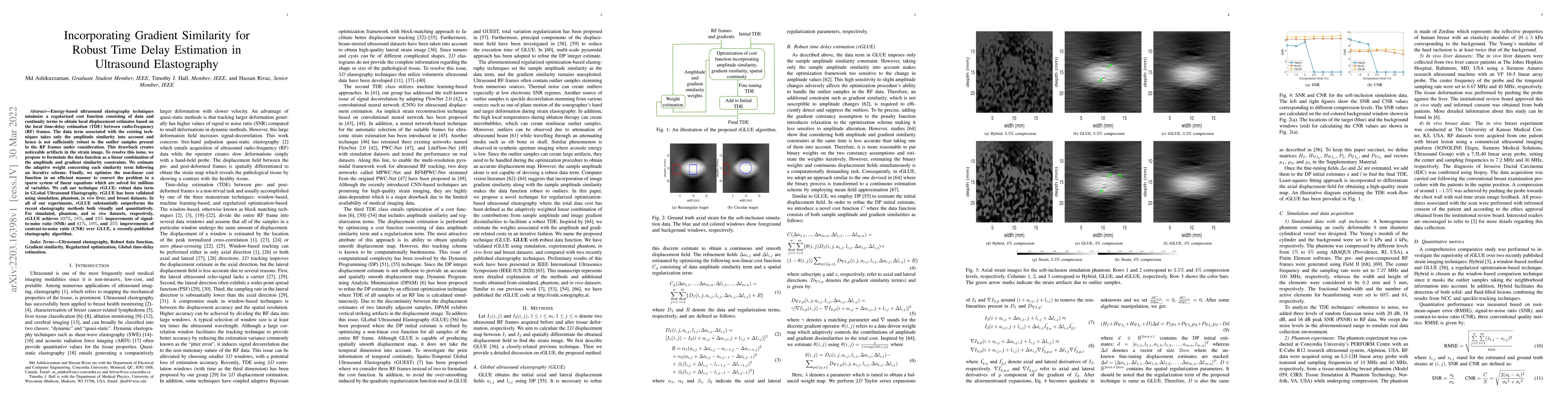
Time delay estimation (TDE) between two radio-frequency (RF) frames is one of the major steps of quasi-static ultrasound elastography, which detects tissue pathology by estimating its mechanical pro...
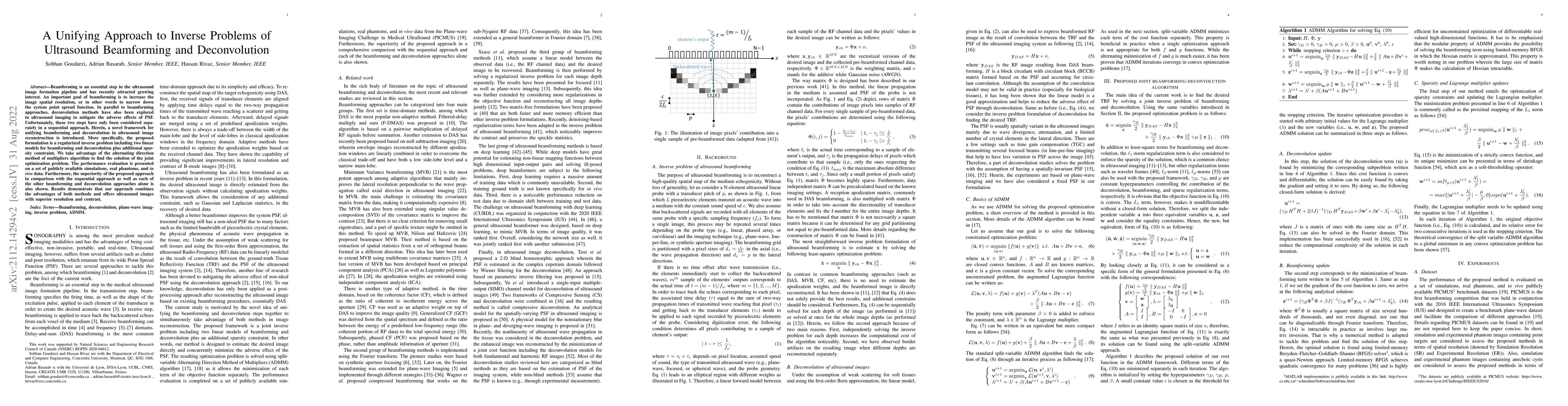
Beamforming is an essential step in the ultrasound image formation pipeline and has recently attracted growing interest. An important goal of beamforming is to increase the image spatial resolution,...
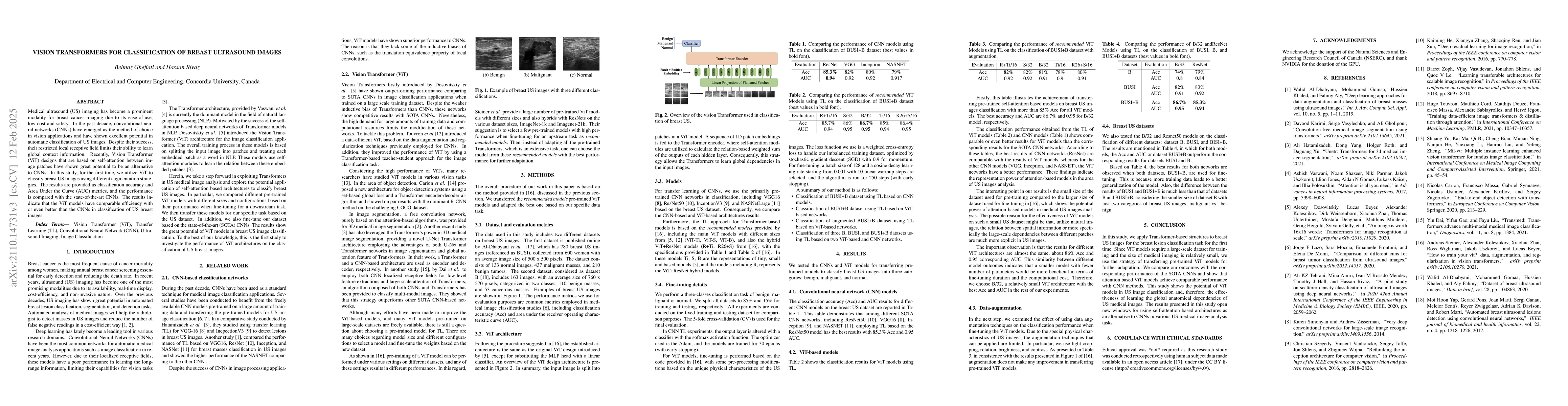
Medical ultrasound (US) imaging has become a prominent modality for breast cancer imaging due to its ease-of-use, low-cost and safety. In the past decade, convolutional neural networks (CNNs) have e...
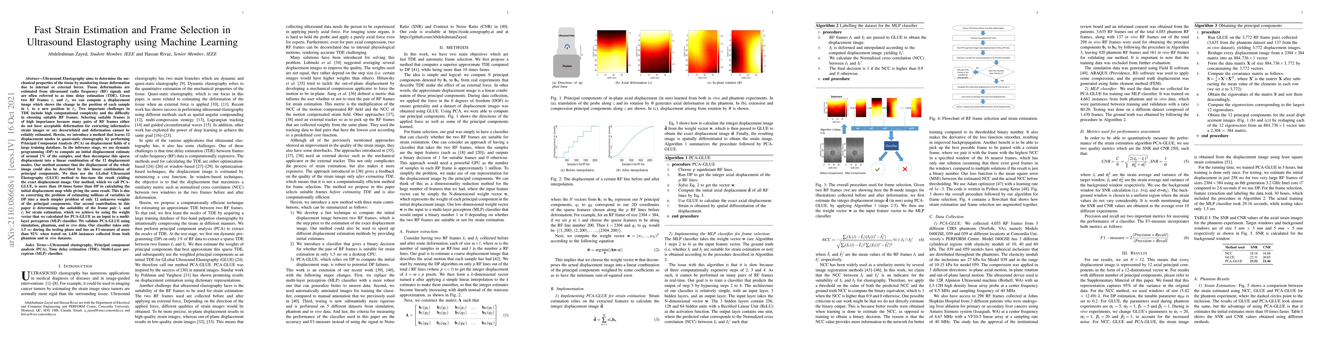
Ultrasound Elastography aims to determine the mechanical properties of the tissue by monitoring tissue deformation due to internal or external forces. Tissue deformations are estimated from ultrasou...
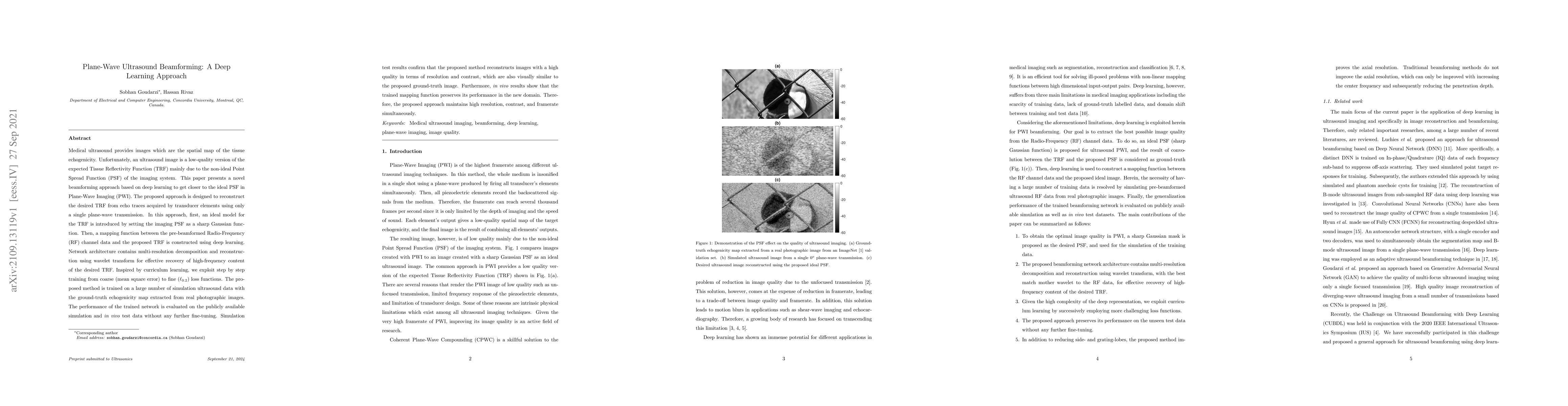
Medical ultrasound provides images which are the spatial map of the tissue echogenicity. Unfortunately, an ultrasound image is a low-quality version of the expected Tissue Reflectivity Function (TRF...
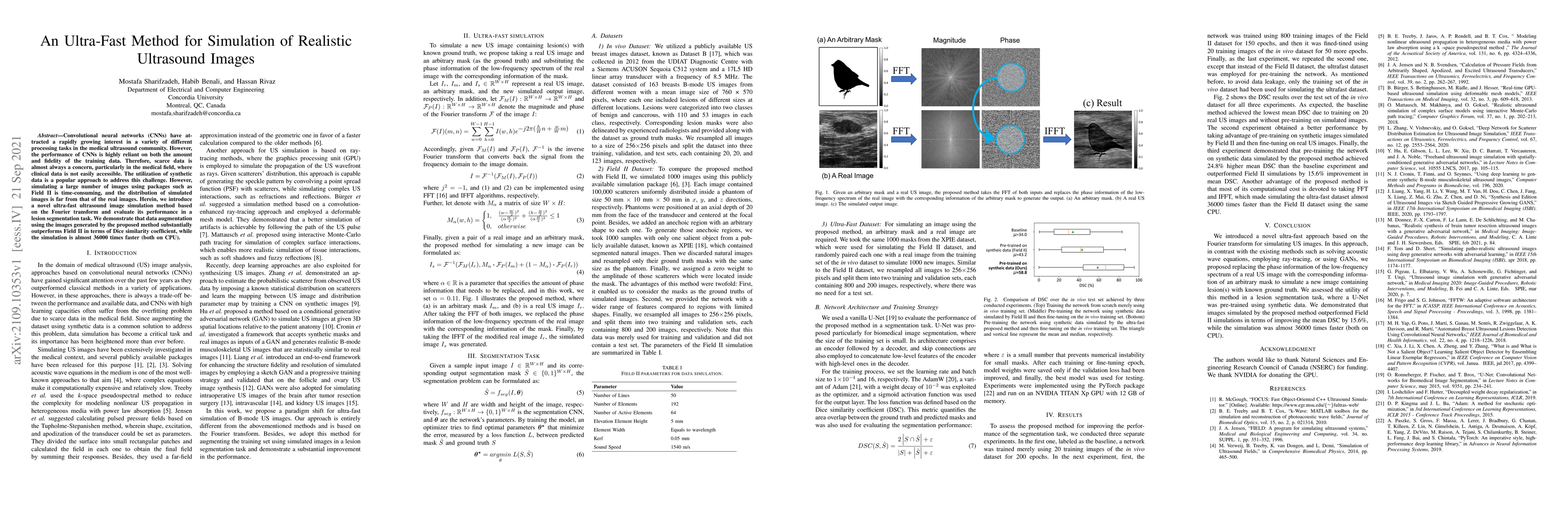
Convolutional neural networks (CNNs) have attracted a rapidly growing interest in a variety of different processing tasks in the medical ultrasound community. However, the performance of CNNs is hig...
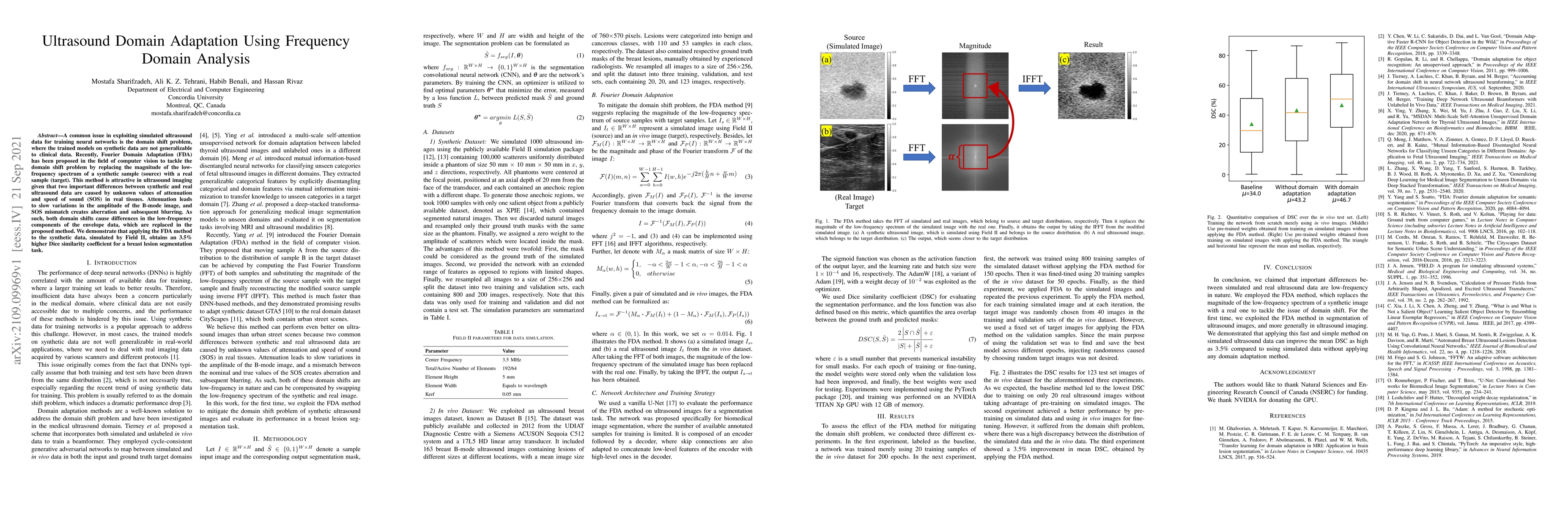
A common issue in exploiting simulated ultrasound data for training neural networks is the domain shift problem, where the trained models on synthetic data are not generalizable to clinical data. Re...
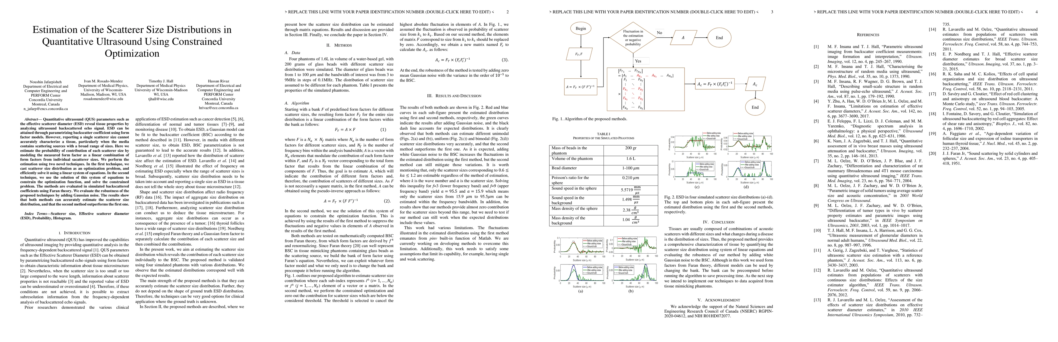
Quantitative ultrasound (QUS) parameters such as the effective scatterer diameter (ESD) reveal tissue properties by analyzing ultrasound backscattered echo signal. ESD can be attained through parame...
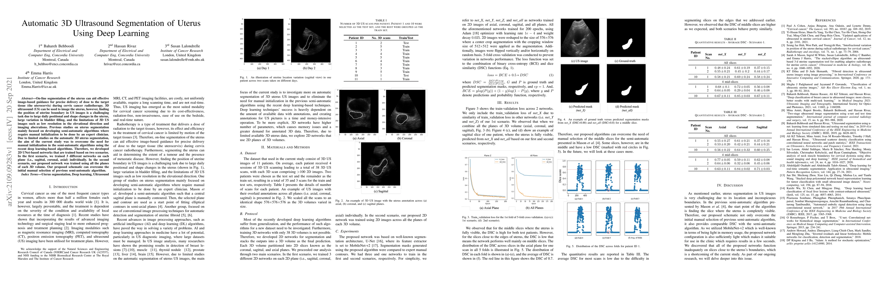
On-line segmentation of the uterus can aid effective image-based guidance for precise delivery of dose to the target tissue (the uterocervix) during cervix cancer radiotherapy. 3D ultrasound (US) ca...
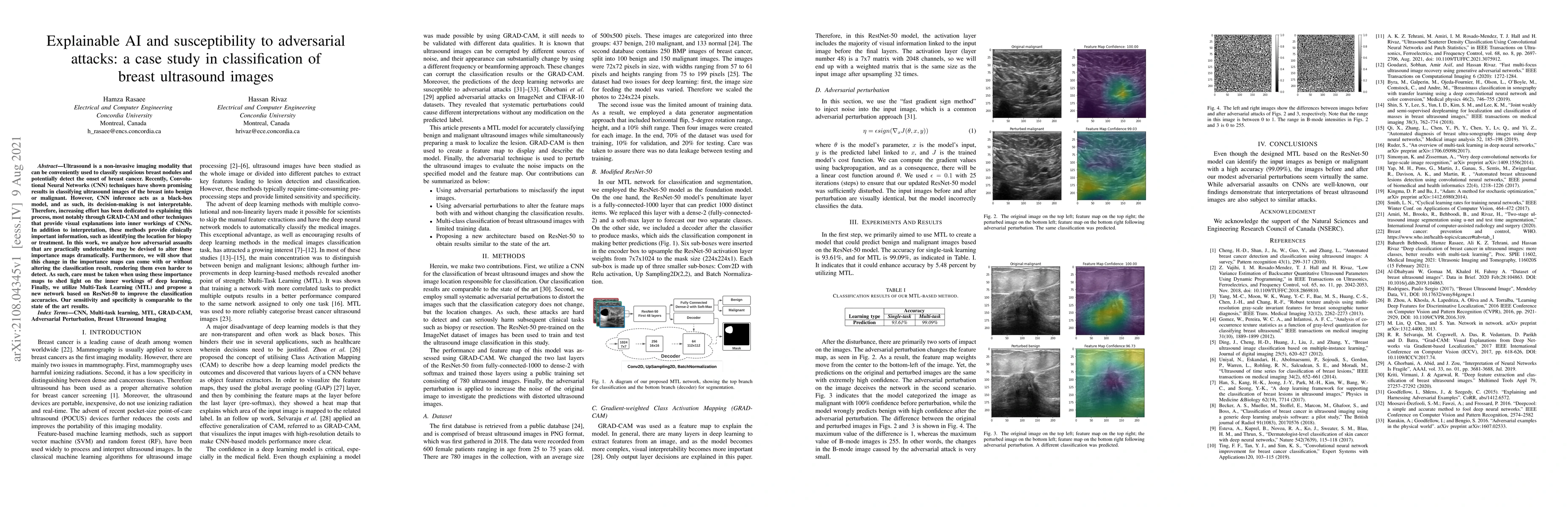
Ultrasound is a non-invasive imaging modality that can be conveniently used to classify suspicious breast nodules and potentially detect the onset of breast cancer. Recently, Convolutional Neural Ne...
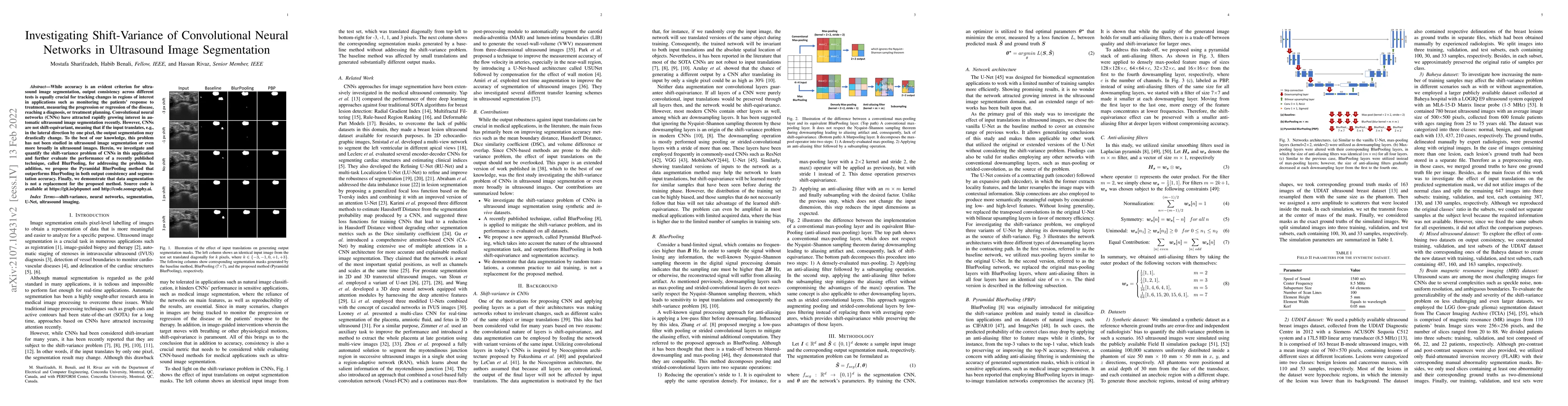
While accuracy is an evident criterion for ultrasound image segmentation, output consistency across different tests is equally crucial for tracking changes in regions of interest in applications suc...
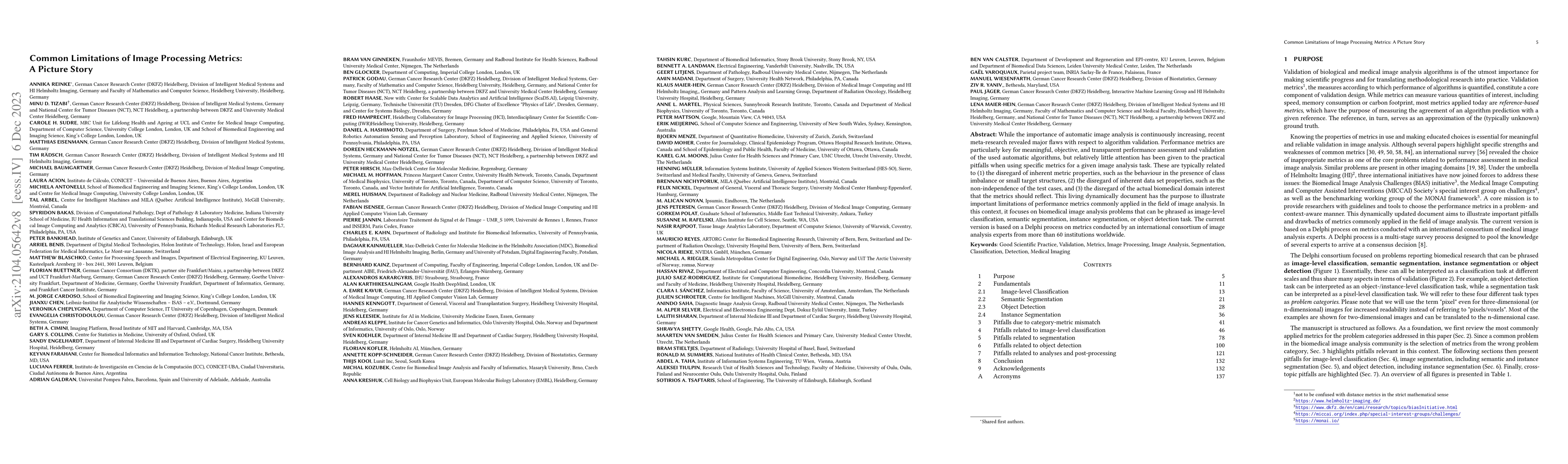
While the importance of automatic image analysis is continuously increasing, recent meta-research revealed major flaws with respect to algorithm validation. Performance metrics are particularly key ...
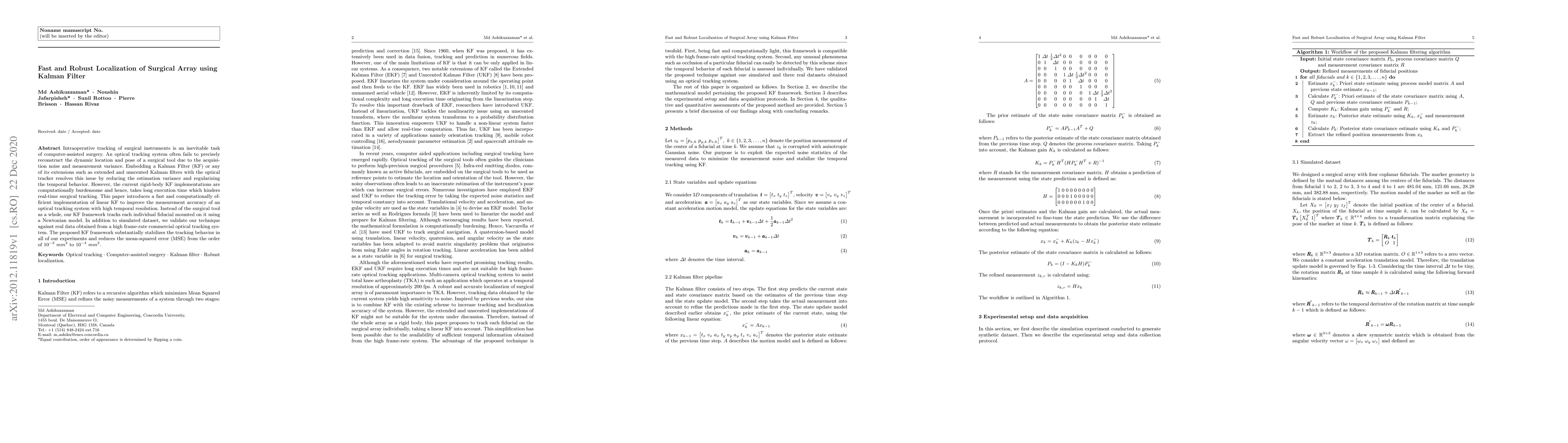
Intraoperative tracking of surgical instruments is an inevitable task of computer-assisted surgery. An optical tracking system often fails to precisely reconstruct the dynamic location and pose of a...
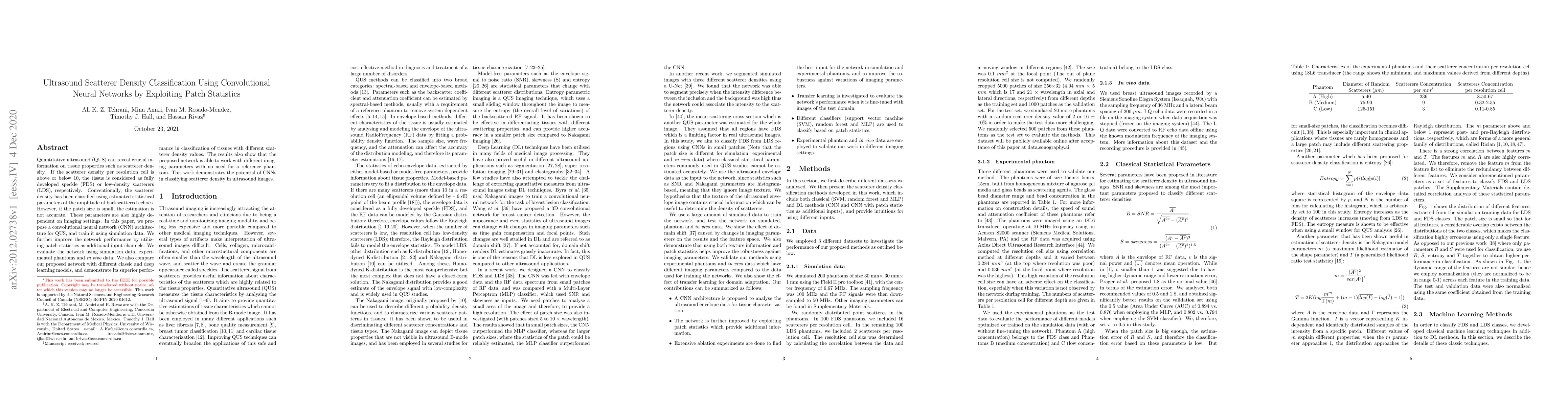
Quantitative ultrasound (QUS) can reveal crucial information on tissue properties such as scatterer density. If the scatterer density per resolution cell is above or below 10, the tissue is consider...
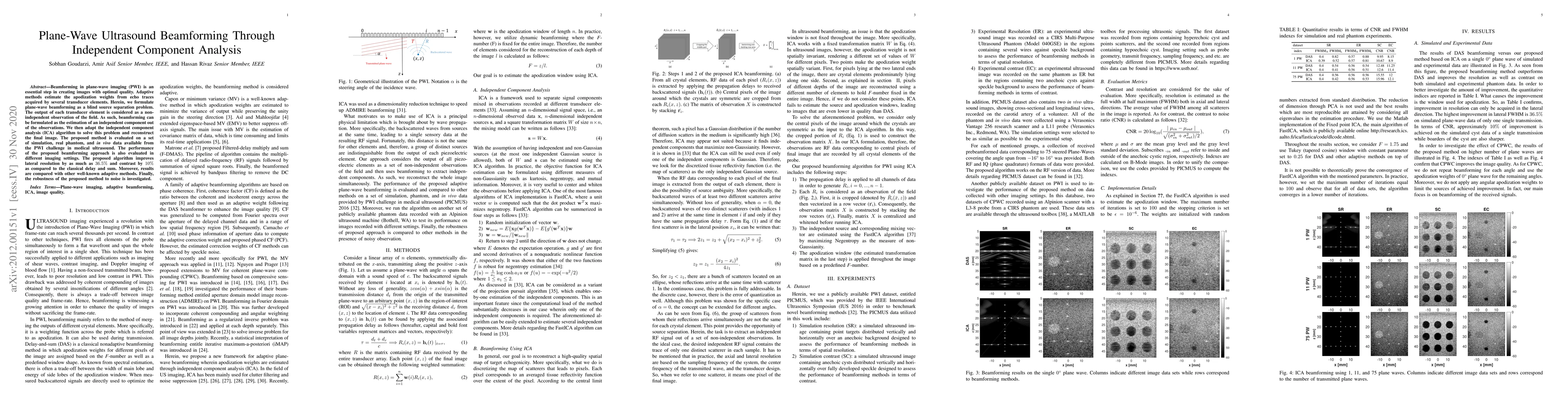
Beamforming in plane-wave imaging (PWI) is an essential step in creating images with optimal quality. Adaptive methods estimate the apodization weights from echo traces acquired by several transduce...
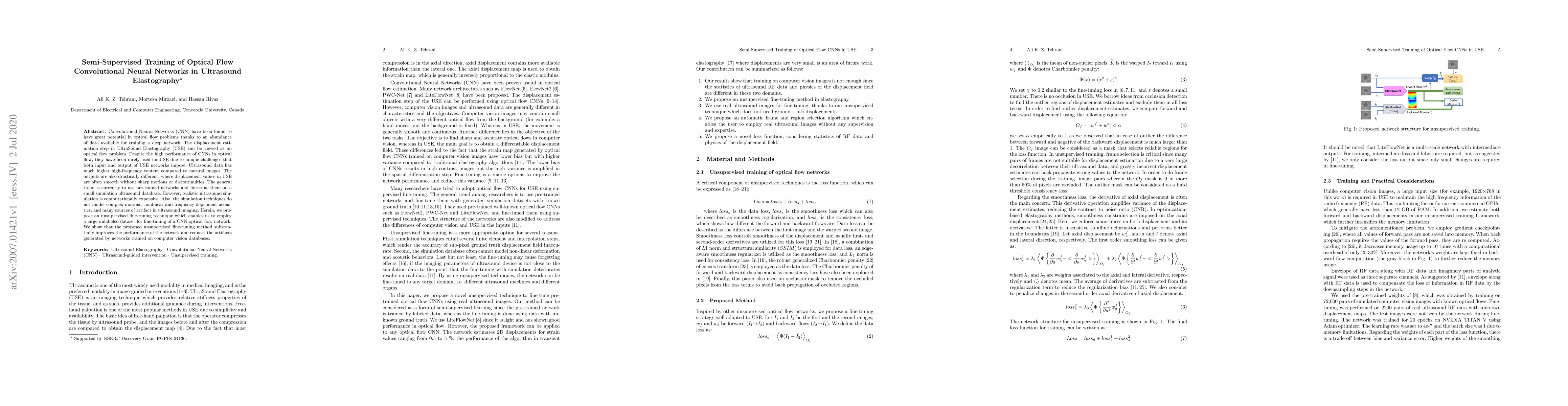
Convolutional Neural Networks (CNN) have been found to have great potential in optical flow problems thanks to an abundance of data available for training a deep network. The displacement estimation...
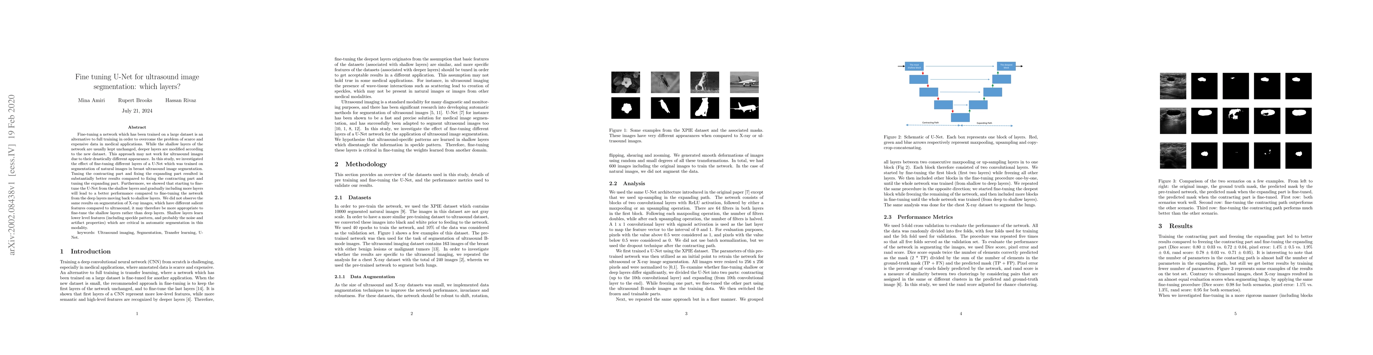
Fine-tuning a network which has been trained on a large dataset is an alternative to full training in order to overcome the problem of scarce and expensive data in medical applications. While the sh...
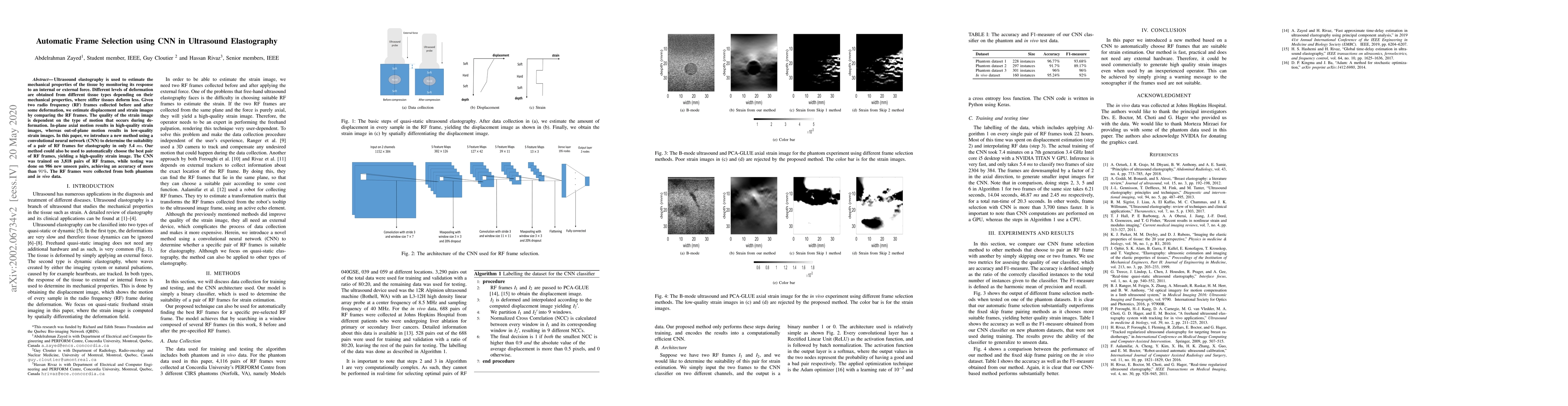
Ultrasound elastography is used to estimate the mechanical properties of the tissue by monitoring its response to an internal or external force. Different levels of deformation are obtained from dif...
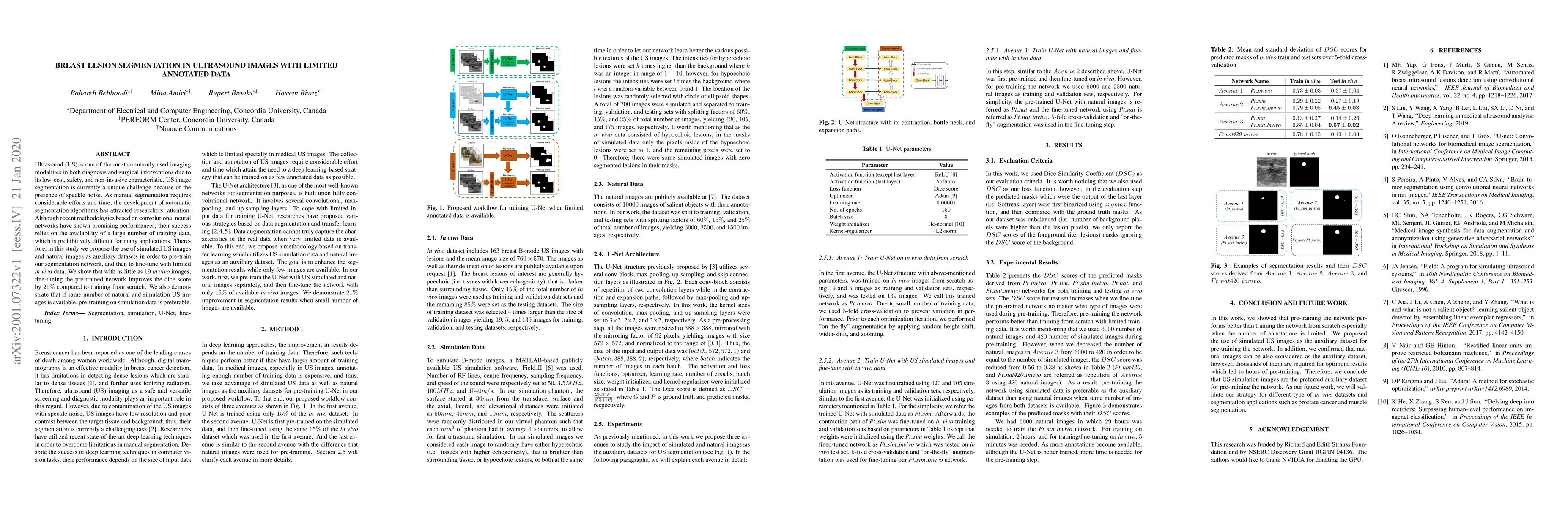
Ultrasound (US) is one of the most commonly used imaging modalities in both diagnosis and surgical interventions due to its low-cost, safety, and non-invasive characteristic. US image segmentation i...
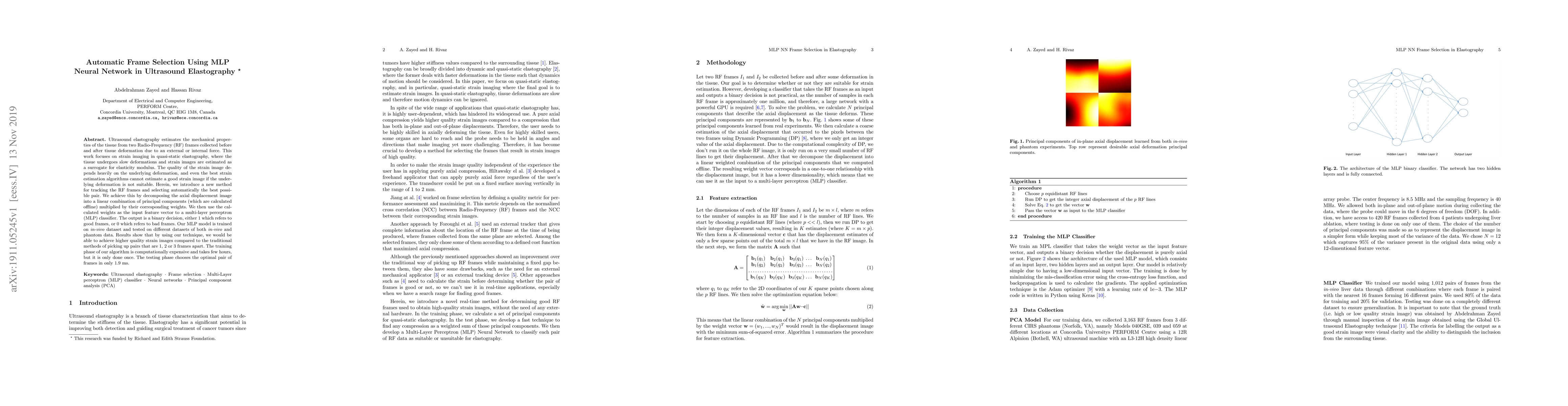
Ultrasound elastography estimates the mechanical properties of the tissue from two Radio-Frequency (RF) frames collected before and after tissue deformation due to an external or internal force. Thi...
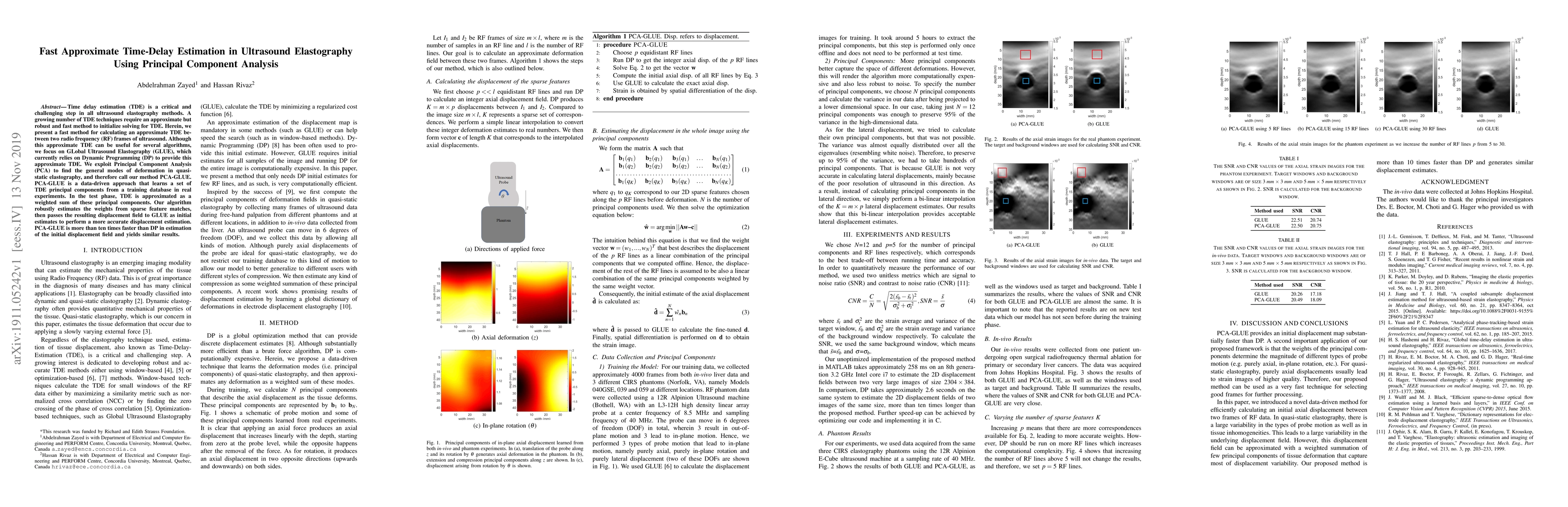
Time delay estimation (TDE) is a critical and challenging step in all ultrasound elastography methods. A growing number of TDE techniques require an approximate but robust and fast method to initial...
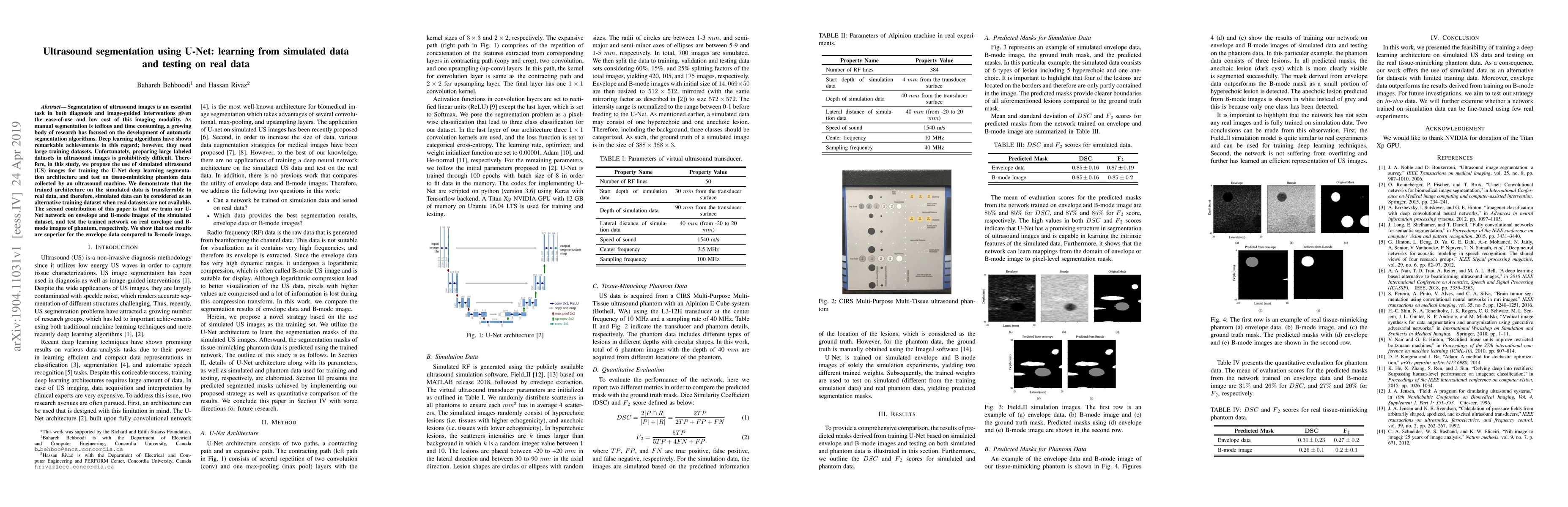
Segmentation of ultrasound images is an essential task in both diagnosis and image-guided interventions given the ease-of-use and low cost of this imaging modality. As manual segmentation is tedious...
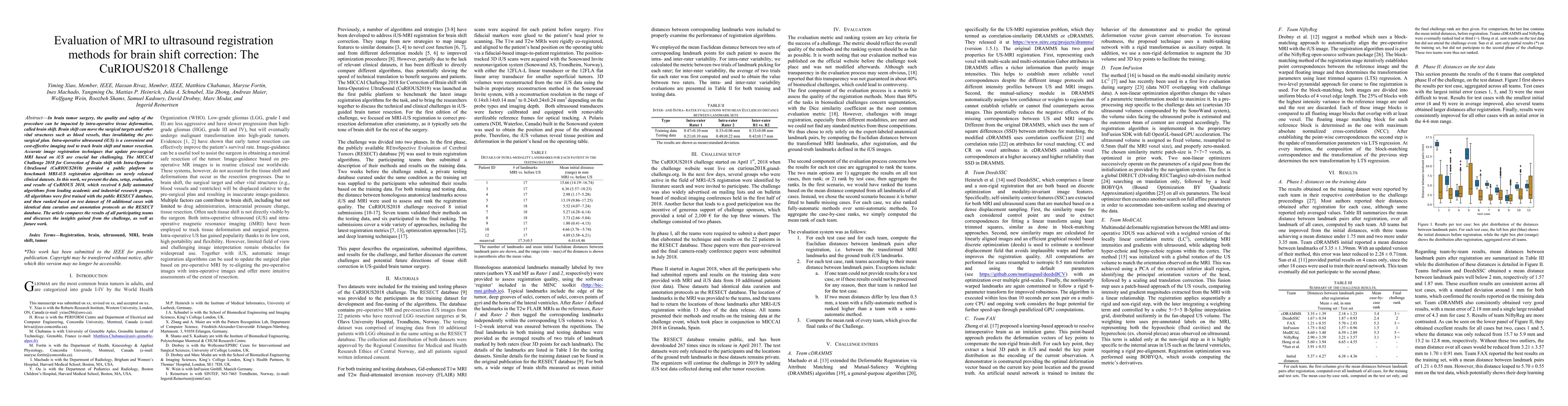
In brain tumor surgery, the quality and safety of the procedure can be impacted by intra-operative tissue deformation, called brain shift. Brain shift can move the surgical targets and other vital s...
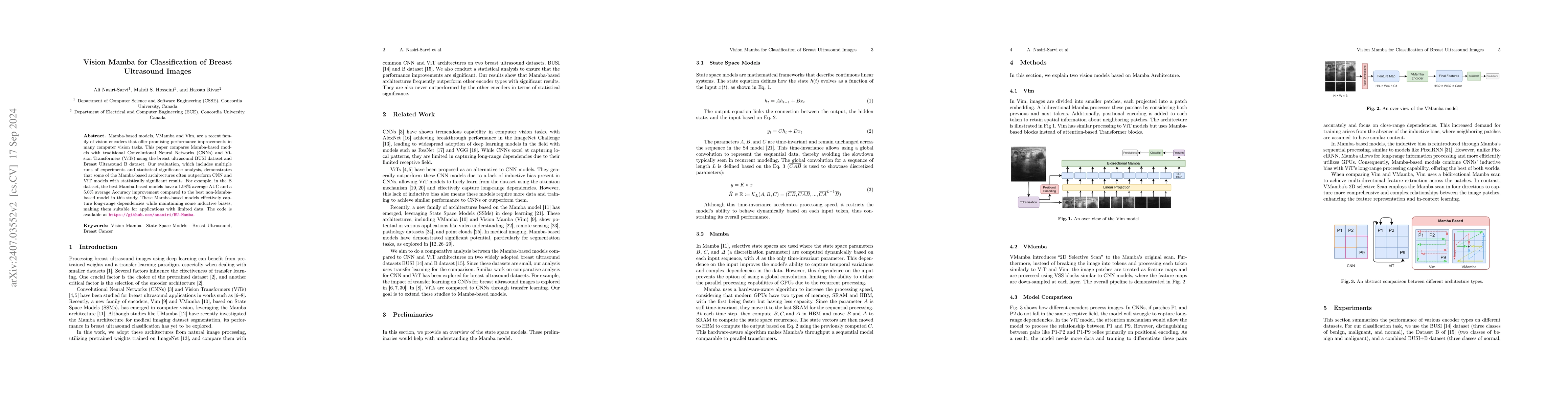
Mamba-based models, VMamba and Vim, are a recent family of vision encoders that offer promising performance improvements in many computer vision tasks. This paper compares Mamba-based models with trad...
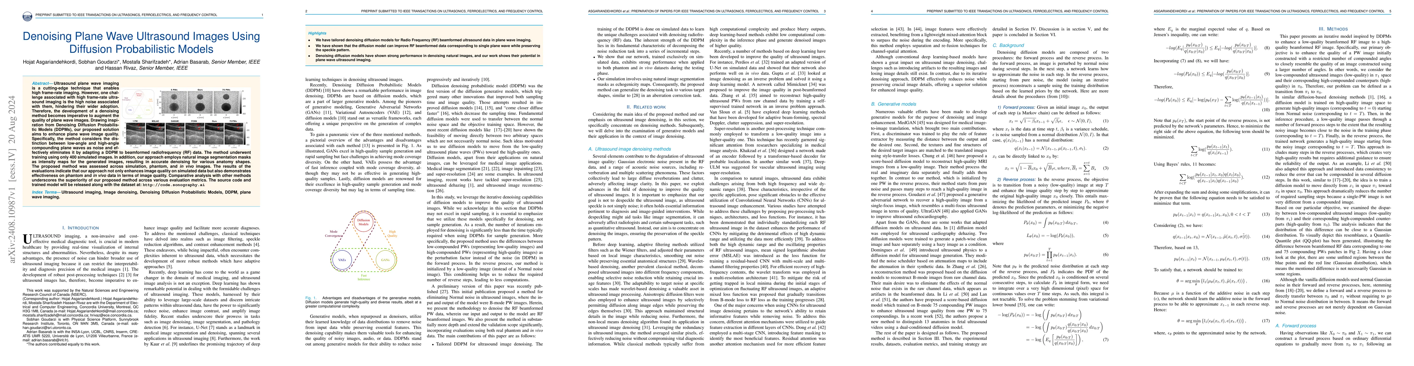
Ultrasound plane wave imaging is a cutting-edge technique that enables high frame-rate imaging. However, one challenge associated with high frame-rate ultrasound imaging is the high noise associated w...
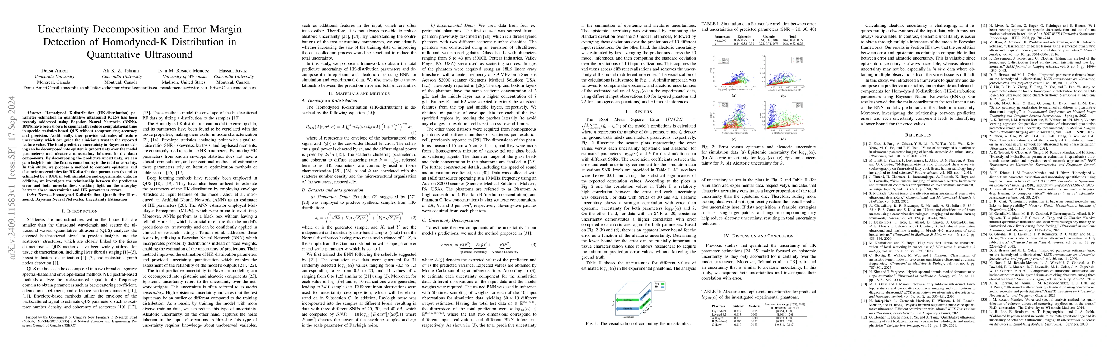
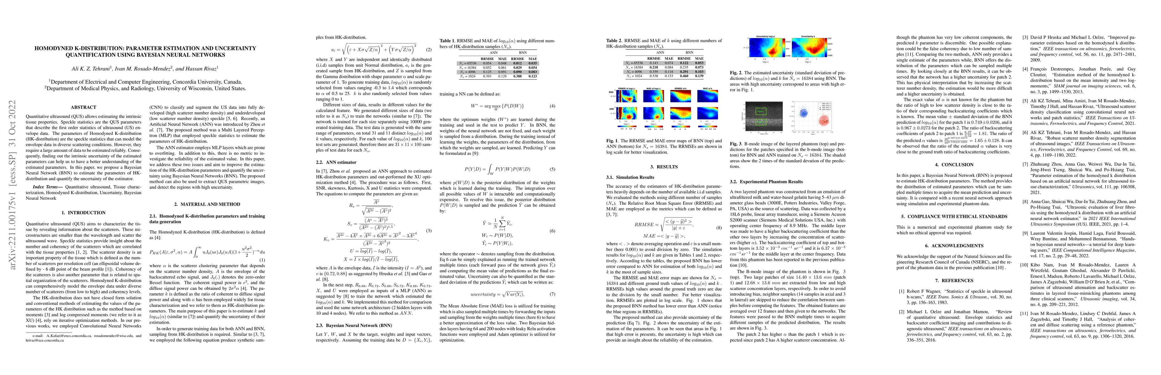
Homodyned K-distribution (HK-distribution) parameter estimation in quantitative ultrasound (QUS) has been recently addressed using Bayesian Neural Networks (BNNs). BNNs have been shown to significantl...
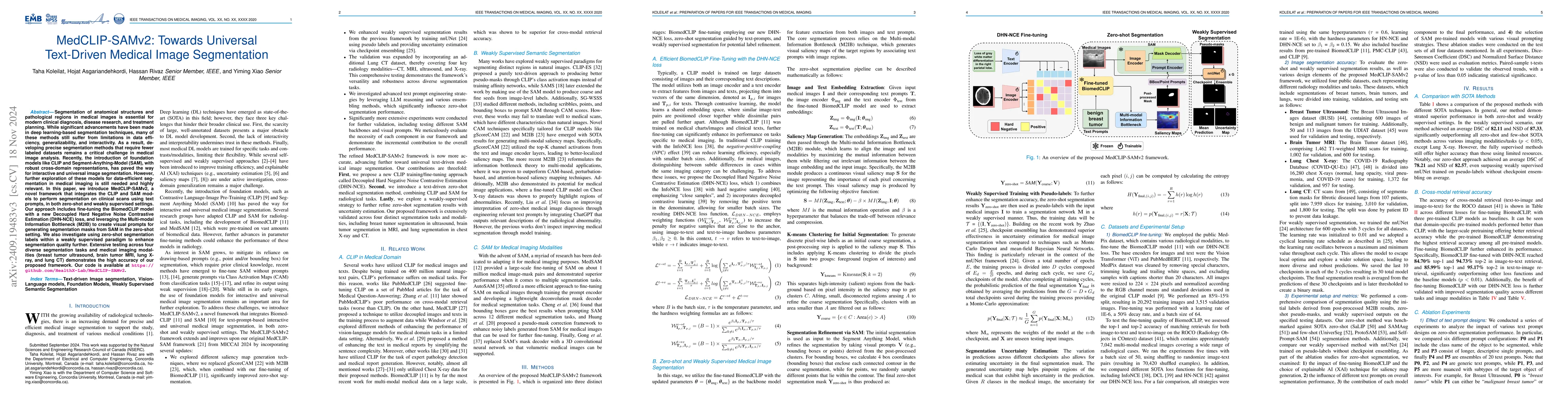
Segmentation of anatomical structures and pathological regions in medical images is essential for modern clinical diagnosis, disease research, and treatment planning. While significant advancements ha...
Super-resolution ultrasound imaging with ultrasound localization microscopy (ULM) offers a high-resolution view of microvascular structures. Yet, ULM image quality heavily relies on precise microbubbl...
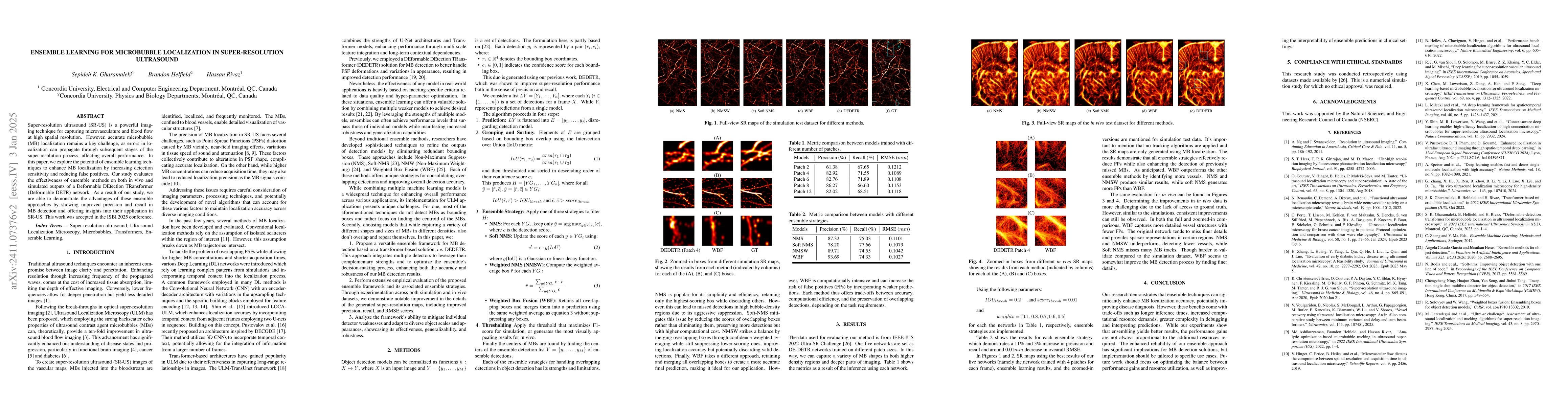
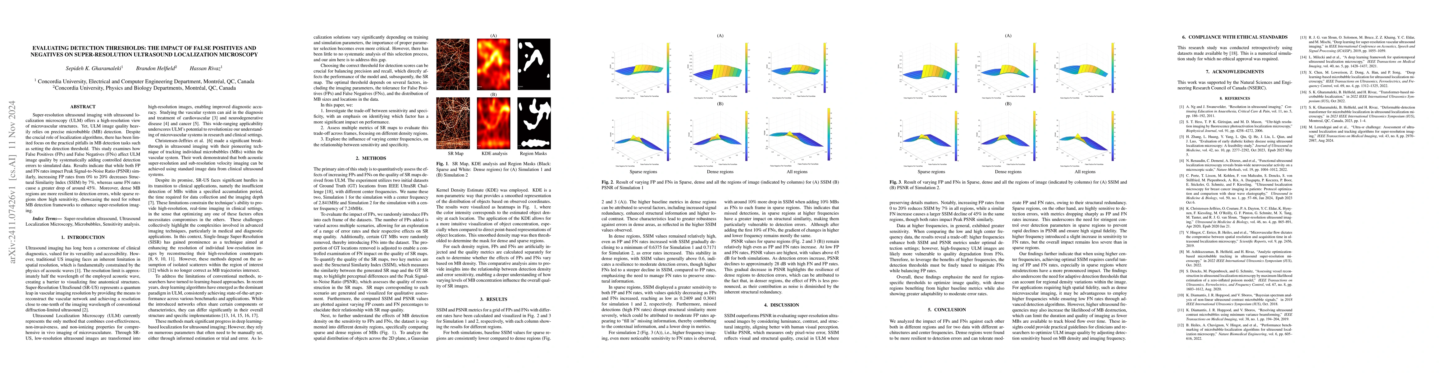
Super-resolution ultrasound (SR-US) is a powerful imaging technique for capturing microvasculature and blood flow at high spatial resolution. However, accurate microbubble (MB) localization remains a ...
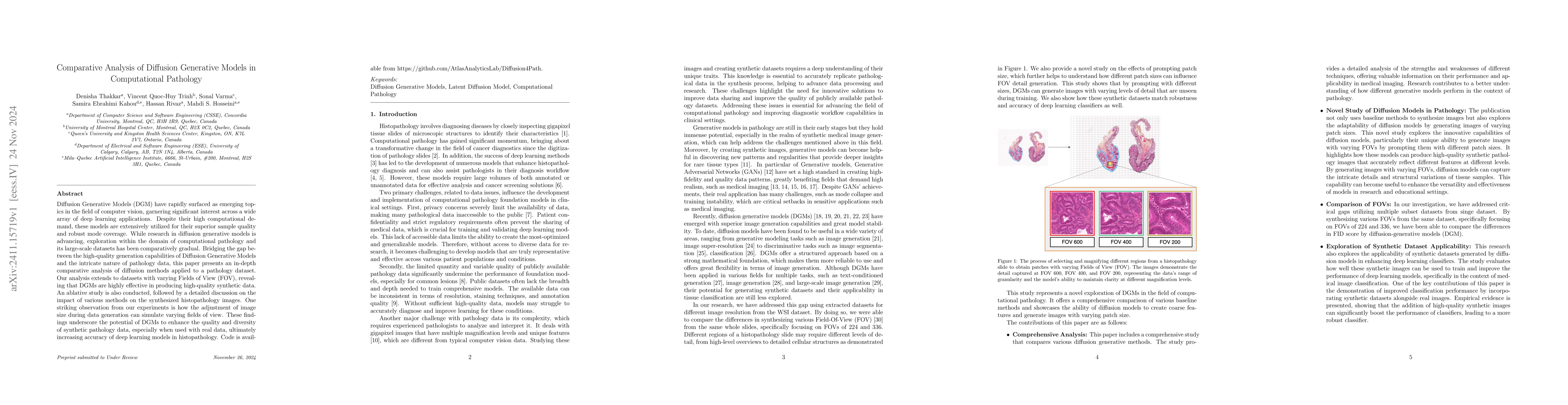
Diffusion Generative Models (DGM) have rapidly surfaced as emerging topics in the field of computer vision, garnering significant interest across a wide array of deep learning applications. Despite th...
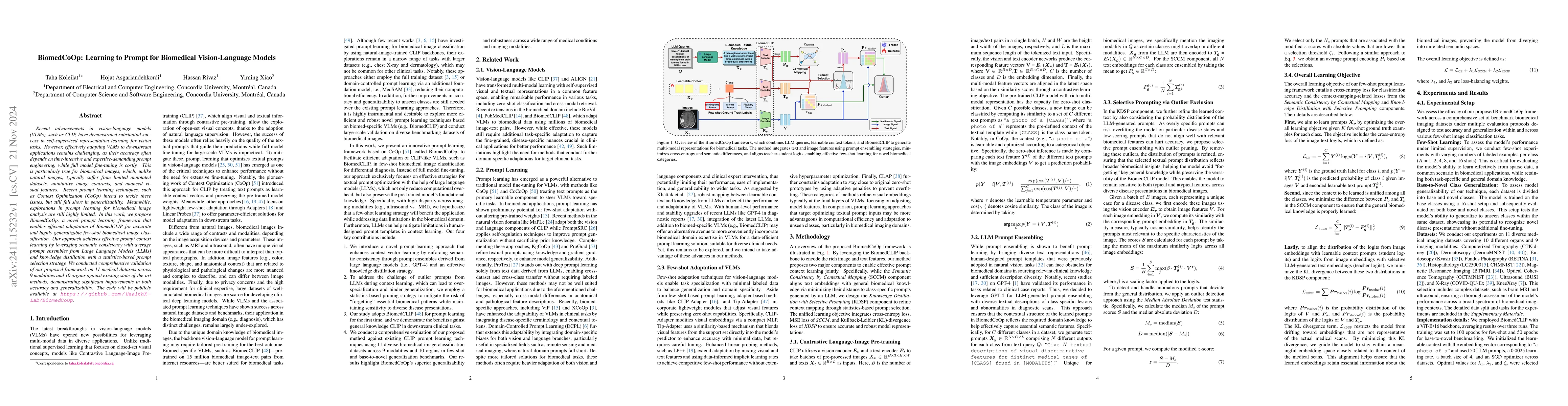
Recent advancements in vision-language models (VLMs), such as CLIP, have demonstrated substantial success in self-supervised representation learning for vision tasks. However, effectively adapting VLM...
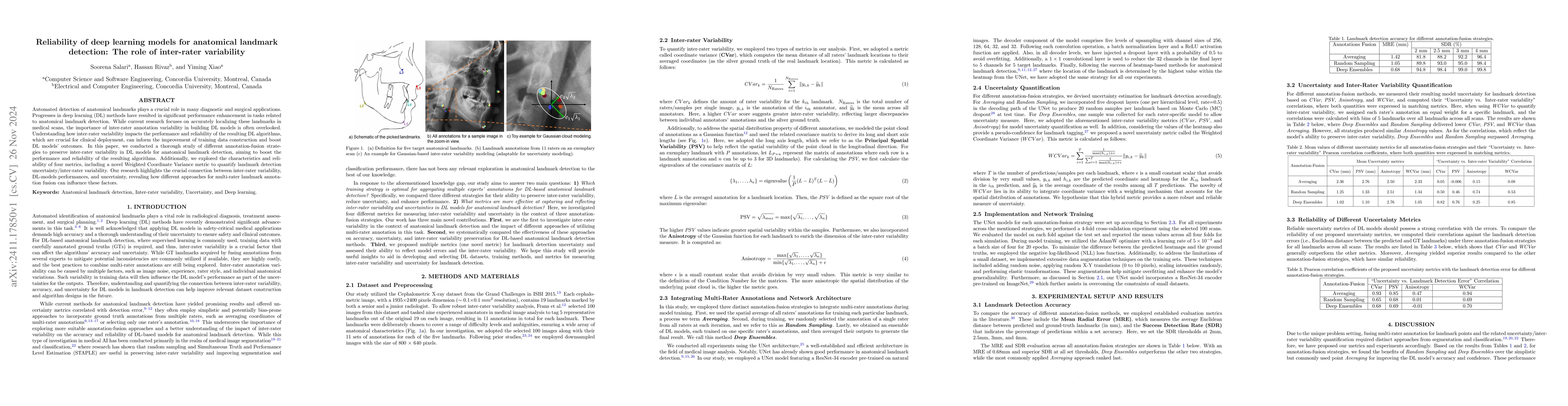
Automated detection of anatomical landmarks plays a crucial role in many diagnostic and surgical applications. Progresses in deep learning (DL) methods have resulted in significant performance enhance...
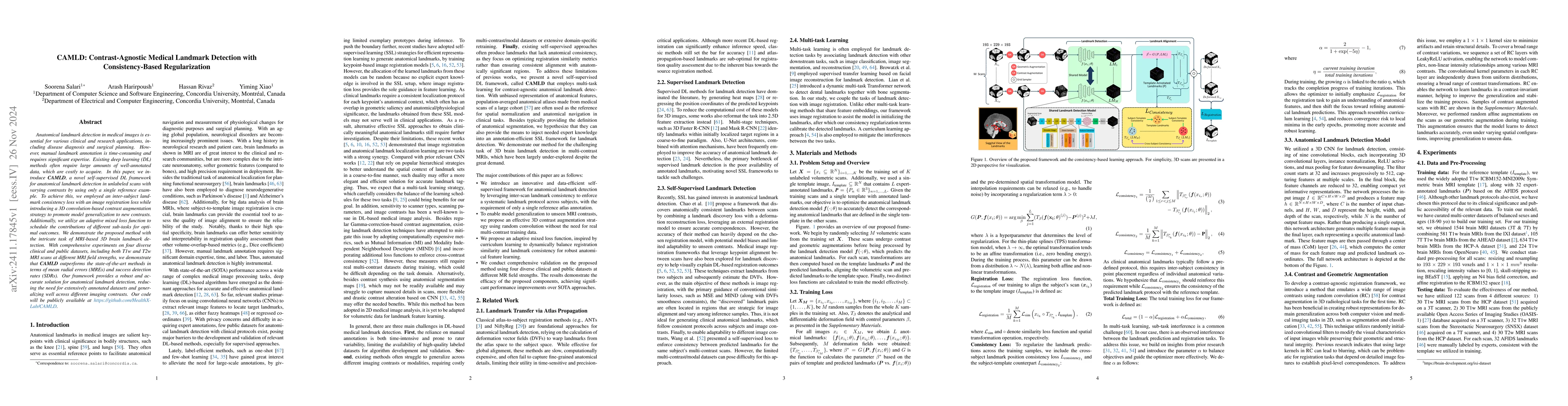
Anatomical landmark detection in medical images is essential for various clinical and research applications, including disease diagnosis and surgical planning. However, manual landmark annotation is t...
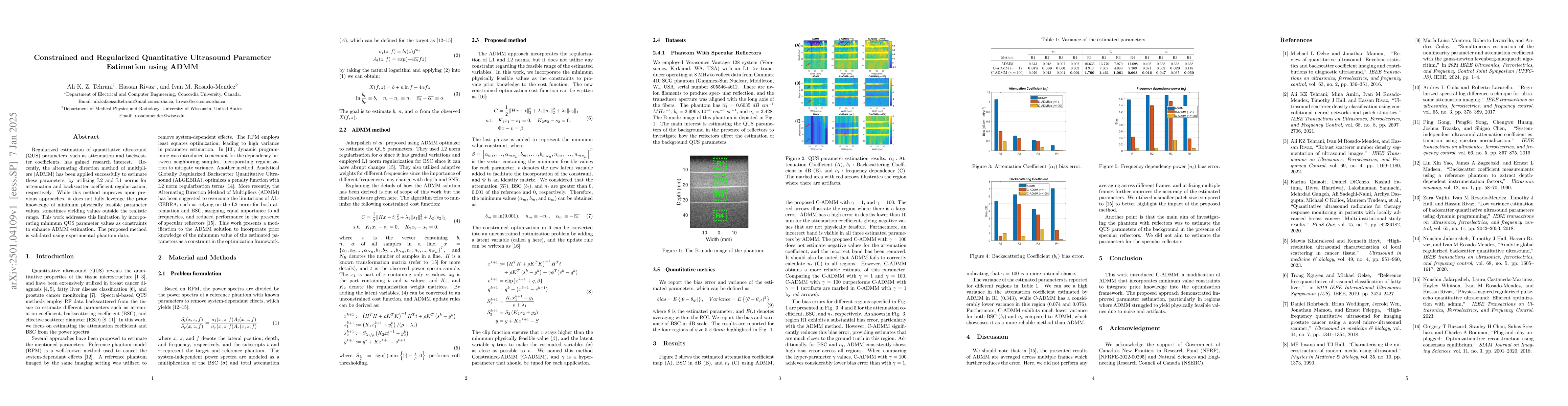
Regularized estimation of quantitative ultrasound (QUS) parameters, such as attenuation and backscatter coefficients, has gained research interest. Recently, the alternating direction method of multip...
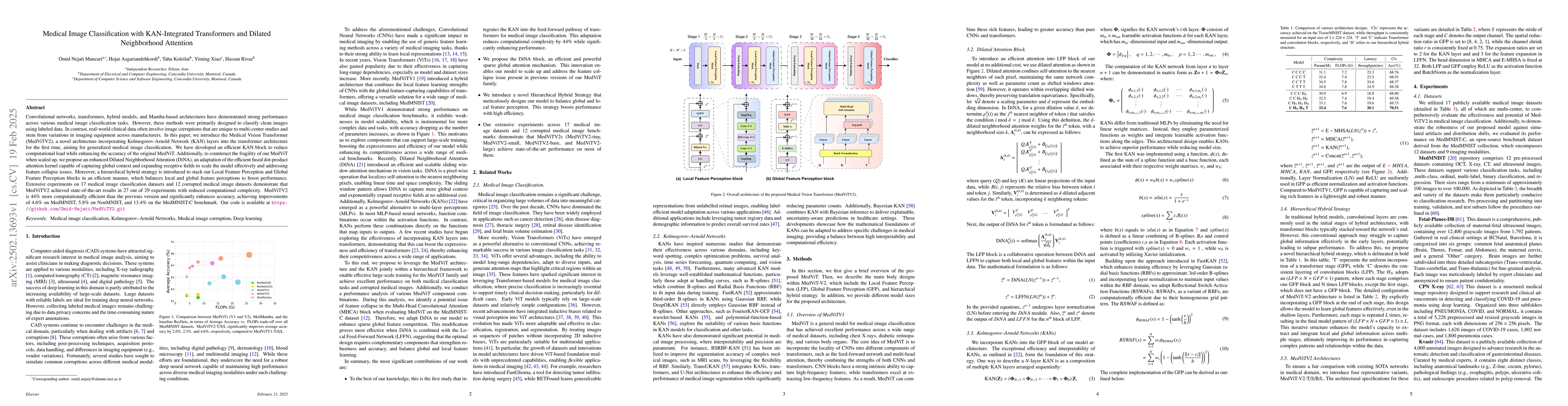
Convolutional networks, transformers, hybrid models, and Mamba-based architectures have demonstrated strong performance across various medical image classification tasks. However, these methods were p...
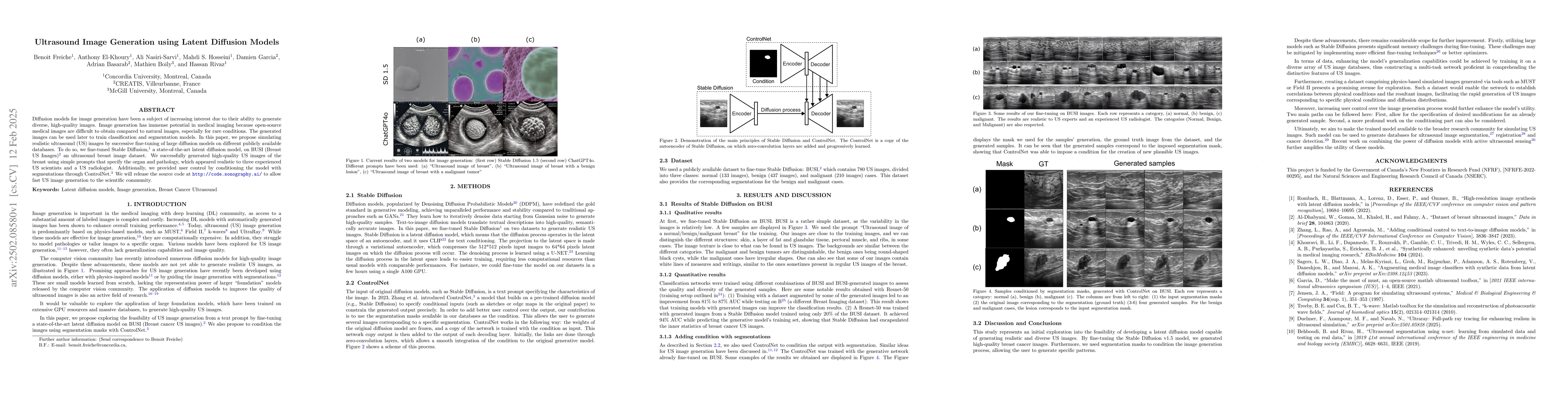
Diffusion models for image generation have been a subject of increasing interest due to their ability to generate diverse, high-quality images. Image generation has immense potential in medical imagin...
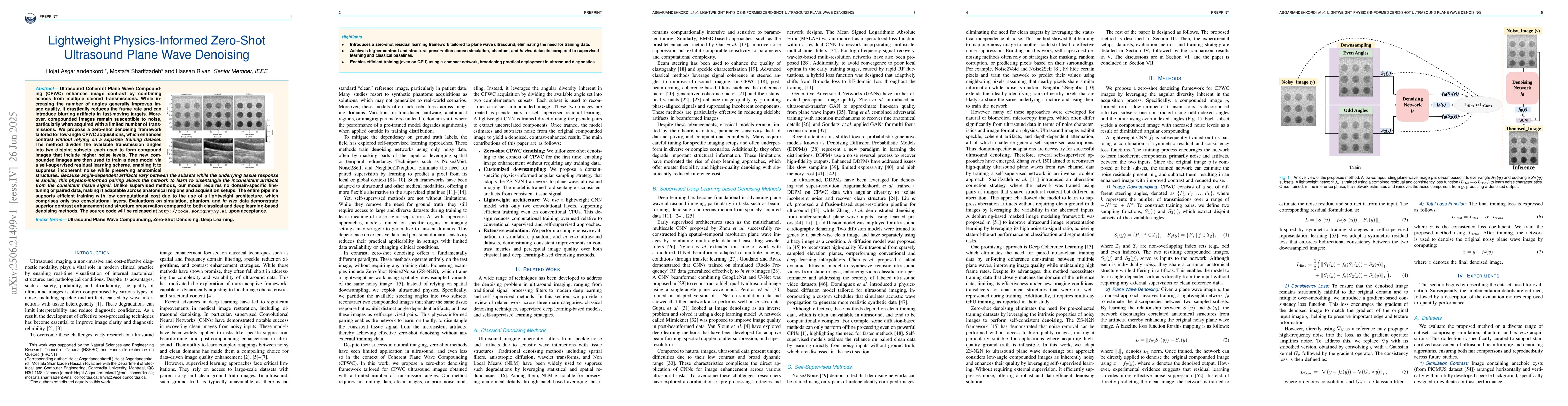
Ultrasound Coherent Plane Wave Compounding (CPWC) enhances image contrast by combining echoes from multiple steered transmissions. While increasing the number of angles generally improves image qualit...
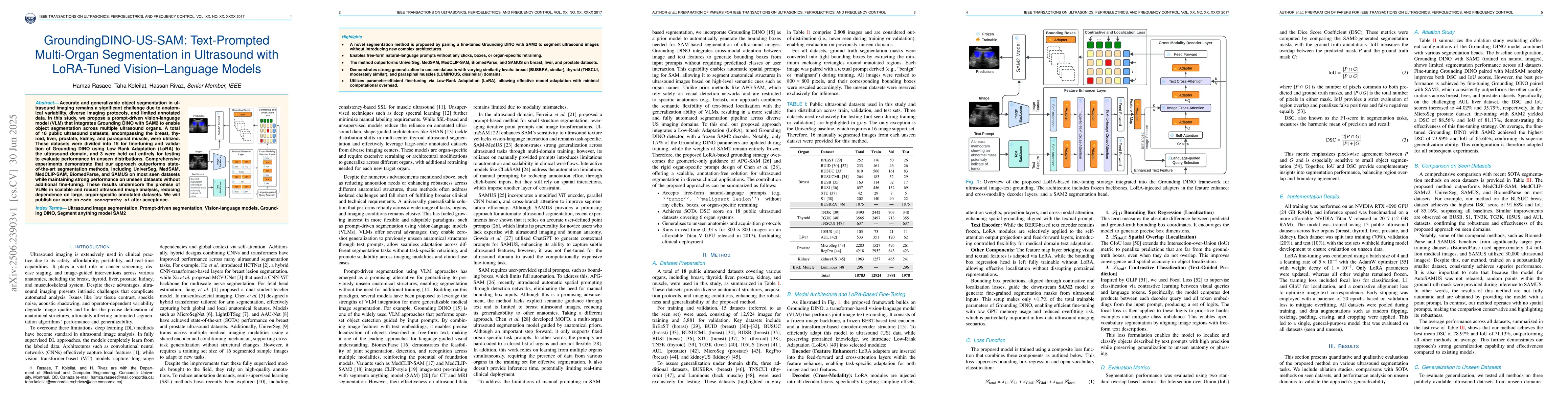
Accurate and generalizable object segmentation in ultrasound imaging remains a significant challenge due to anatomical variability, diverse imaging protocols, and limited annotated data. In this study...
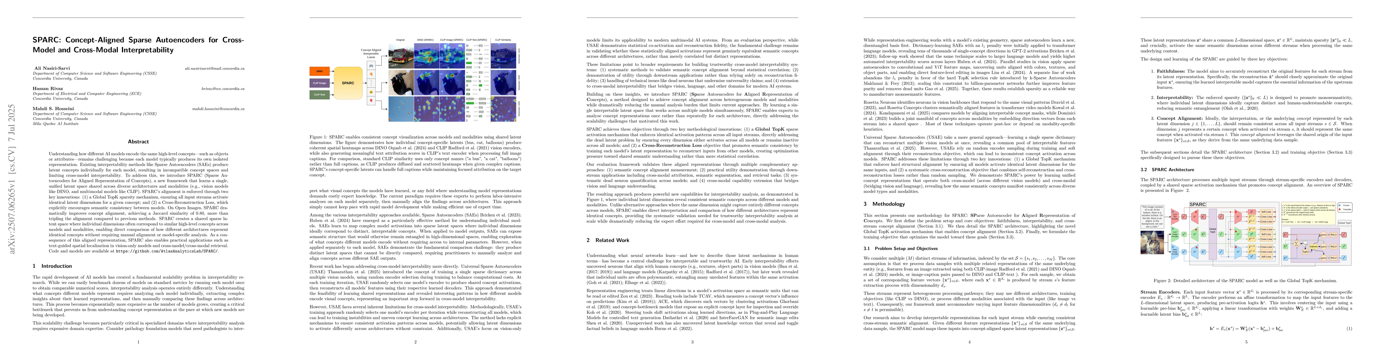
Understanding how different AI models encode the same high-level concepts, such as objects or attributes, remains challenging because each model typically produces its own isolated representation. Exi...
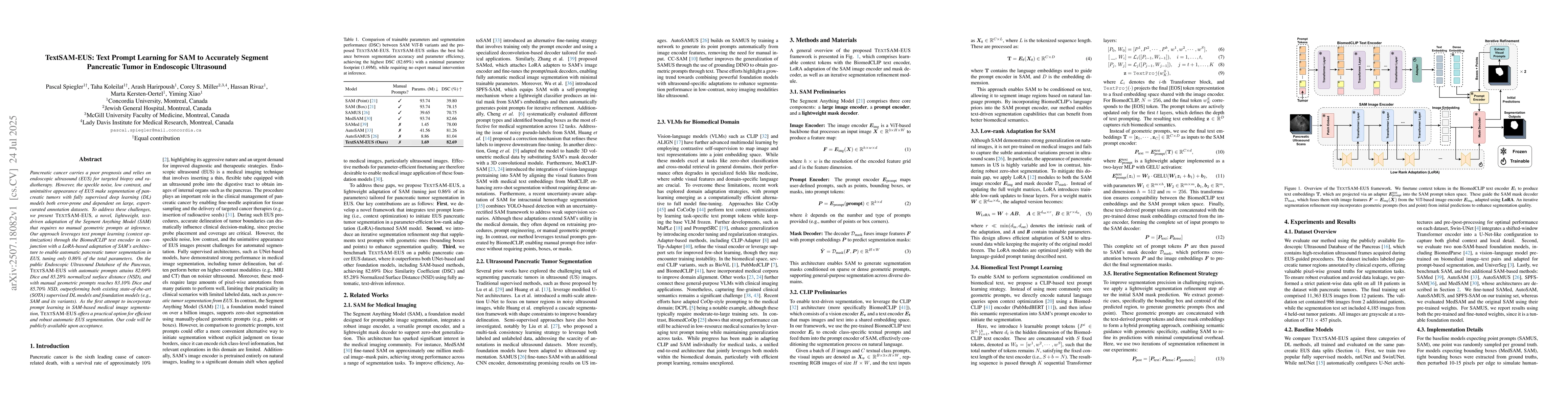
Pancreatic cancer carries a poor prognosis and relies on endoscopic ultrasound (EUS) for targeted biopsy and radiotherapy. However, the speckle noise, low contrast, and unintuitive appearance of EUS m...
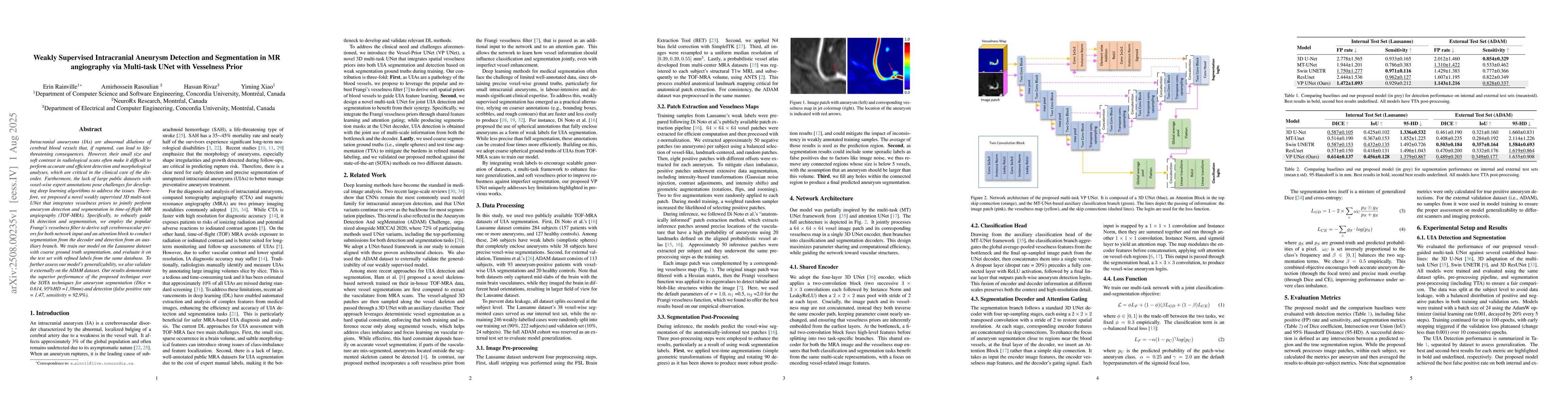
Intracranial aneurysms (IAs) are abnormal dilations of cerebral blood vessels that, if ruptured, can lead to life-threatening consequences. However, their small size and soft contrast in radiological ...
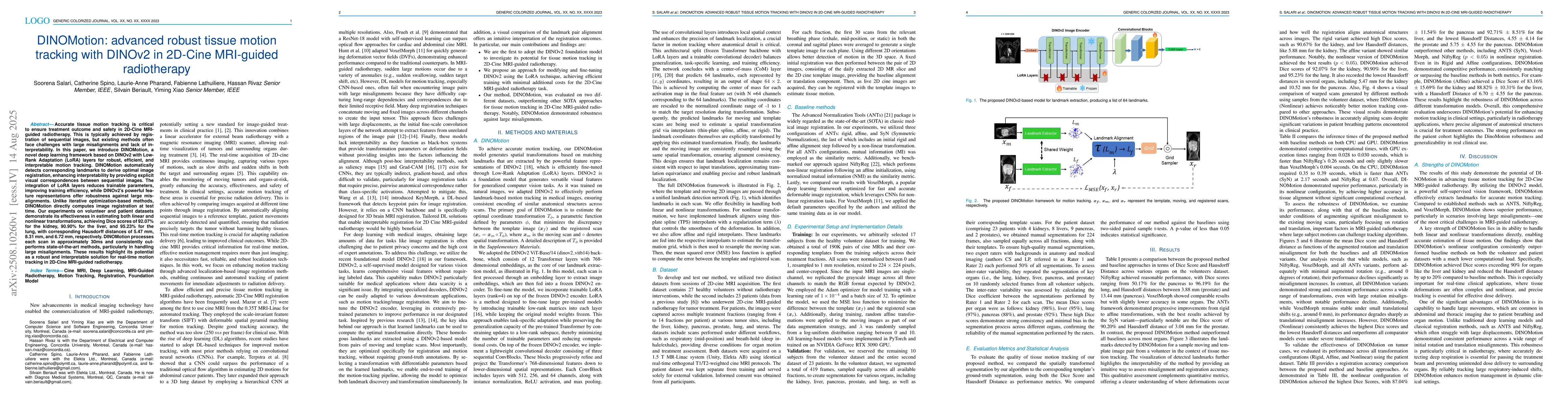
Accurate tissue motion tracking is critical to ensure treatment outcome and safety in 2D-Cine MRI-guided radiotherapy. This is typically achieved by registration of sequential images, but existing met...
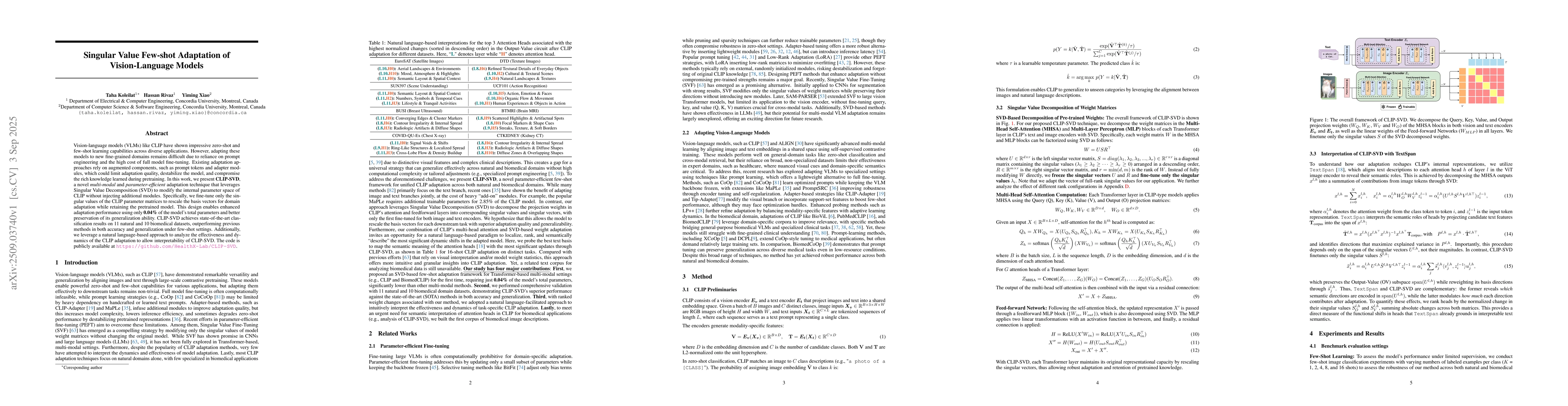
Vision-language models (VLMs) like CLIP have shown impressive zero-shot and few-shot learning capabilities across diverse applications. However, adapting these models to new fine-grained domains remai...

Conventional pulse-echo ultrasound suffers when low-cost probes deliver only narrow fractional bandwidths, elongating pulses and erasing high-frequency detail. We address this limitation by learning a...
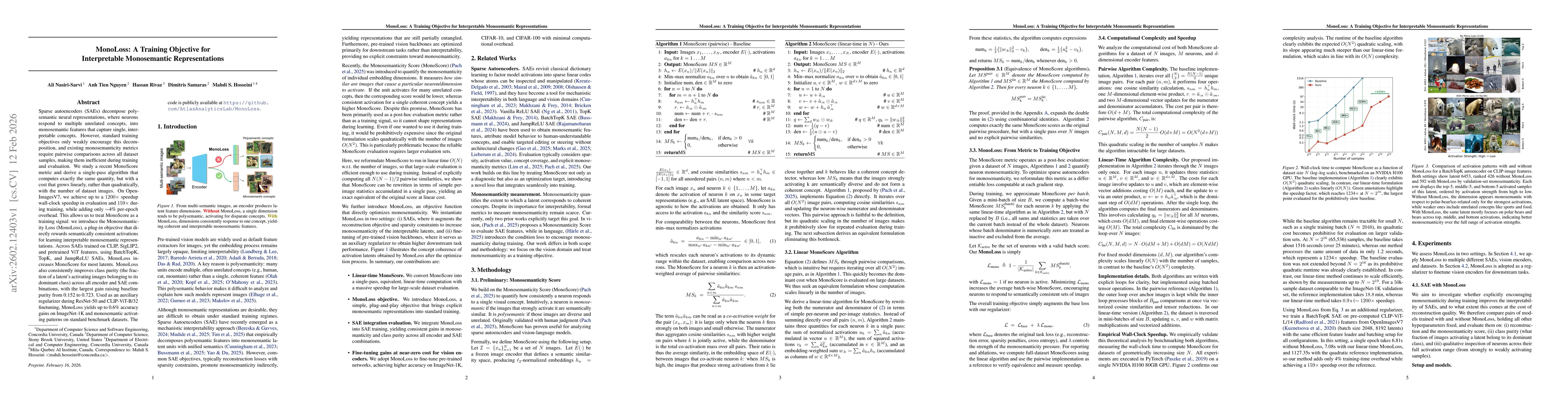
Sparse autoencoders (SAEs) decompose polysemantic neural representations, where neurons respond to multiple unrelated concepts, into monosemantic features that capture single, interpretable concepts. ...

Medical image segmentation remains challenging due to limited annotations for training, ambiguous anatomical features, and domain shifts. While vision-language models such as CLIP offer strong cross-m...
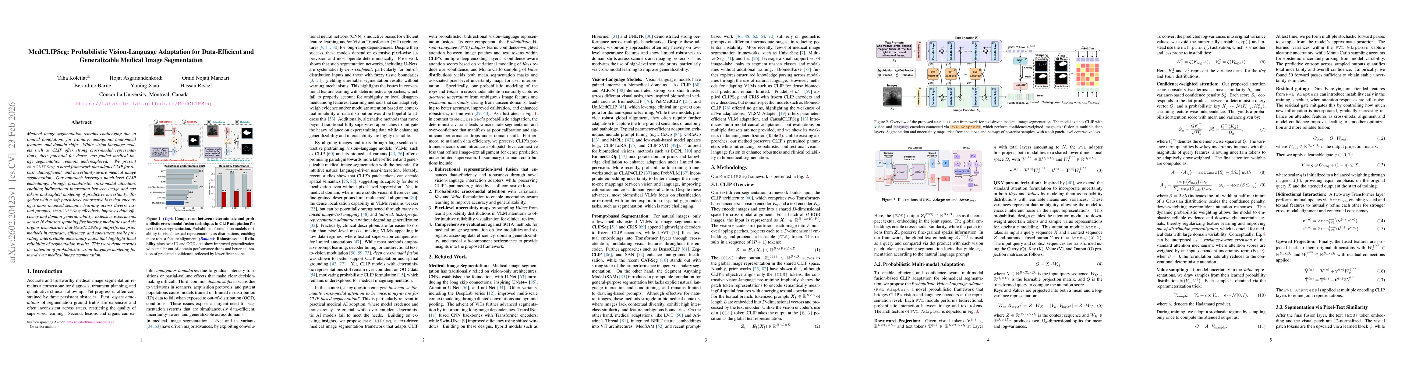
Medical image segmentation remains challenging due to limited annotations for training, ambiguous anatomical features, and domain shifts. While vision-language models such as CLIP offer strong cross-m...
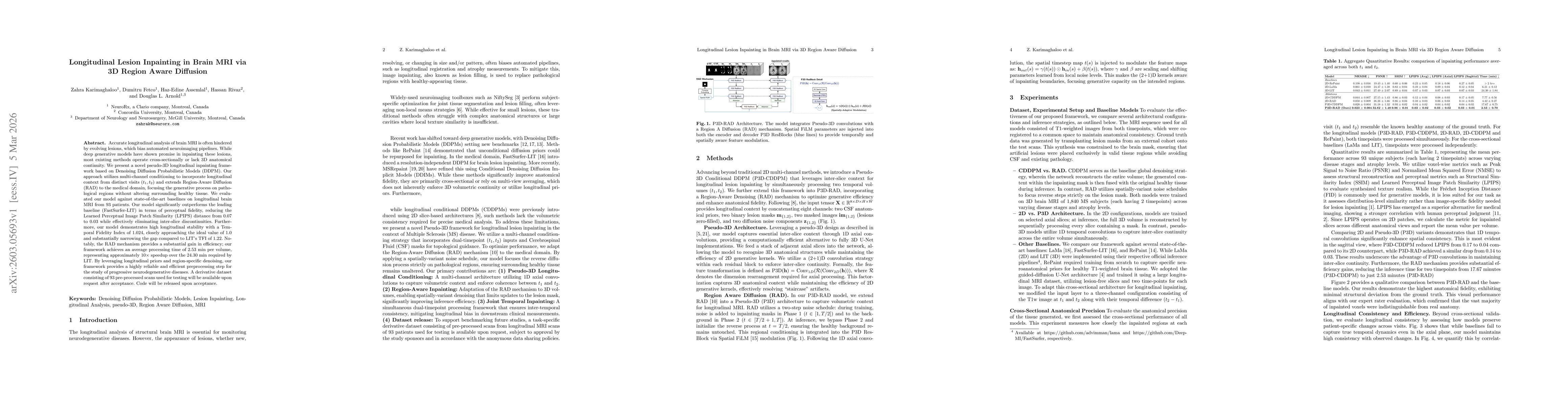
Accurate longitudinal analysis of brain MRI is often hindered by evolving lesions, which bias automated neuroimaging pipelines. While deep generative models have shown promise in inpainting these lesi...
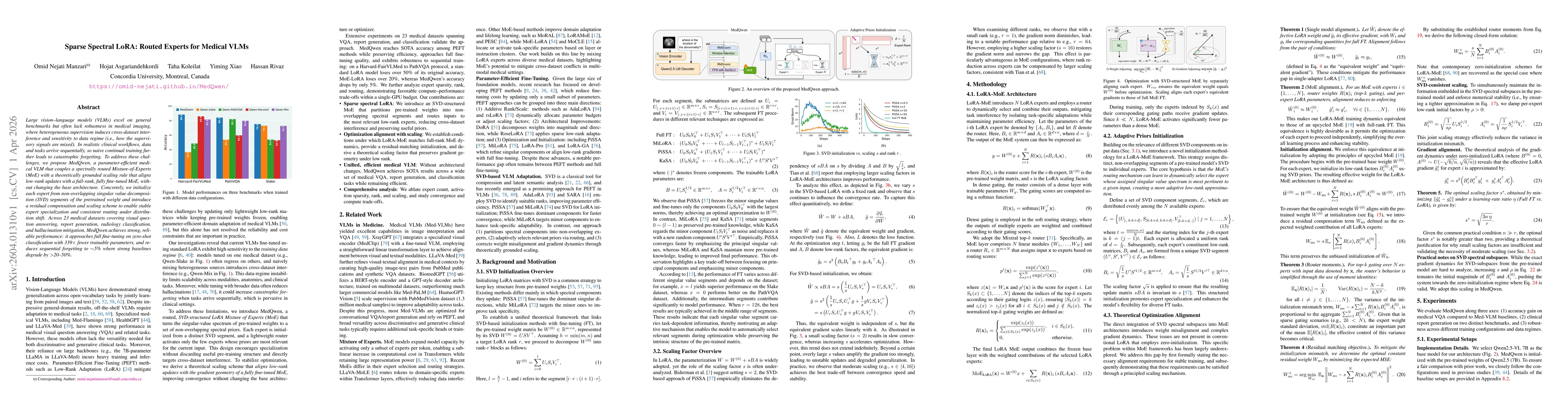
Large vision-language models (VLMs) excel on general benchmarks but often lack robustness in medical imaging, where heterogeneous supervision induces cross-dataset interference and sensitivity to data...
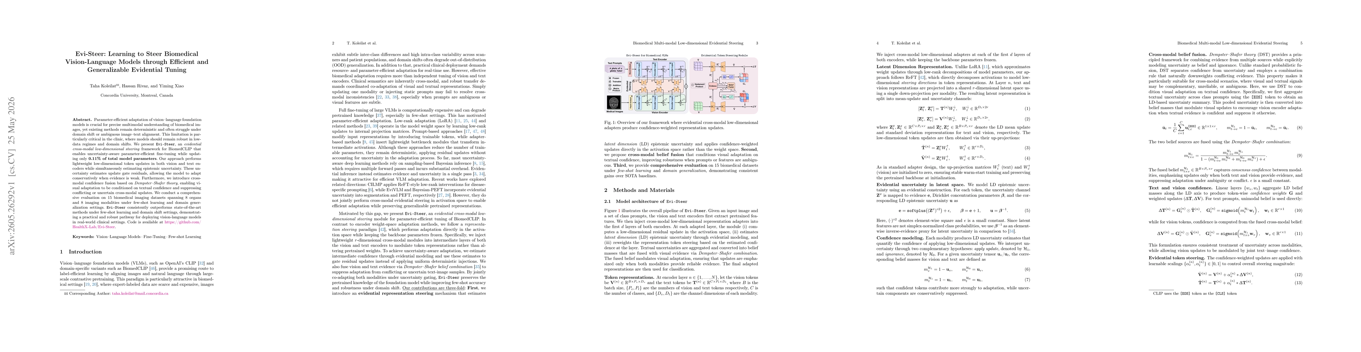
Parameter-efficient adaptation of vision-language foundation models is crucial for precise multimodal understanding of biomedical images, yet existing methods remain deterministic and often struggle u...