Publication
Metrics
Quick Actions
AI Quick Summary
This research introduces a novel, lightweight, zero-shot denoising framework for ultrasound Coherent Plane Wave Compounding (CPWC). It uses a self-supervised deep learning model trained on disjoint compound images from limited transmission angles, preserving anatomical structures and enhancing contrast without requiring domain-specific fine-tuning or paired data. Evaluations show superior performance over classical and deep learning methods.
Quick Answers
What is "Lightweight Physics-Informed Zero-Shot Ultrasound Plane Wave Denoising" about?
This research introduces a novel, lightweight, zero-shot denoising framework for ultrasound Coherent Plane Wave Compounding (CPWC). It uses a self-supervised deep learning model trained on disjoint compound images from limited transmission angles, preserving anatomical structures and enhancing contrast without requiring domain-specific fine-tuning or paired data. Evaluations show superior performance over classical and deep learning methods.
What methodology did the authors use?
The research introduces a zero-shot self-supervised ultrasound denoising framework that leverages angle-based data decomposition and a lightweight convolutional neural network trained with residual and consistency loss functions, without requiring external datasets. More in Methodology →
What are the key results?
The proposed method outperforms classical and zero-shot denoising techniques in contrast-to-noise ratio (CNR), generalized CNR (gCNR), and statistical distribution preservation across simulation, phantom, and in vivo datasets. — It effectively reduces noise while preserving anatomical structures, achieving high contrast and detail in challenging deep regions. More in Key Results →
Why is this work significant?
This work provides a practical, data-efficient, and adaptable ultrasound denoising solution that can be deployed in real-world clinical settings, especially where training data is scarce or unavailable, potentially improving image quality and diagnostic accuracy. More in Significance →
What are the main limitations?
The current implementation is not optimized for real-time processing, with inference times around 6 seconds per image. — The method's performance on larger, more diverse clinical datasets and various probe geometries remains to be validated. More in Limitations →
Paper Preview
Abstract
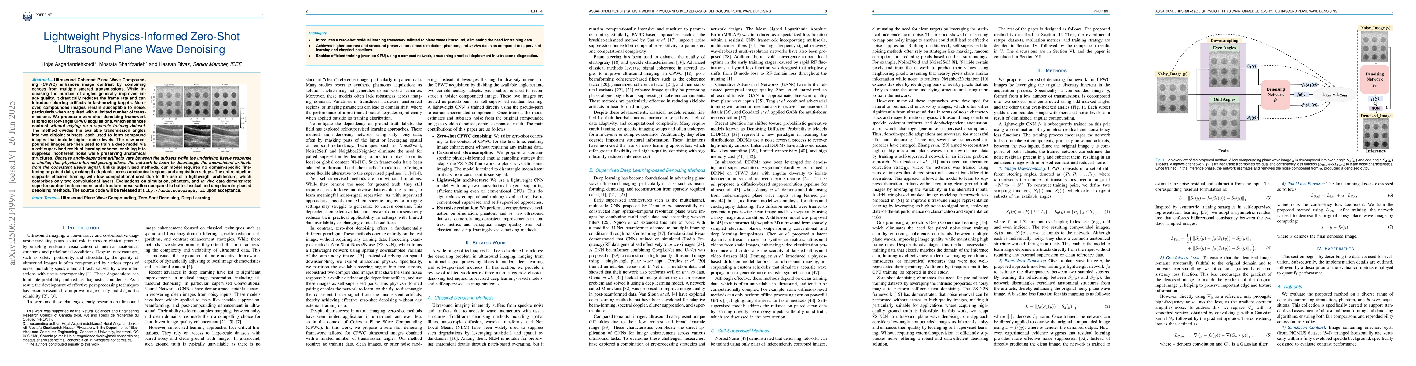
Ultrasound Coherent Plane Wave Compounding (CPWC) enhances image contrast by combining echoes from multiple steered transmissions. While increasing the number of angles generally improves image quality, it drastically reduces the frame rate and can introduce blurring artifacts in fast-moving targets. Moreover, compounded images remain susceptible to noise, particularly when acquired with a limited number of transmissions. We propose a zero-shot denoising framework tailored for low-angle CPWC acquisitions, which enhances contrast without relying on a separate training dataset. The method divides the available transmission angles into two disjoint subsets, each used to form compound images that include higher noise levels. The new compounded images are then used to train a deep model via a self-supervised residual learning scheme, enabling it to suppress incoherent noise while preserving anatomical structures. Because angle-dependent artifacts vary between the subsets while the underlying tissue response is similar, this physics-informed pairing allows the network to learn to disentangle the inconsistent artifacts from the consistent tissue signal. Unlike supervised methods, our model requires no domain-specific fine-tuning or paired data, making it adaptable across anatomical regions and acquisition setups. The entire pipeline supports efficient training with low computational cost due to the use of a lightweight architecture, which comprises only two convolutional layers. Evaluations on simulation, phantom, and in vivo data demonstrate superior contrast enhancement and structure preservation compared to both classical and deep learning-based denoising methods.
AI Key Findings
Generated Jul 01, 2025
Methodology — What approach did the authors take?
The research introduces a zero-shot self-supervised ultrasound denoising framework that leverages angle-based data decomposition and a lightweight convolutional neural network trained with residual and consistency loss functions, without requiring external datasets.
Key Results — What are the main findings?
- The proposed method outperforms classical and zero-shot denoising techniques in contrast-to-noise ratio (CNR), generalized CNR (gCNR), and statistical distribution preservation across simulation, phantom, and in vivo datasets.
- It effectively reduces noise while preserving anatomical structures, achieving high contrast and detail in challenging deep regions.
- The approach demonstrates robustness and generalizability without domain-specific fine-tuning or paired training data, even under different imaging conditions.
Significance — Why does this research matter?
This work provides a practical, data-efficient, and adaptable ultrasound denoising solution that can be deployed in real-world clinical settings, especially where training data is scarce or unavailable, potentially improving image quality and diagnostic accuracy.
Technical Contribution — What is the technical contribution?
The main technical contribution is a physics-inspired, angle-based self-supervised training scheme that enables effective ultrasound image denoising without external datasets, combined with a highly efficient lightweight CNN architecture.
Novelty — What is new about this work?
This work is novel in applying a zero-shot, physics-informed self-supervised learning approach specifically tailored for ultrasound plane wave imaging, eliminating the need for paired or large-scale training data and demonstrating superior performance across multiple datasets.
Limitations — What are the limitations of this study?
- The current implementation is not optimized for real-time processing, with inference times around 6 seconds per image.
- The method's performance on larger, more diverse clinical datasets and various probe geometries remains to be validated.
Future Work — What did the authors propose for future work?
- Developing strategies for online, real-time denoising to enable clinical deployment.
- Training on larger, more diverse datasets to improve robustness and extend applicability across different ultrasound modalities and hardware.
How to Cite This Paper
@article{rivaz2025lightweight,
  title         = {Lightweight Physics-Informed Zero-Shot Ultrasound Plane Wave Denoising},
  author        = {Rivaz, Hassan and Asgariandehkordi, Hojat and Sharifzadeh, Mostafa},
  year          = {2025},
  eprint        = {2506.21499},
  archivePrefix = {arXiv},
  primaryClass  = {eess.IV},
}Rivaz, H., Asgariandehkordi, H., & Sharifzadeh, M. (2025). Lightweight Physics-Informed Zero-Shot Ultrasound Plane Wave Denoising. arXiv. https://arxiv.org/abs/2506.21499Rivaz, Hassan, et al. "Lightweight Physics-Informed Zero-Shot Ultrasound Plane Wave Denoising." arXiv, 2025, arxiv.org/abs/2506.21499.PDF Preview
Citation Network
Current paper (gray), citations (green), references (blue)
Display is limited for performance on very large graphs.
Similar Papers
Found 4 papersDenoising Plane Wave Ultrasound Images Using Diffusion Probabilistic Models
Hojat Asgariandehkordi, Sobhan Goudarzi, Mostafa Sharifzadeh et al.
A Zero-Shot Physics-Informed Dictionary Learning Approach for Sound Field Reconstruction
Stefano Damiano, Federico Miotello, Mirco Pezzoli et al.
No citations found for this paper.
Comments (0)