Academic Profile
Statistics
Similar Authors
Papers on arXiv
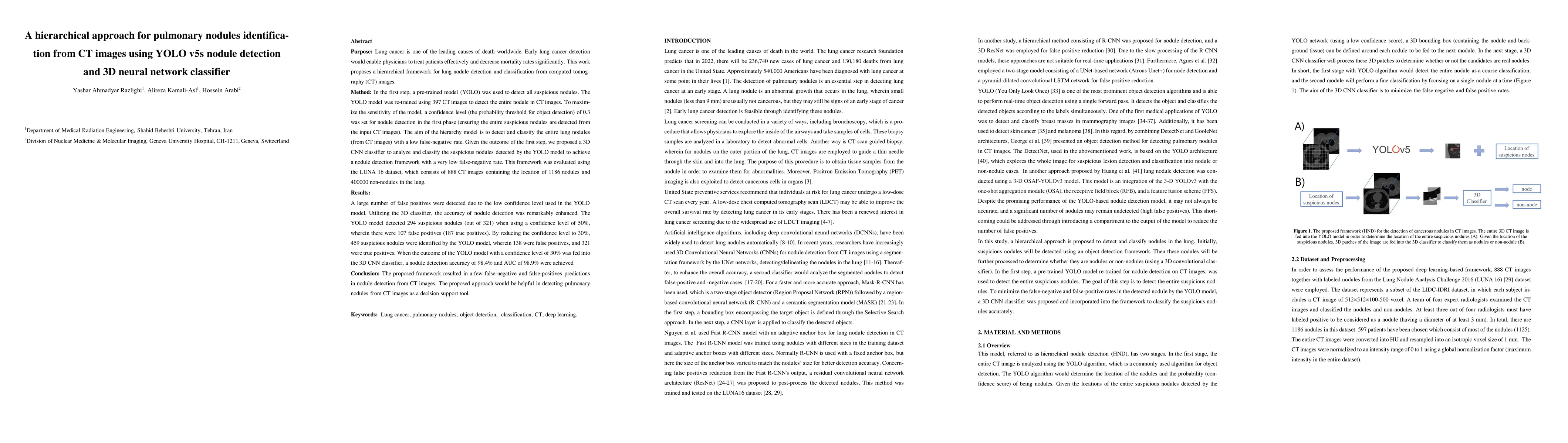
In the first step, a pre-trained model (YOLO) was used to detect all suspicious nod-ules. The YOLO model was re-trained using 397 CT images to detect the entire nodule in CT images. To maximize the ...
A standard dose of radioactive tracer must be delivered into the patients body to obtain high-quality Positron Emission Tomography (PET) images for diagnostic purposes, which raises the risk of radi...
Attenuation and scatter correction (AC) is crucial for quantitative Positron Emission Tomography (PET) imaging. Recently, direct application of AC in the image domain using deep learning approaches ...
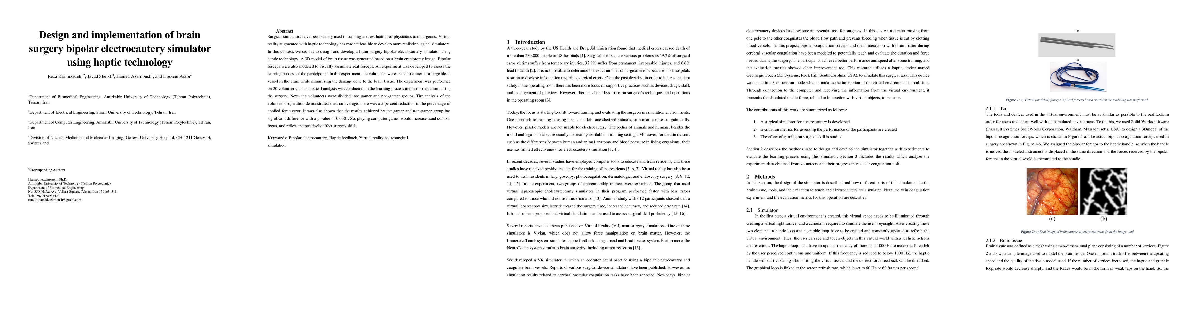
Surgical simulators have been widely used in training and evaluation of physicians and surgeons. Virtual reality augmented with haptic technology has made it feasible to develop more realistic surgi...
Brain tumor segmentation is highly contributive in diagnosing and treatment planning. The manual brain tumor delineation is a time-consuming and tedious task and varies depending on the radiologists...
Automated medical image segmentation is an essential task to aid/speed up diagnosis and treatment procedures in clinical practices. Deep convolutional neural networks have exhibited promising perfor...
This study aimed to evaluate the performance of a novel unsupervised deep learning-based framework for automated infections lesion segmentation from CT images of Covid patients. In the first step, t...
Reducing the injected dose would result in quality degradation and loss of information in PET imaging. To address this issue, deep learning methods have been introduced to predict standard PET image...
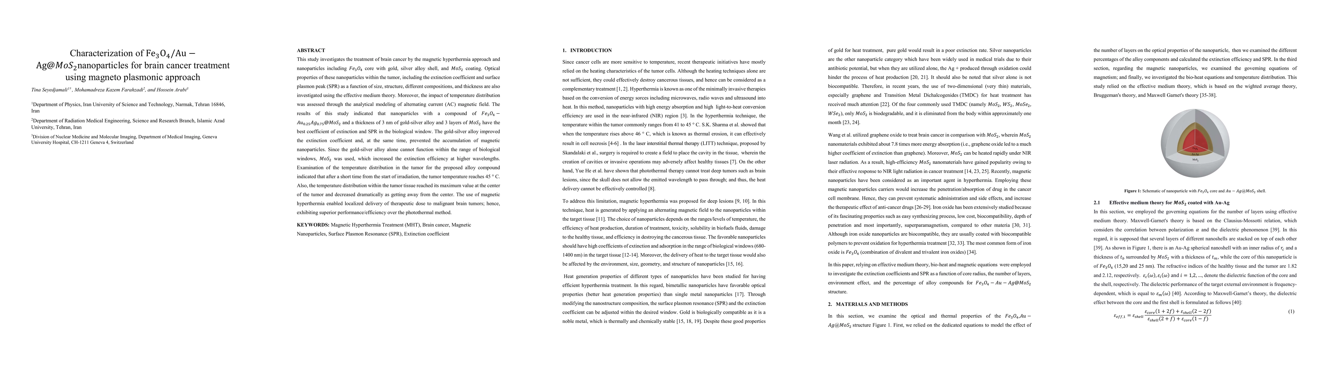
This study investigates the treatment of brain cancer by the magnetic hyperthermia approach and nanoparticles including Fe_3 O_4 core with gold, silver alloy shell, and MoS_2 coating. Optical proper...
99m-Tc Ethyl-Cysteinate-Dimer SPECT and MR imaging play a significant role in diagnosing anosmia. In this study, two-tissue class and three-tissue class attenuation maps (2C-MR and 3CMR) obtained fr...
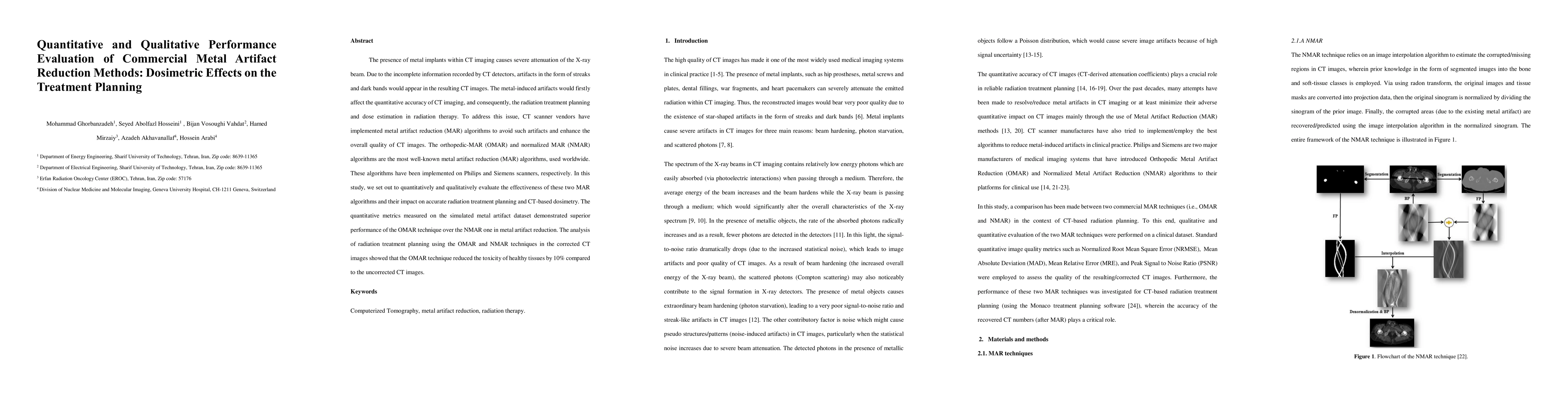
The presence of metal implants within CT imaging causes severe attenuation of the X-ray beam. Due to the incomplete information recorded by CT detectors, artifacts in the form of streaks and dark ba...
MRI-guided radiation treatment planning is widely applied because of its superior soft-tissue contrast and no ionization radiation compared to CT-based planning. In this regard, synthetic CT (sCT) i...
Automated semantic image segmentation is an essential step in quantitative image analysis and disease diagnosis. This study investigates the performance of a deep learning-based model for lung segme...
Clinical SPECT-MPI images of 345 patients acquired from a dedicated cardiac SPECT in list-mode format were retrospectively employed to predict normal-dose images from low-dose data at the half, quar...
In this study, 18F-FDG PET/CT brain scans of 50 patients with head and neck malignant lesions were employed to systematically assess the relationship between the amount of injected dose (10%, 8%, 6%...
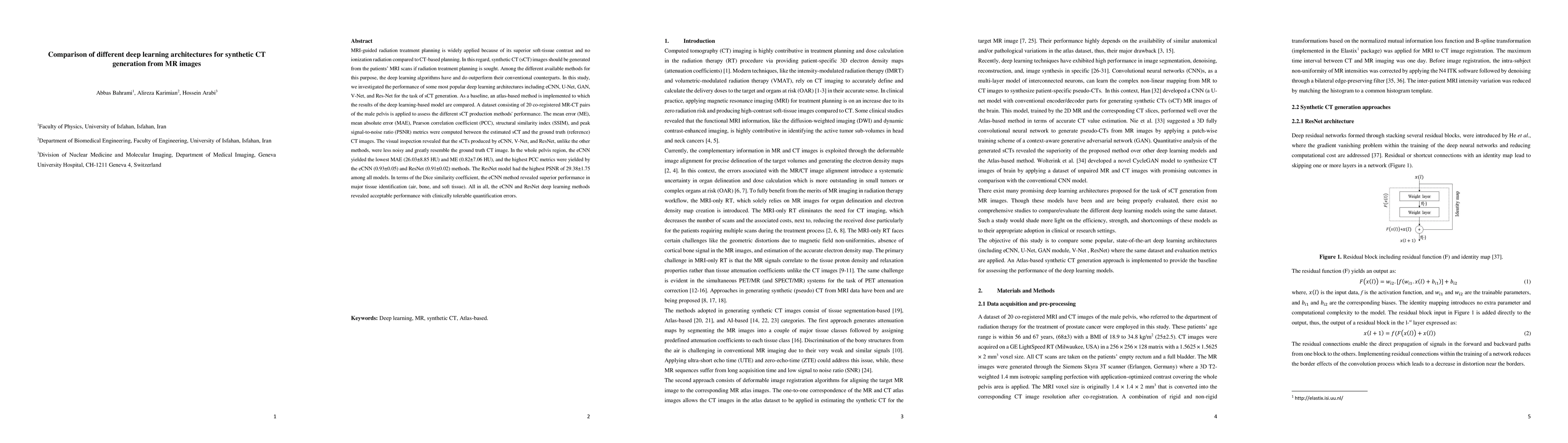
Currently, MRI-only radiotherapy (RT) eliminates some of the concerns about using CT images in RT chains such as the registration of MR images to a separate CT, extra dose delivery, and the addition...
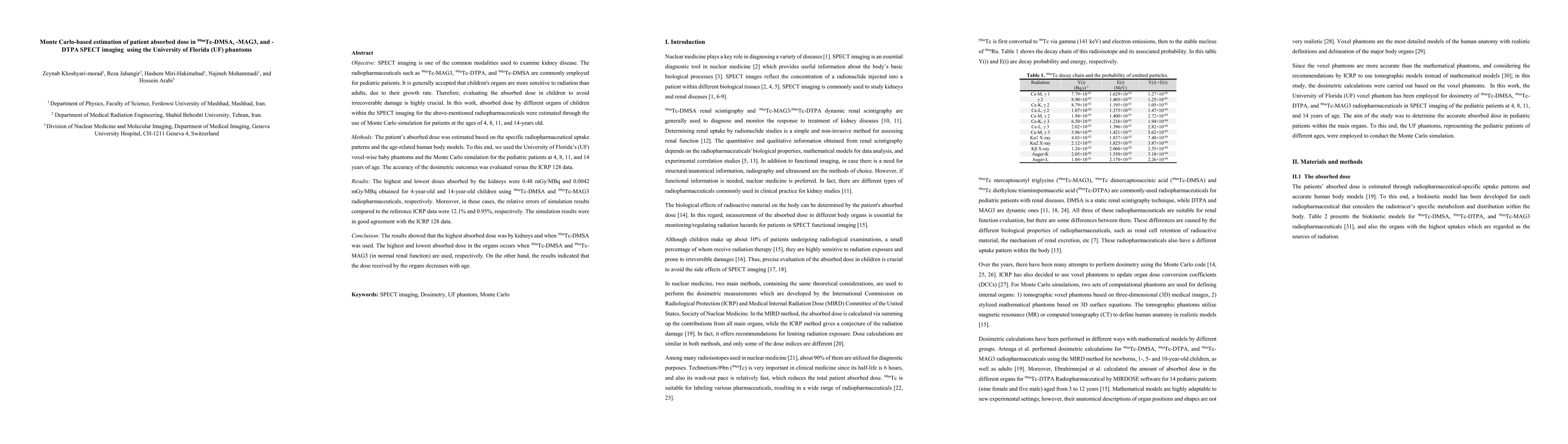
SPECT imaging is one of the common modalities used to examine kidney disease. The radiopharmaceuticals such as 99mTc-MAG3, 99mTc-DTPA, and 99mTc-DMSA are commonly employed for pediatric patients. It...
We set out to simulate four reduced dose-levels (60%-dose, 40%-dose, 20%-dose, and 10%-dose) of standard CT imaging using Beer-Lambert's law across 49 patients infected with COVID-19. Then, three de...
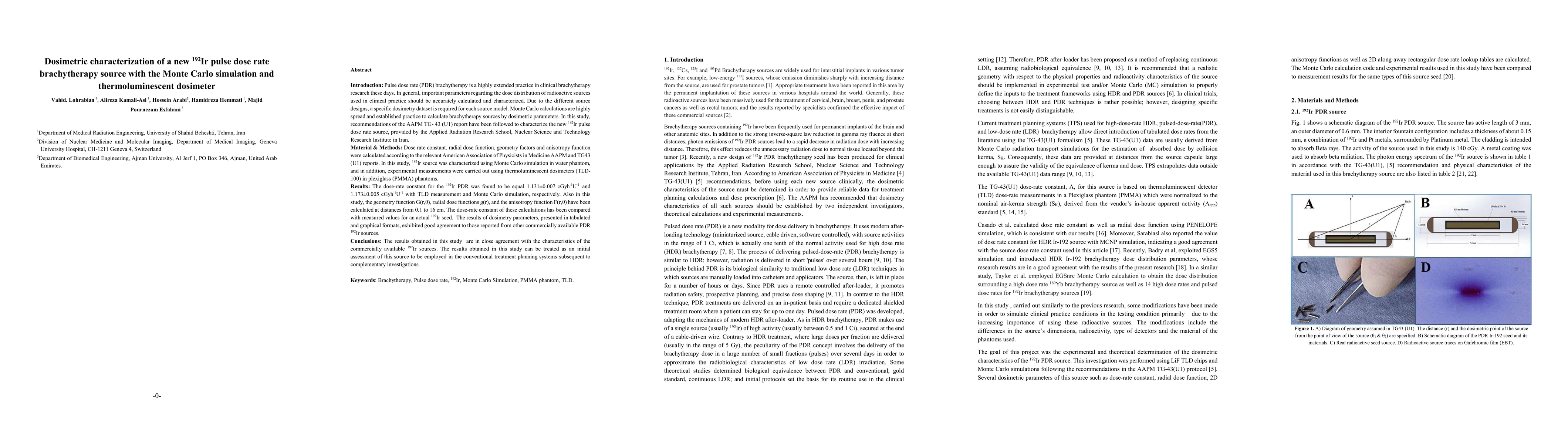
In this study, recommendations of the AAPM TG- 43 (U1) report have been followed to characterize the new 192Ir pulse dose rate source, provided by the Applied Radiation Research School, Nuclear Scie...
The aim of this study is to estimate the intrinsic efficiency and energy resolution of different types of solid-state gamma-ray detectors in order to generate a precise dual-energy x-ray beam from t...
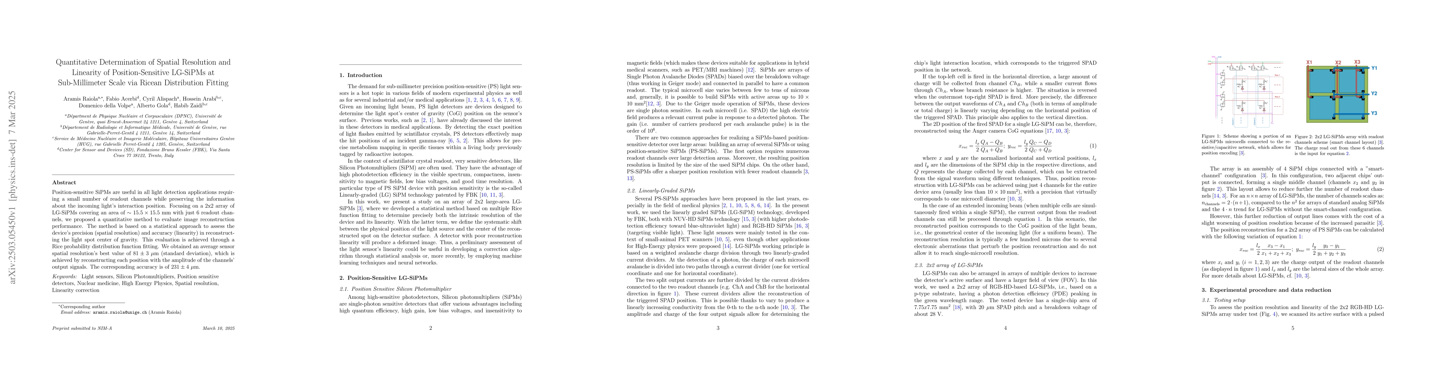
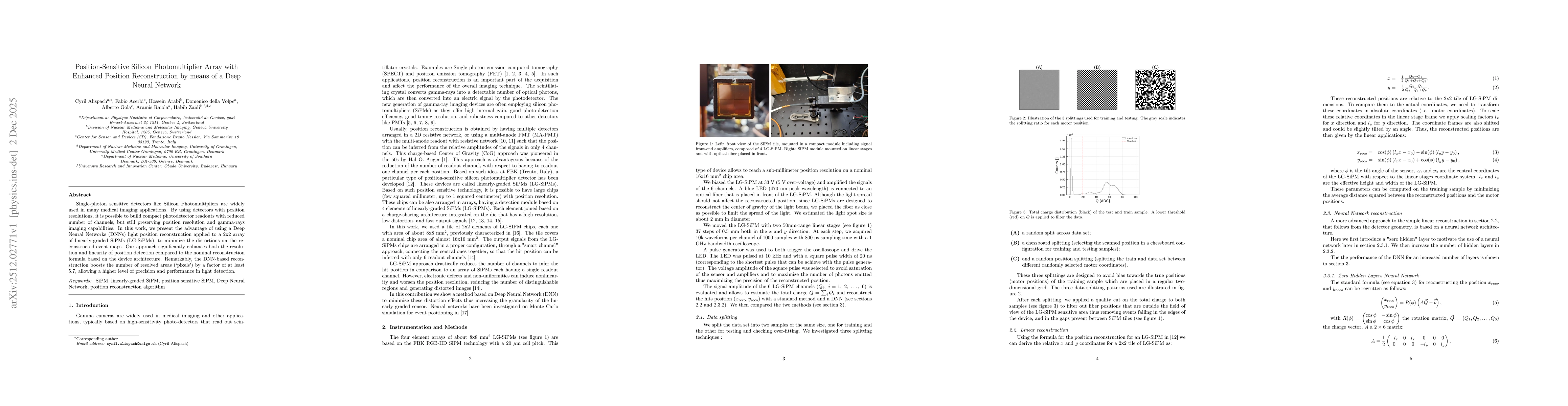
Position-sensitive SiPMs are useful in all light detection applications requiring a small number of readout channels while preserving the information about the incoming light's interaction position. F...
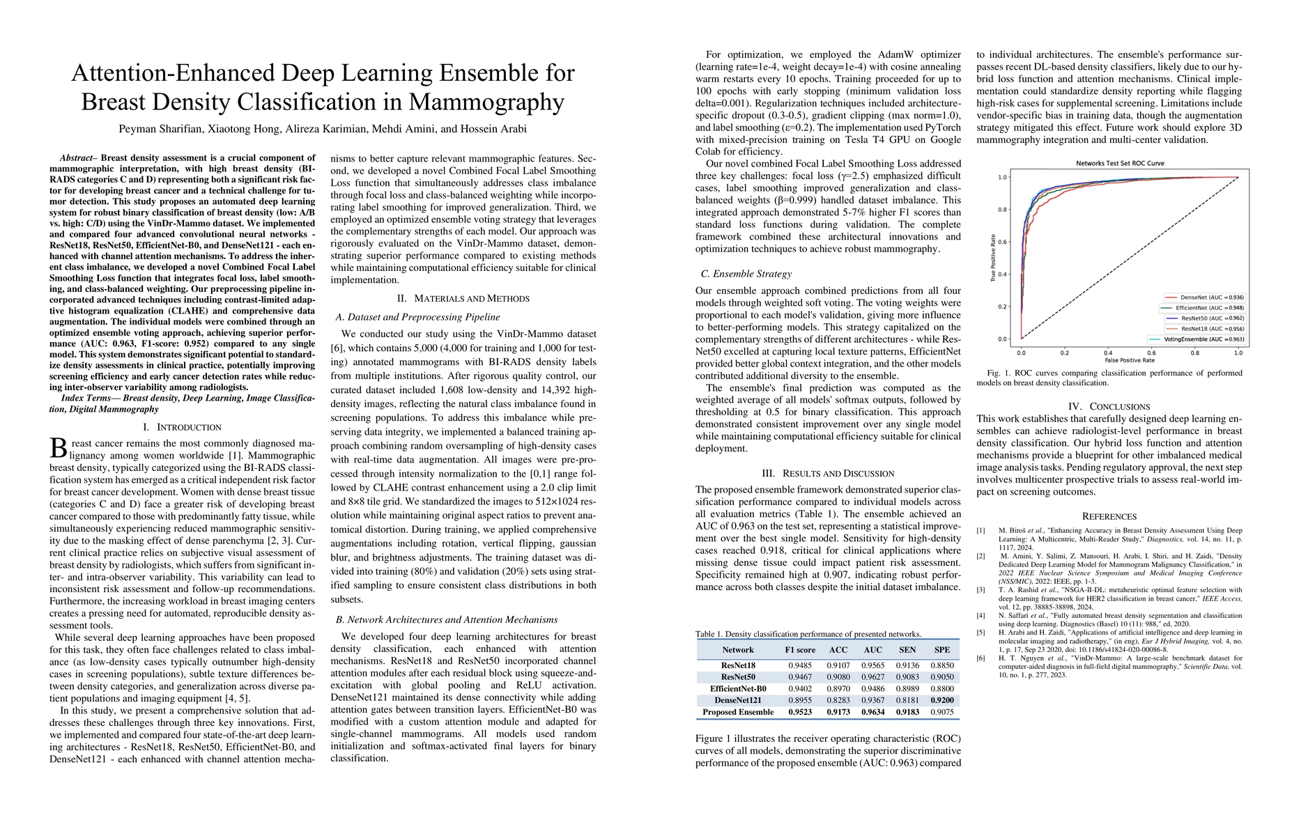
Breast density assessment is a crucial component of mammographic interpretation, with high breast density (BI-RADS categories C and D) representing both a significant risk factor for developing breast...
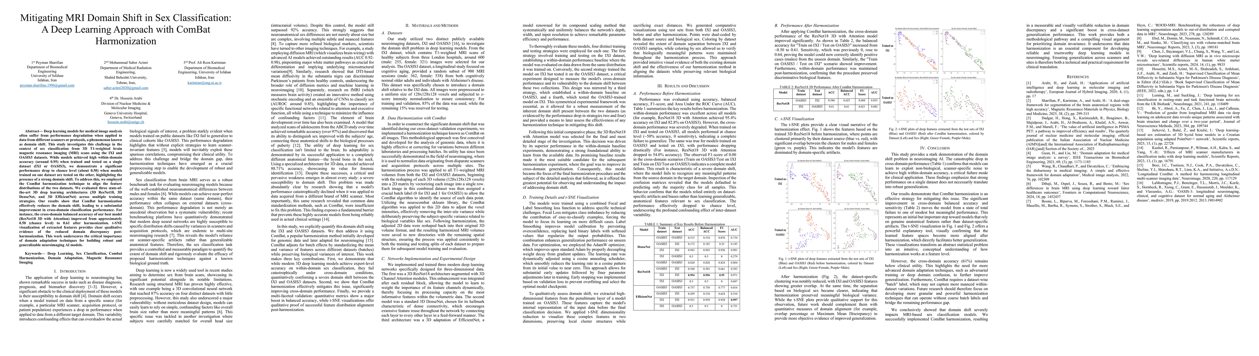
Deep learning models for medical image analysis often suffer from performance degradation when applied to data from different scanners or protocols, a phenomenon known as domain shift. This study inve...
To evaluate how partial volume correction (PVC) affects the reproducibility of 18F-FDG PET radiomic features in lymphoma lesions, with respect to lesion volume and tissue type. This single-center retr...
This pilot study compares per-lesion radiomics features of [68Ga]-DOTA FAPI-46 and [18F]-FDG PET/CT in non-small cell lung cancer (NSCLC) to explore complementary insights into intratumoral heterogene...
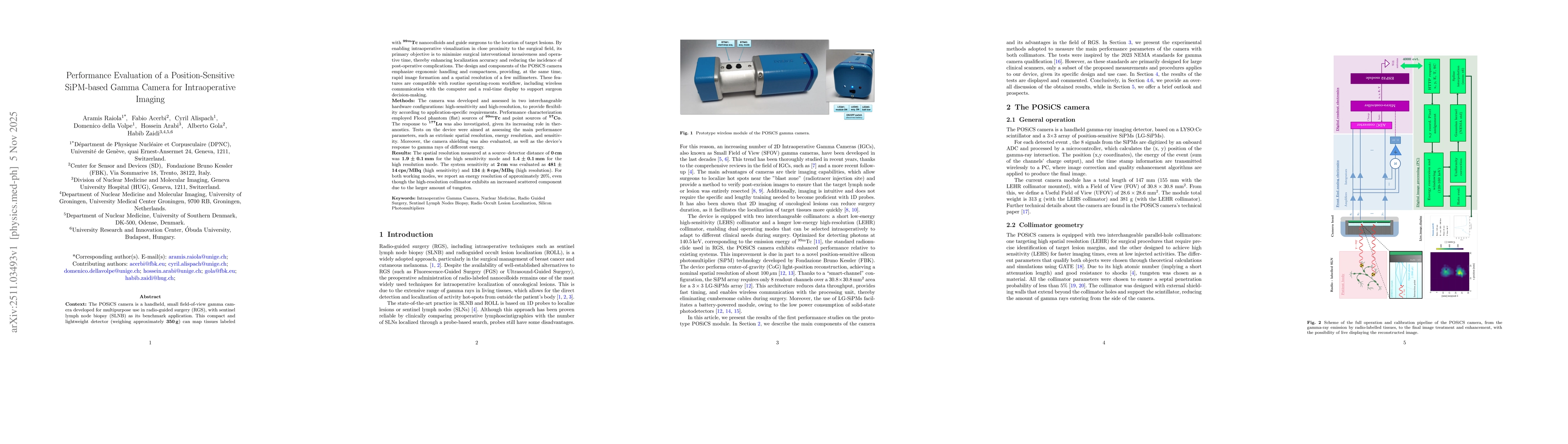
The POSiCS camera is a handheld, small field-of-view gamma camera developed for multipurpose use in radio-guided surgery (RGS), with sentinel lymph node biopsy (SLNB) as its benchmark application. Thi...
Single-photon sensitive detectors like Silicon Photomultipliers are widely used in many medical imaging applications. By using detectors with position resolutions, it is possible to build compact phot...