Deep Learning-Based Partial Volume Correction in Standard and Low-Dose PET-CT Imaging
Publication
Metrics
AI Quick Summary
This paper proposes a deep learning framework to correct partial volume effects in PET-CT imaging, aiming to enhance image quality and reduce noise from low-dose PET scans without requiring additional anatomical data. The method predicts full-dose PET images from either standard or low-dose inputs, addressing both PVC and denoising challenges.
Paper Preview
Abstract
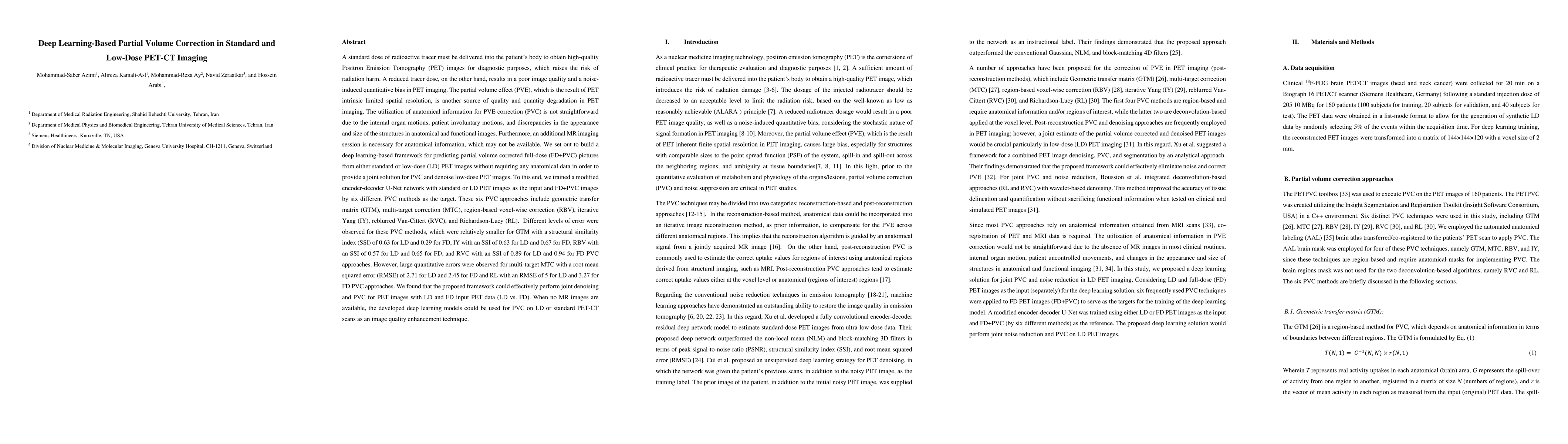
A standard dose of radioactive tracer must be delivered into the patients body to obtain high-quality Positron Emission Tomography (PET) images for diagnostic purposes, which raises the risk of radiation harm. A reduced tracer dose, on the other hand, results in poor image quality and a noise-induced quantitative bias in PET imaging. The partial volume effect (PVE), which is the result of PET intrinsic limited spatial resolution, is another source of quality and quantity degradation in PET imaging. The utilization of anatomical information for PVE correction (PVC) is not straightforward due to the internal organ motions, patient involuntary motions, and discrepancies in the appearance and size of the structures in anatomical and functional images. Furthermore, an additional MR imaging session is necessary for anatomical information, which may not be available. We set out to build a deep learning-based framework for predicting partial volume corrected full-dose (FD-PVC) pictures from either standard or low-dose (LD) PET images without requiring any anatomical data in order to provide a joint solution for PVC and denoise low-dose PET images.
AI Key Findings
Get AI-generated insights about this paper's methodology, results, significance, and more — seven facets brought into focus.
Impact
Paper Details
Authors
PDF Preview
Key Terms
Citation Network
Current paper (gray), citations (green), references (blue)
Display is limited for performance on very large graphs.
Discussion 0