Academic Profile
Statistics
Similar Authors
Papers on arXiv
Positron Emission Tomography (PET) is an important clinical imaging tool but inevitably introduces radiation hazards to patients and healthcare providers. Reducing the tracer injection dose and elim...
As PET imaging is accompanied by radiation exposure and potentially increased cancer risk, reducing radiation dose in PET scans without compromising the image quality is an important topic. Deep lea...
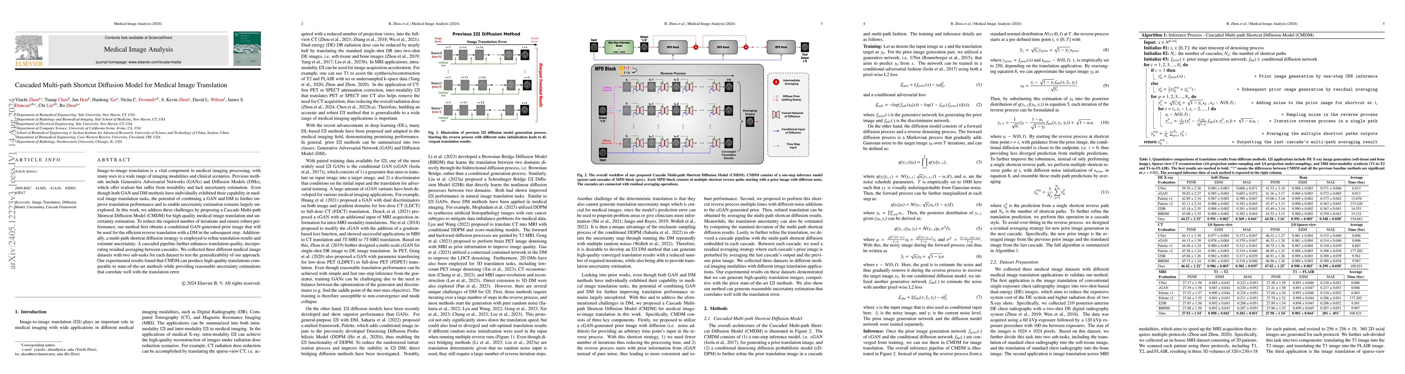
Image-to-image translation is a vital component in medical imaging processing, with many uses in a wide range of imaging modalities and clinical scenarios. Previous methods include Generative Advers...
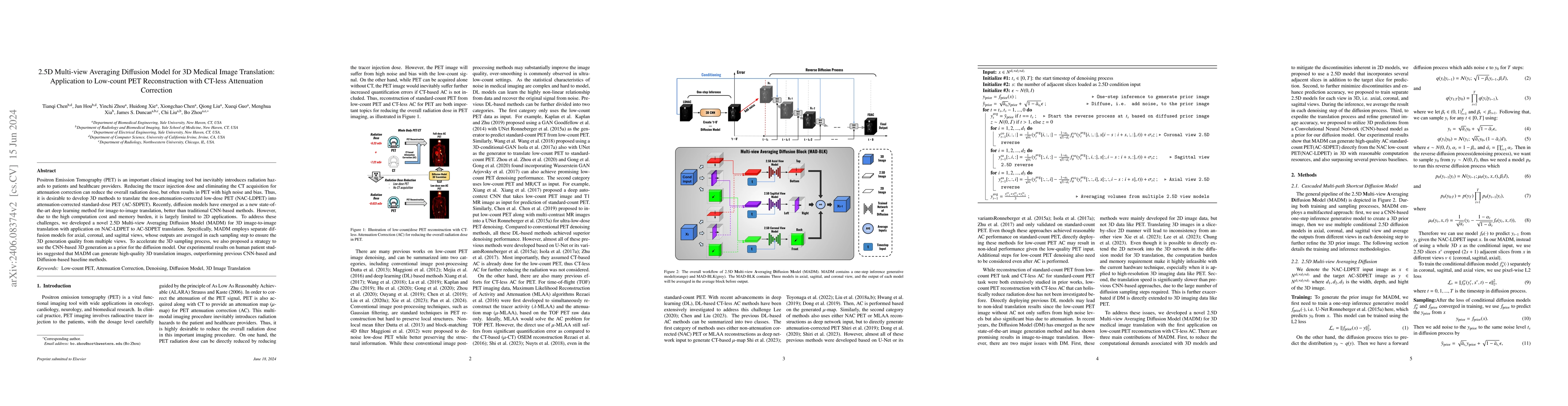
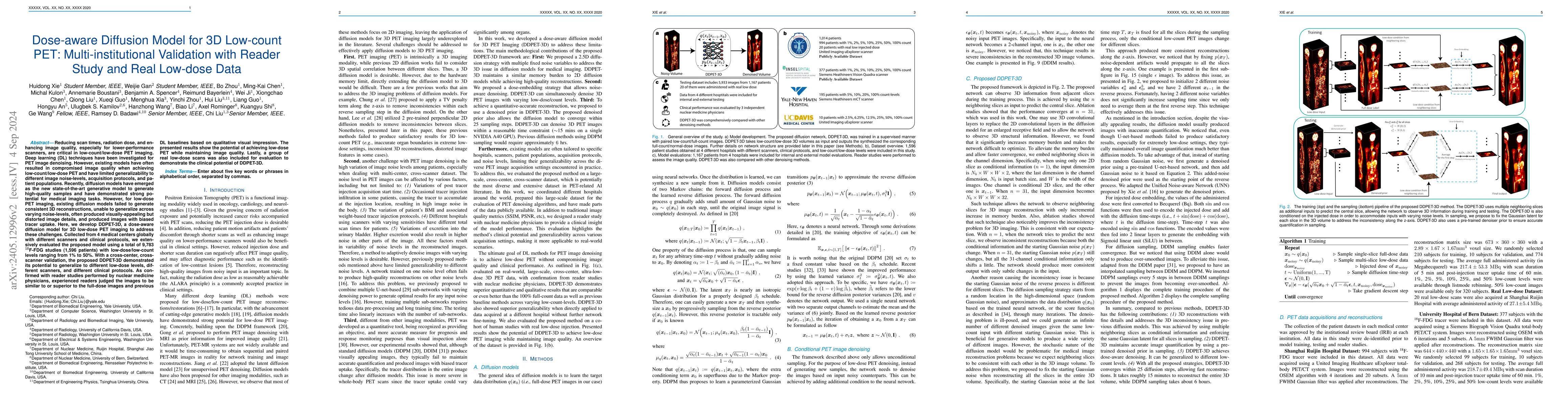
Deep learning-based positron emission tomography (PET) image denoising offers the potential to reduce radiation exposure and scanning time by transforming low-count images into high-count equivalent...
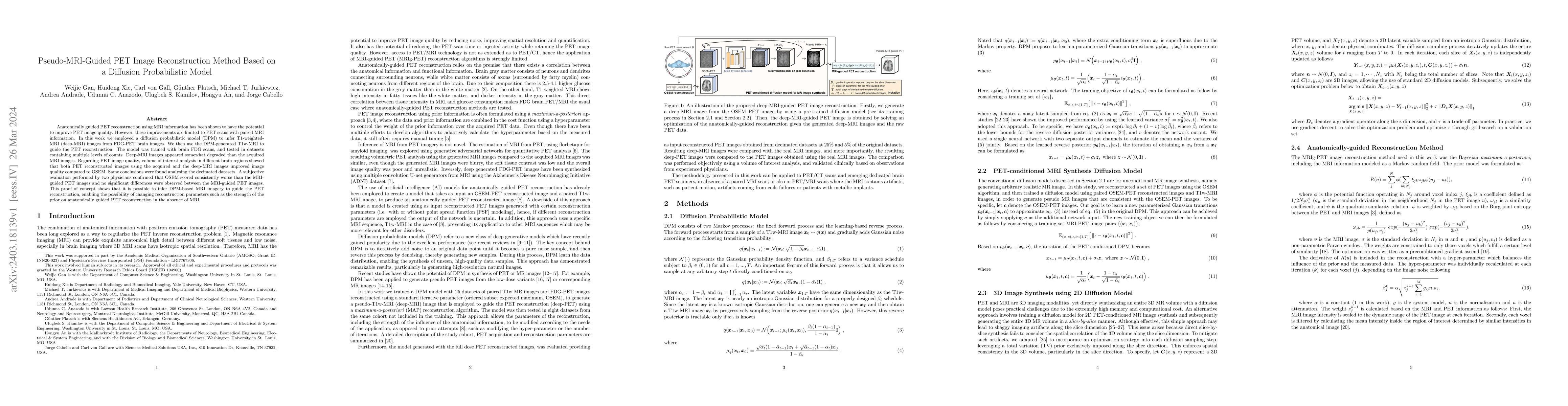
Anatomically guided PET reconstruction using MRI information has been shown to have the potential to improve PET image quality. However, these improvements are limited to PET scans with paired MRI i...
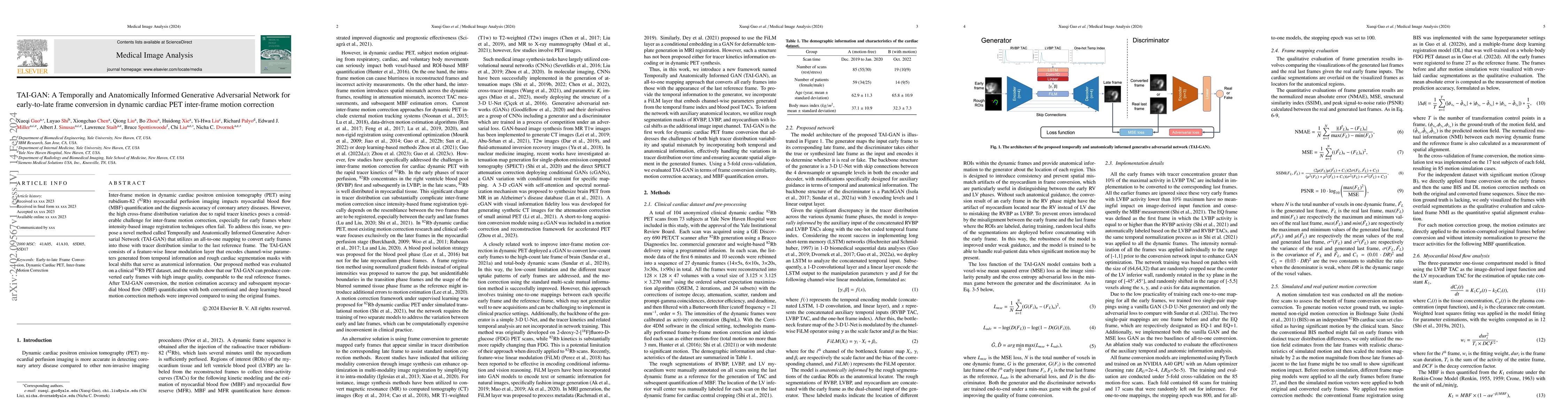
Inter-frame motion in dynamic cardiac positron emission tomography (PET) using rubidium-82 (82-Rb) myocardial perfusion imaging impacts myocardial blood flow (MBF) quantification and the diagnosis a...
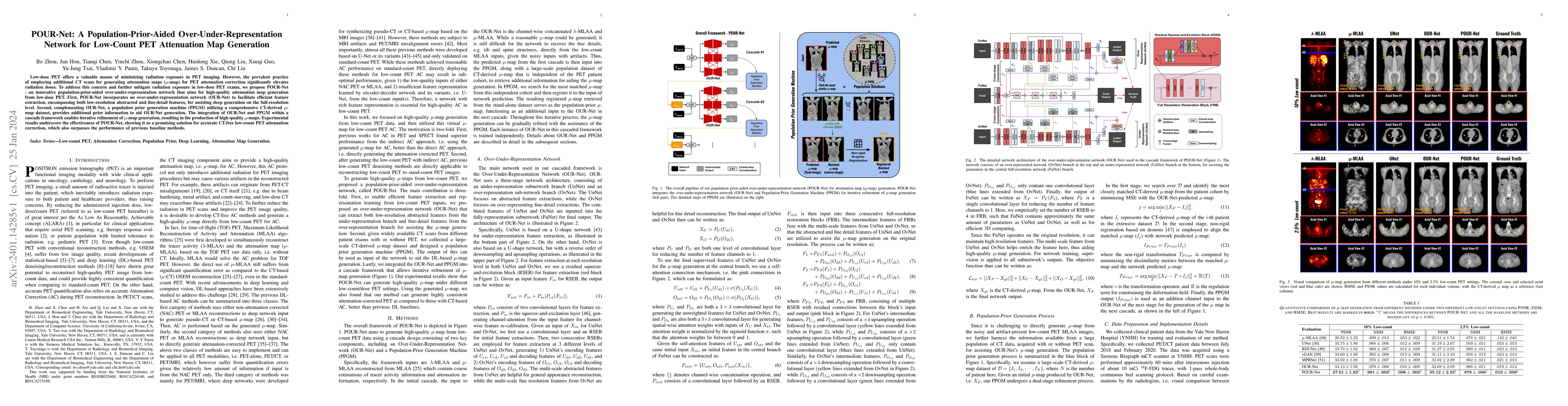
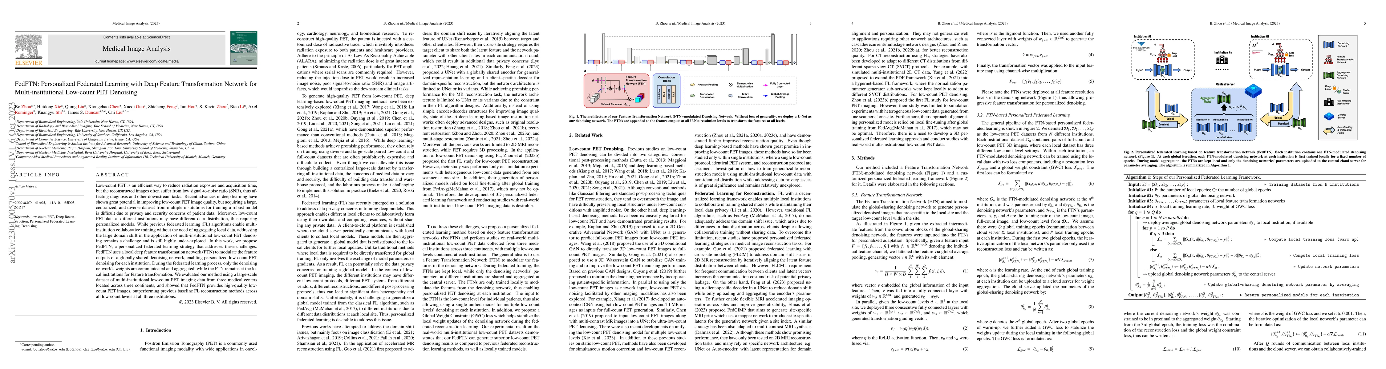
Low-dose PET offers a valuable means of minimizing radiation exposure in PET imaging. However, the prevalent practice of employing additional CT scans for generating attenuation maps (u-map) for PET...
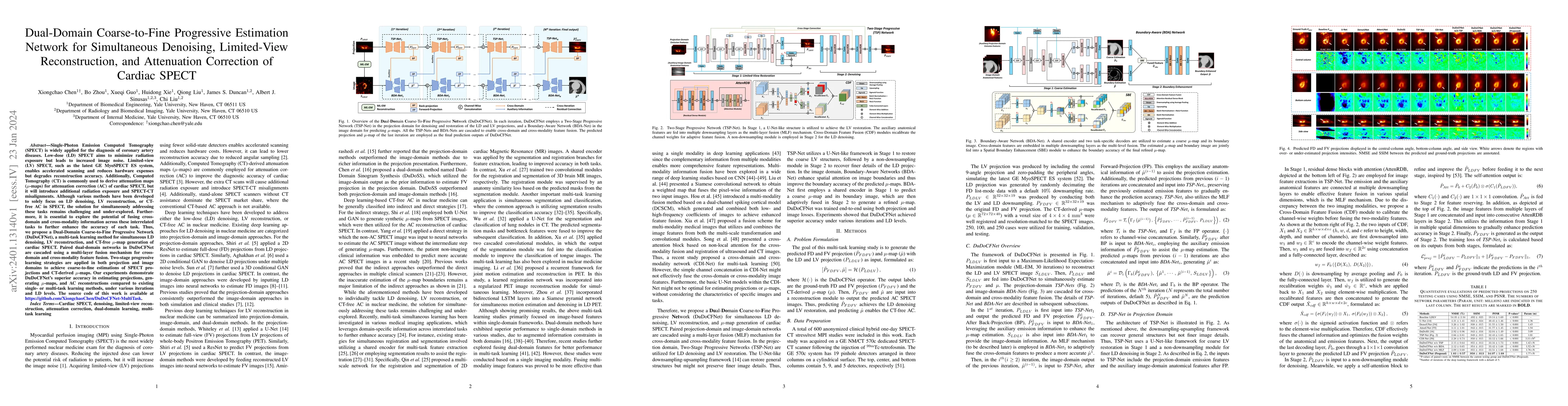
Single-Photon Emission Computed Tomography (SPECT) is widely applied for the diagnosis of coronary artery diseases. Low-dose (LD) SPECT aims to minimize radiation exposure but leads to increased ima...
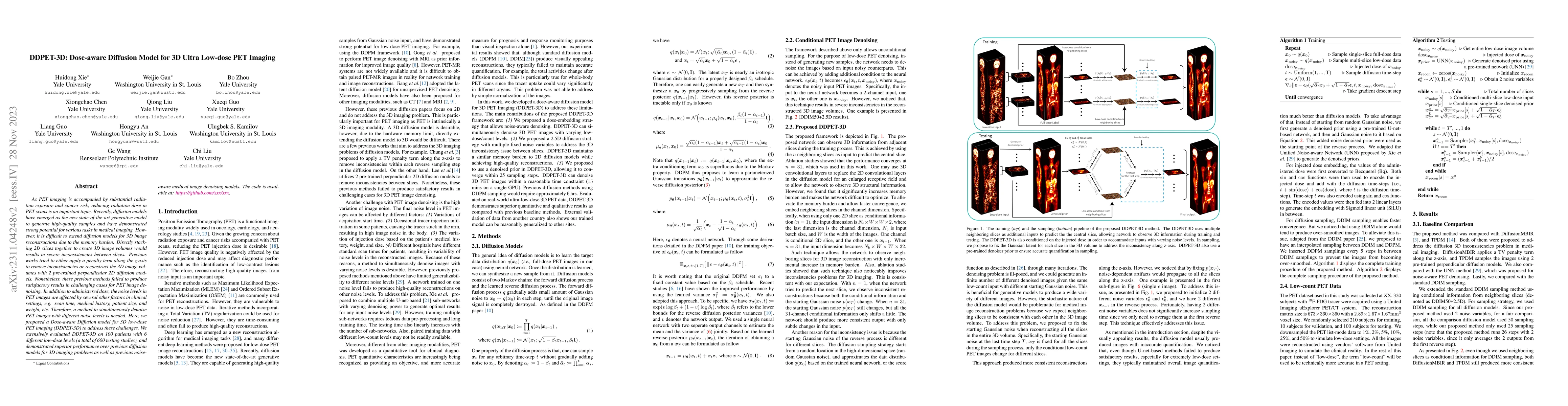
As PET imaging is accompanied by substantial radiation exposure and cancer risk, reducing radiation dose in PET scans is an important topic. Recently, diffusion models have emerged as the new state-...
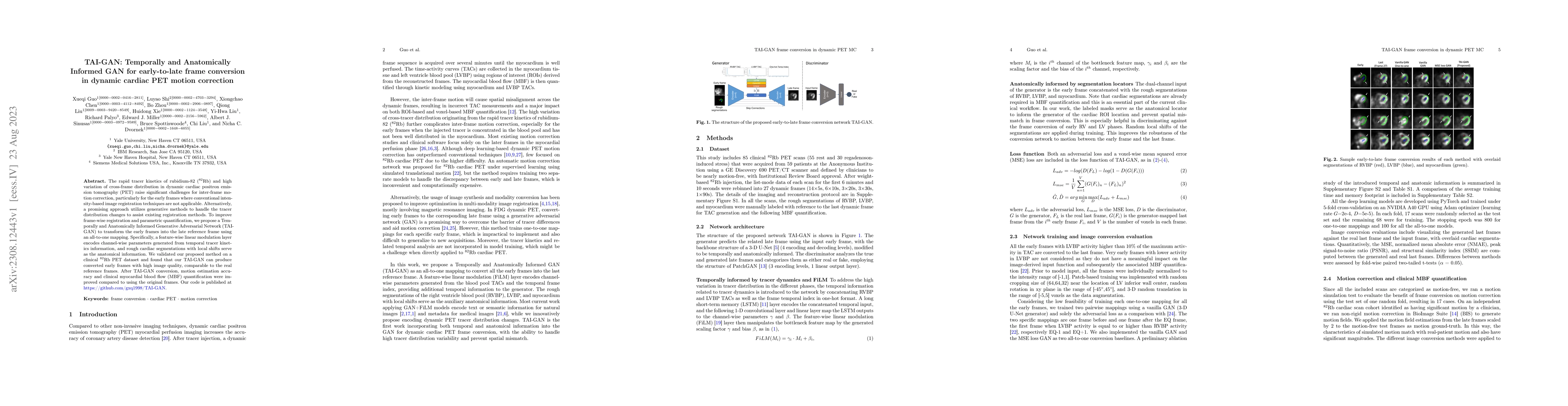
The rapid tracer kinetics of rubidium-82 ($^{82}$Rb) and high variation of cross-frame distribution in dynamic cardiac positron emission tomography (PET) raise significant challenges for inter-frame...
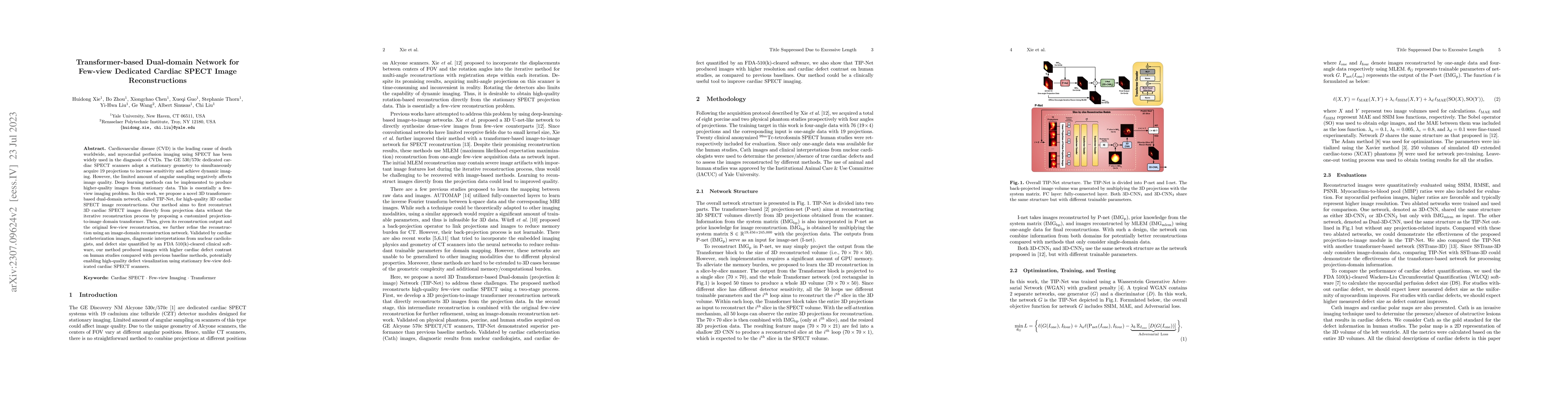
Cardiovascular disease (CVD) is the leading cause of death worldwide, and myocardial perfusion imaging using SPECT has been widely used in the diagnosis of CVDs. The GE 530/570c dedicated cardiac SP...
Myocardial perfusion imaging (MPI) by single-photon emission computed tomography (SPECT) is widely applied for the diagnosis of cardiovascular diseases. Reducing the dose of the injected tracer is e...
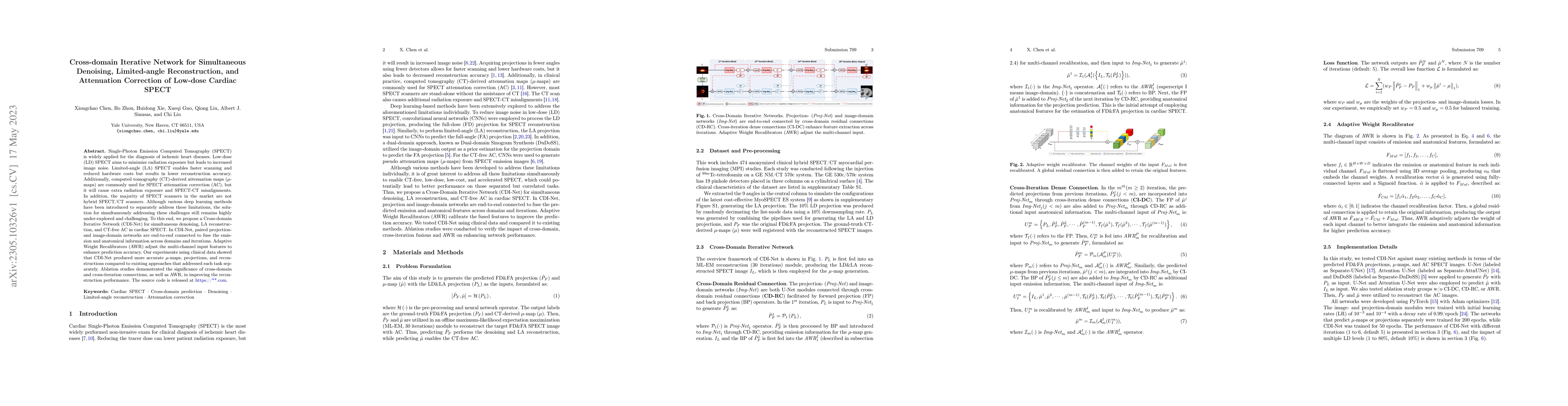
Single-Photon Emission Computed Tomography (SPECT) is widely applied for the diagnosis of ischemic heart diseases. Low-dose (LD) SPECT aims to minimize radiation exposure but leads to increased imag...
As PET imaging is accompanied by substantial radiation exposure and cancer risk, reducing radiation dose in PET scans is an important topic. However, low-count PET scans often suffer from high image...
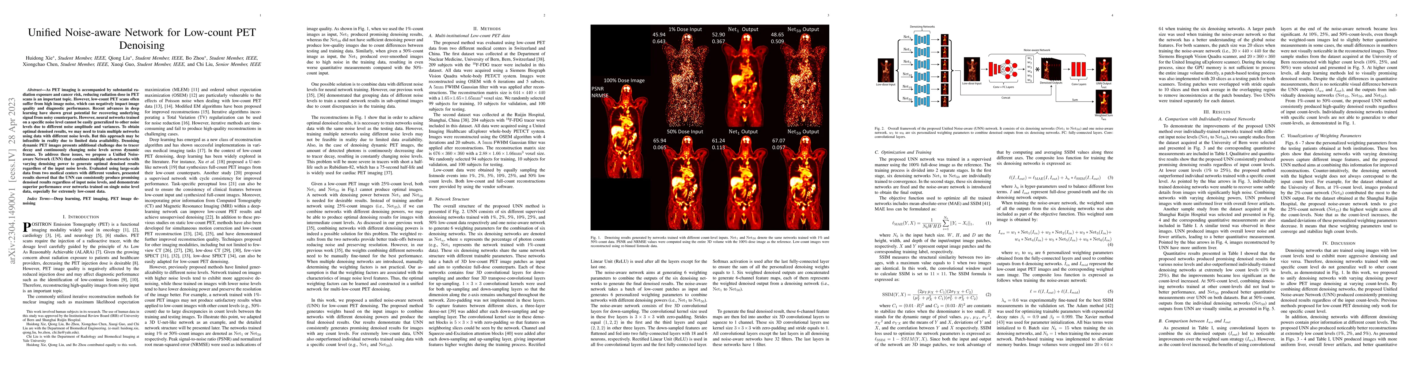
Low-count PET is an efficient way to reduce radiation exposure and acquisition time, but the reconstructed images often suffer from low signal-to-noise ratio (SNR), thus affecting diagnosis and othe...
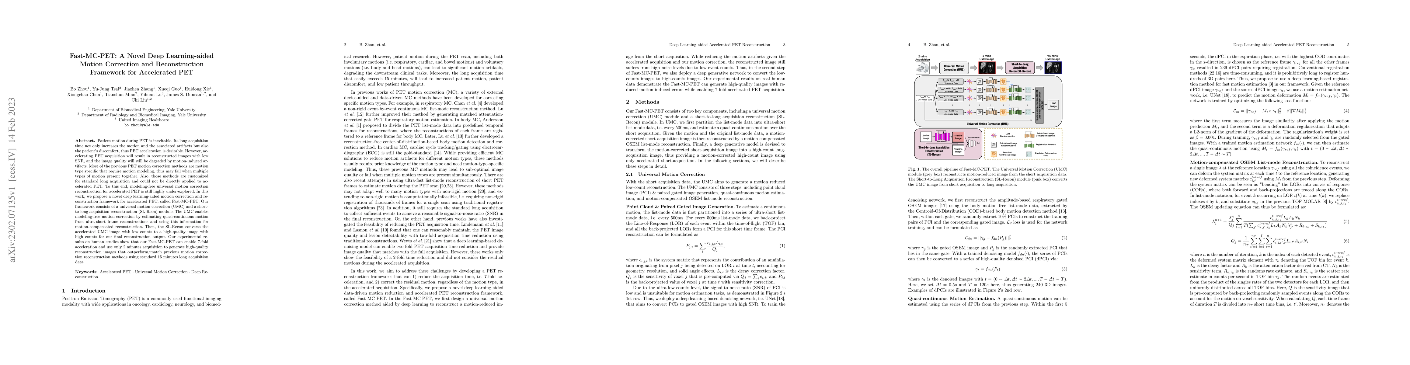
Patient motion during PET is inevitable. Its long acquisition time not only increases the motion and the associated artifacts but also the patient's discomfort, thus PET acceleration is desirable. H...
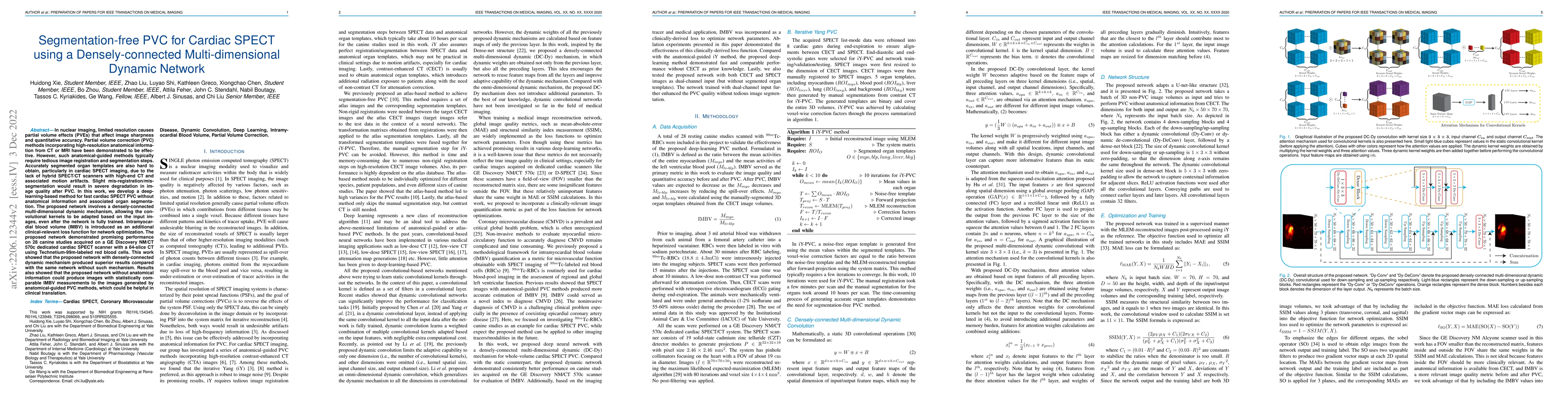
In nuclear imaging, limited resolution causes partial volume effects (PVEs) that affect image sharpness and quantitative accuracy. Partial volume correction (PVC) methods incorporating high-resoluti...
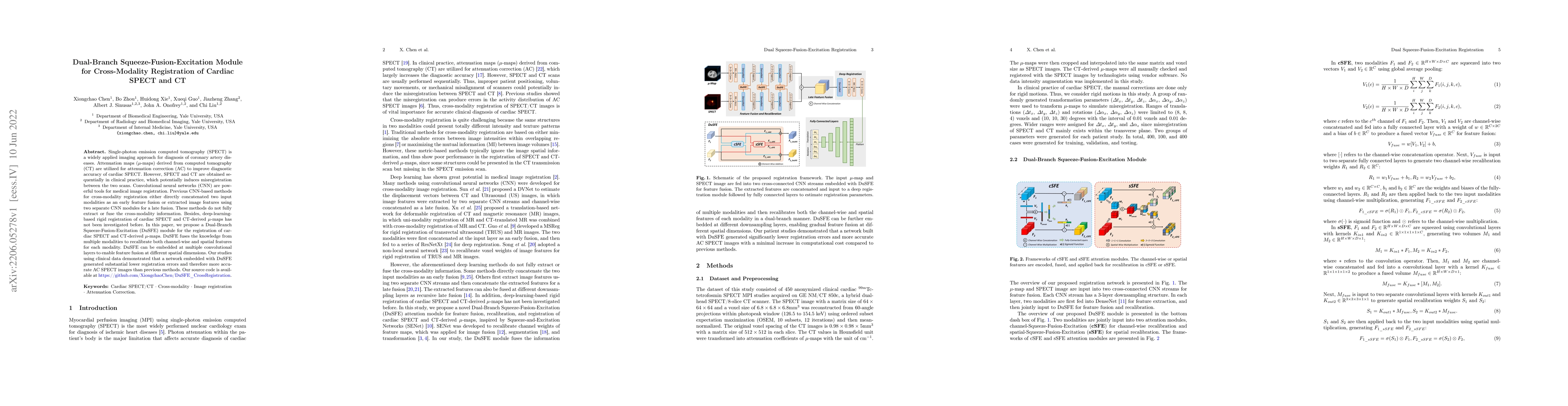
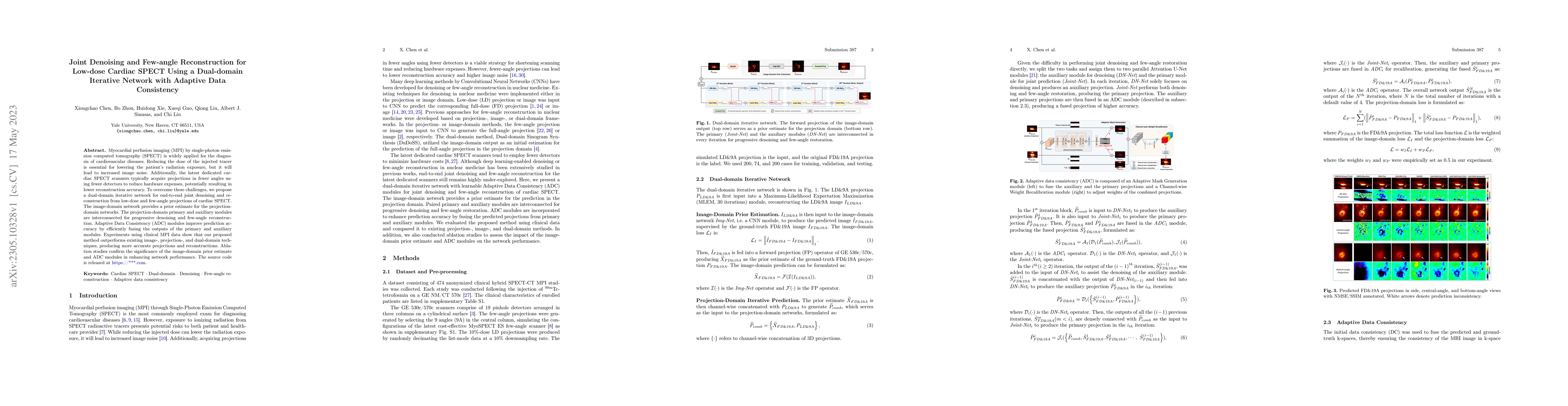
Single-photon emission computed tomography (SPECT) is a widely applied imaging approach for diagnosis of coronary artery diseases. Attenuation maps (u-maps) derived from computed tomography (CT) are...
Rb-82 is a radioactive isotope widely used for cardiac PET imaging. Despite numerous benefits of 82-Rb, there are several factors that limits its image quality and quantitative accuracy. First, the sh...
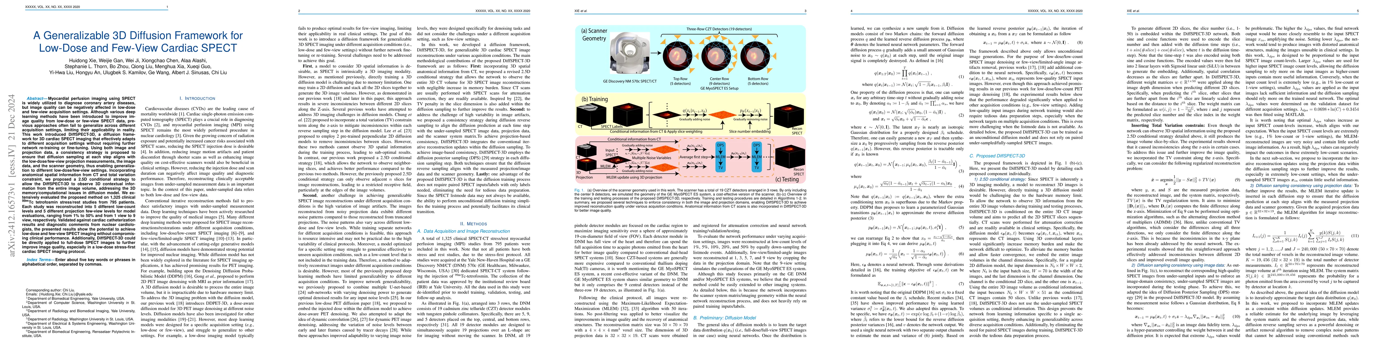
Myocardial perfusion imaging using SPECT is widely utilized to diagnose coronary artery diseases, but image quality can be negatively affected in low-dose and few-view acquisition settings. Although v...
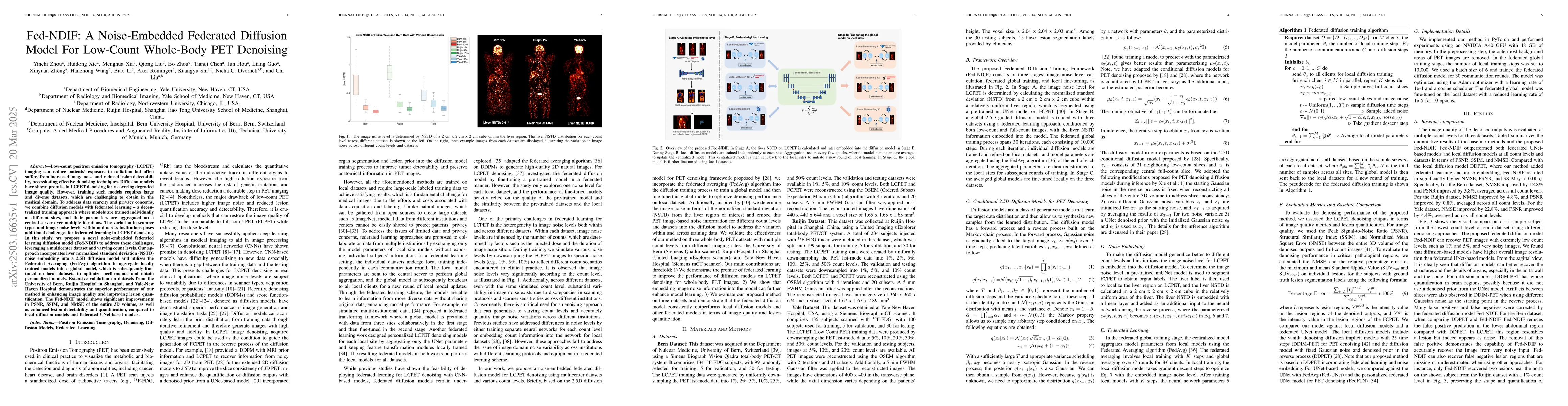
Low-count positron emission tomography (LCPET) imaging can reduce patients' exposure to radiation but often suffers from increased image noise and reduced lesion detectability, necessitating effective...

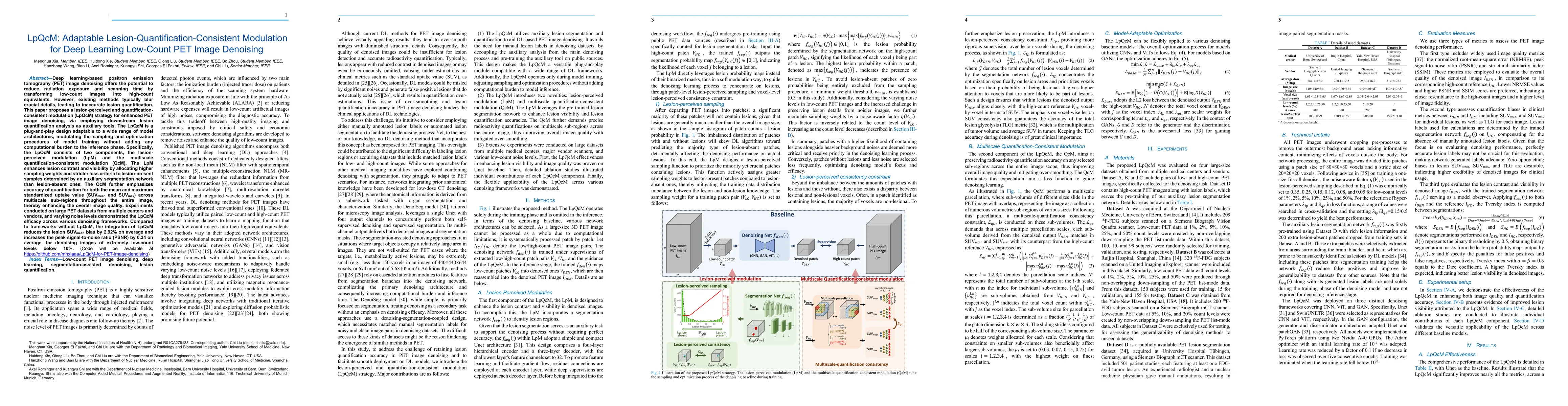
Positron emission tomography (PET) image denoising, along with lesion and organ segmentation, are critical steps in PET-aided diagnosis. However, existing methods typically treat these tasks independe...
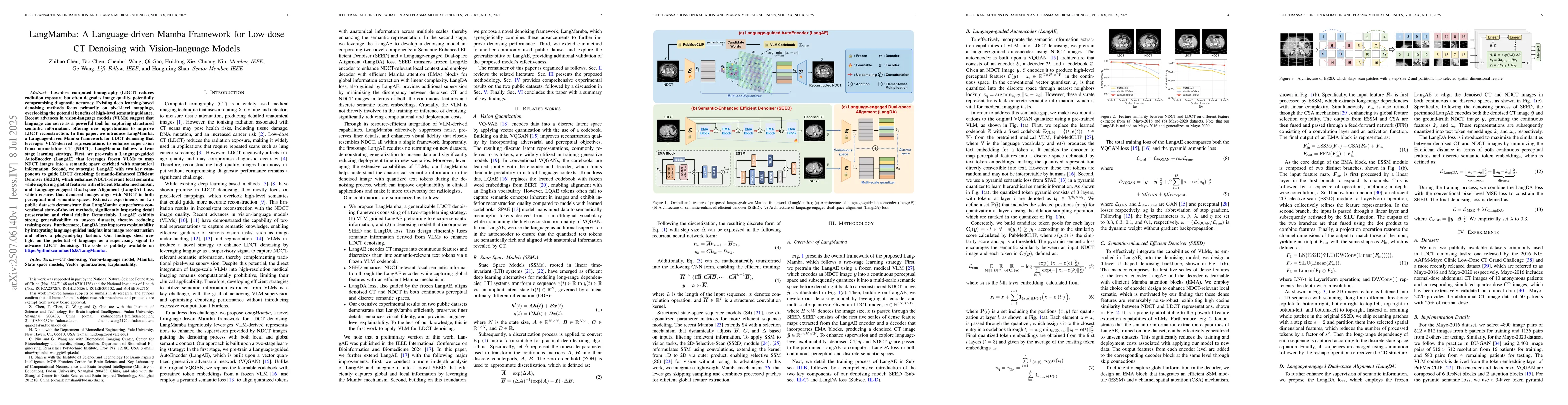
Low-dose computed tomography (LDCT) reduces radiation exposure but often degrades image quality, potentially compromising diagnostic accuracy. Existing deep learning-based denoising methods focus prim...
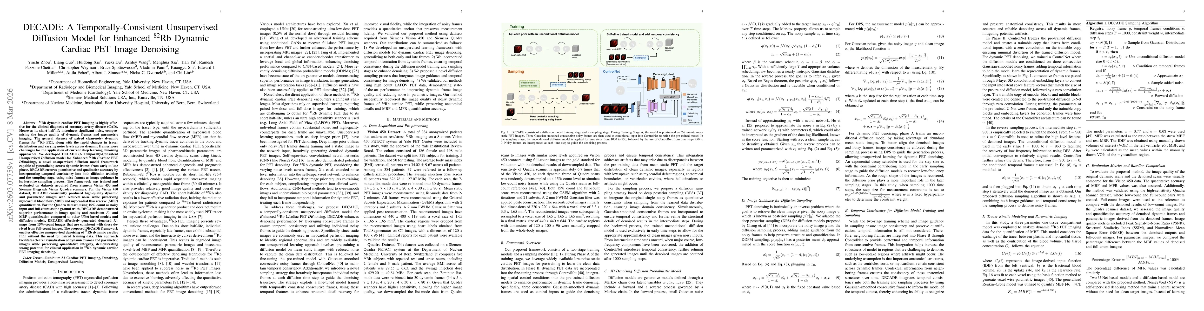
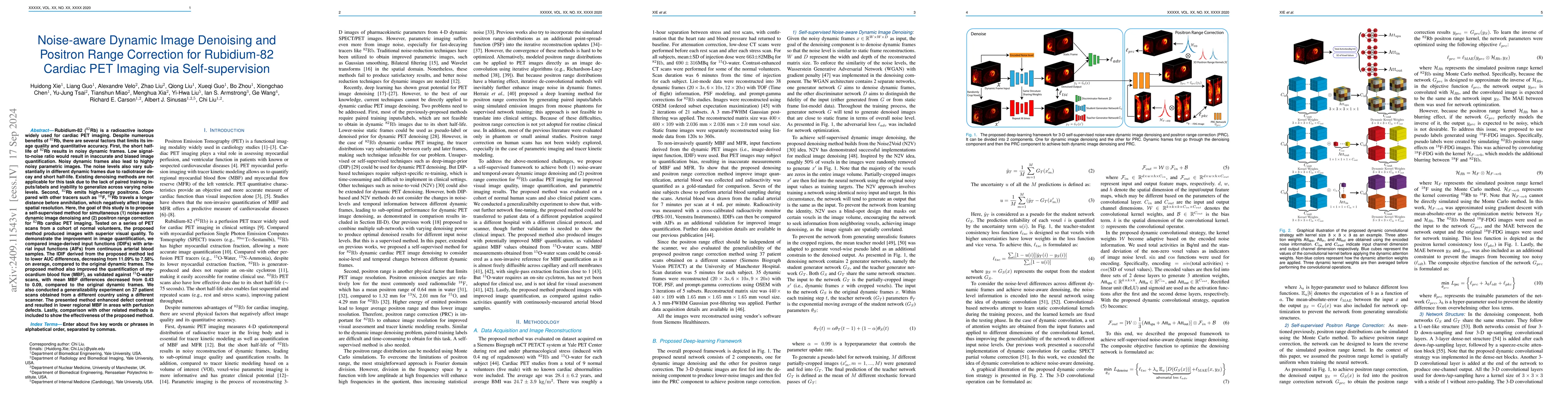
Rb-82 dynamic cardiac PET imaging is widely used for the clinical diagnosis of coronary artery disease (CAD), but its short half-life results in high noise levels that degrade dynamic frame quality an...
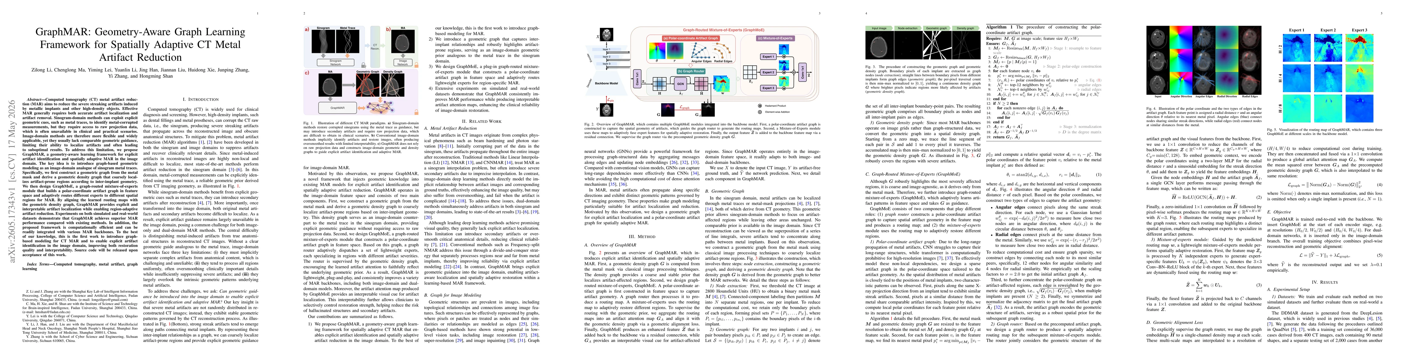
Computed tomography (CT) metal artifact reduction (MAR) aims to reduce the severe streaking artifacts induced by metallic implants and other high-density objects. Effective MAR generally requires both...