Dose-aware Diffusion Model for 3D Low-dose PET: Multi-institutional Validation with Reader Study and Real Low-dose Data
Publication
Metrics
AI Quick Summary
This research proposes DDPET-3D, a dose-aware diffusion model for generating high-quality 3D low-dose PET images, addressing limitations in existing models' generalizability and image quality. Extensive multi-institutional validation across various scanners and protocols confirmed the model's superior denoising performance, as evaluated by nuclear medicine physicians, showing its potential to maintain image quality at significantly reduced radiation doses.
Paper Preview
Abstract
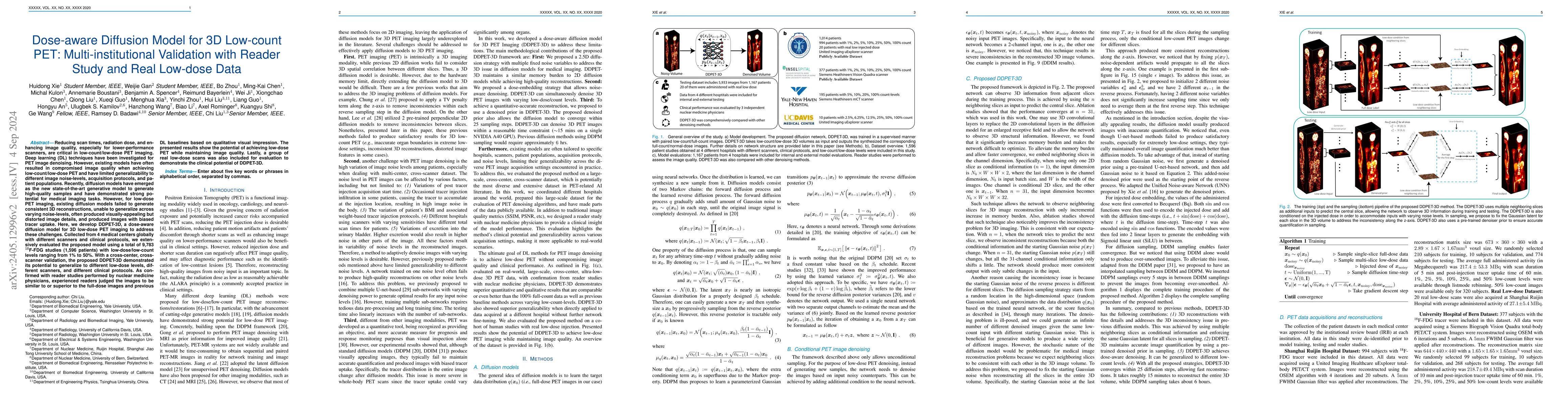
As PET imaging is accompanied by radiation exposure and potentially increased cancer risk, reducing radiation dose in PET scans without compromising the image quality is an important topic. Deep learning (DL) techniques have been investigated for low-dose PET imaging. However, existing models have often resulted in compromised image quality when achieving low-dose PET and have limited generalizability to different image noise-levels, acquisition protocols, patient populations, and hospitals. Recently, diffusion models have emerged as the new state-of-the-art generative model to generate high-quality samples and have demonstrated strong potential for medical imaging tasks. However, for low-dose PET imaging, existing diffusion models failed to generate consistent 3D reconstructions, unable to generalize across varying noise-levels, often produced visually-appealing but distorted image details, and produced images with biased tracer uptake. Here, we develop DDPET-3D, a dose-aware diffusion model for 3D low-dose PET imaging to address these challenges. Collected from 4 medical centers globally with different scanners and clinical protocols, we extensively evaluated the proposed model using a total of 9,783 18F-FDG studies (1,596 patients) with low-dose/low-count levels ranging from 1% to 50%. With a cross-center, cross-scanner validation, the proposed DDPET-3D demonstrated its potential to generalize to different low-dose levels, different scanners, and different clinical protocols. As confirmed with reader studies performed by nuclear medicine physicians, the proposed method produced superior denoised results that are comparable to or even better than the 100% full-count images as well as previous DL baselines. The presented results show the potential of achieving low-dose PET while maintaining image quality. Lastly, a group of real low-dose scans was also included for evaluation.
AI Key Findings
Get AI-generated insights about this paper's methodology, results, significance, and more — seven facets brought into focus.
Impact
Paper Details
Authors
PDF Preview
Key Terms
Citation Network
Current paper (gray), citations (green), references (blue)
Display is limited for performance on very large graphs.
Discussion 0