Academic Profile
Statistics
Similar Authors
Papers on arXiv
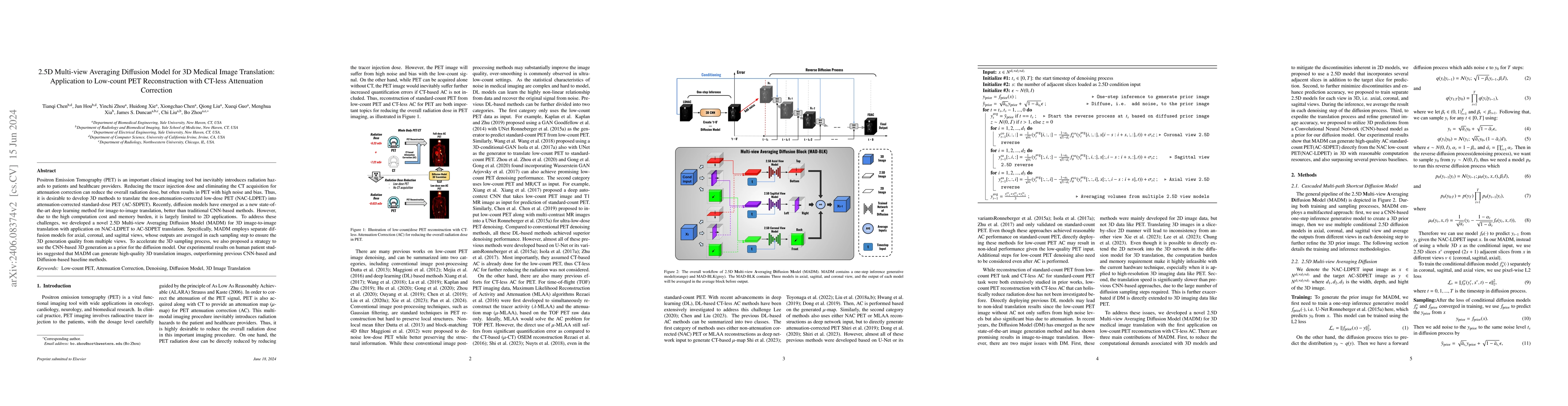
Positron Emission Tomography (PET) is an important clinical imaging tool but inevitably introduces radiation hazards to patients and healthcare providers. Reducing the tracer injection dose and elim...
As PET imaging is accompanied by radiation exposure and potentially increased cancer risk, reducing radiation dose in PET scans without compromising the image quality is an important topic. Deep lea...
Deep learning-based positron emission tomography (PET) image denoising offers the potential to reduce radiation exposure and scanning time by transforming low-count images into high-count equivalent...
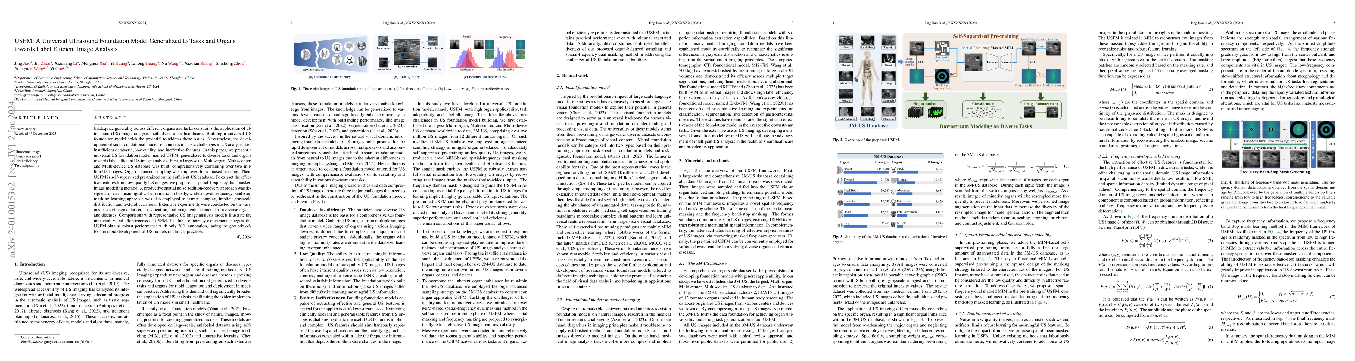
Inadequate generality across different organs and tasks constrains the application of ultrasound (US) image analysis methods in smart healthcare. Building a universal US foundation model holds the p...
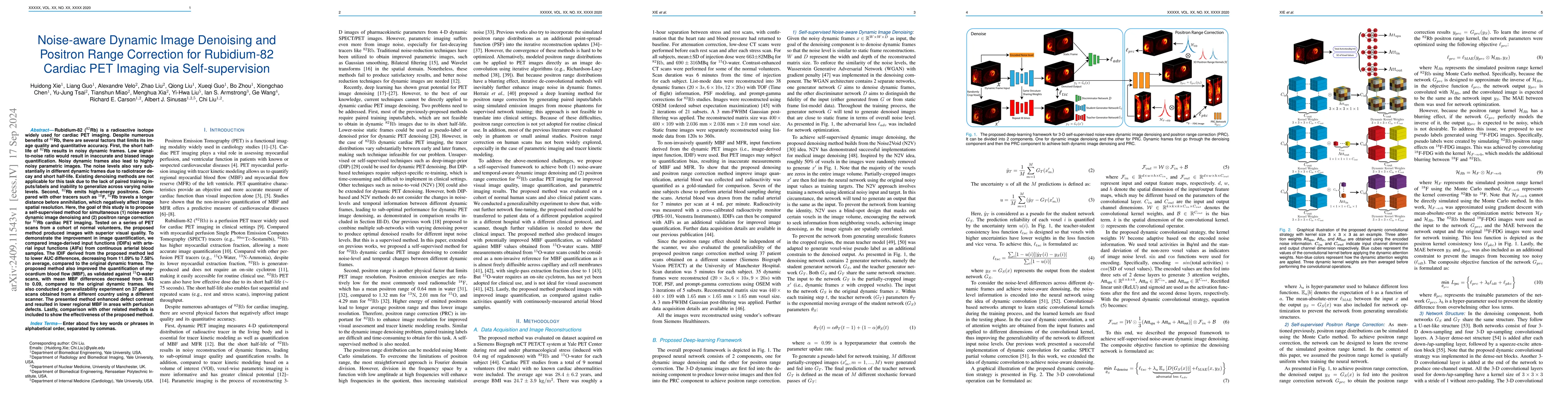
Rb-82 is a radioactive isotope widely used for cardiac PET imaging. Despite numerous benefits of 82-Rb, there are several factors that limits its image quality and quantitative accuracy. First, the sh...
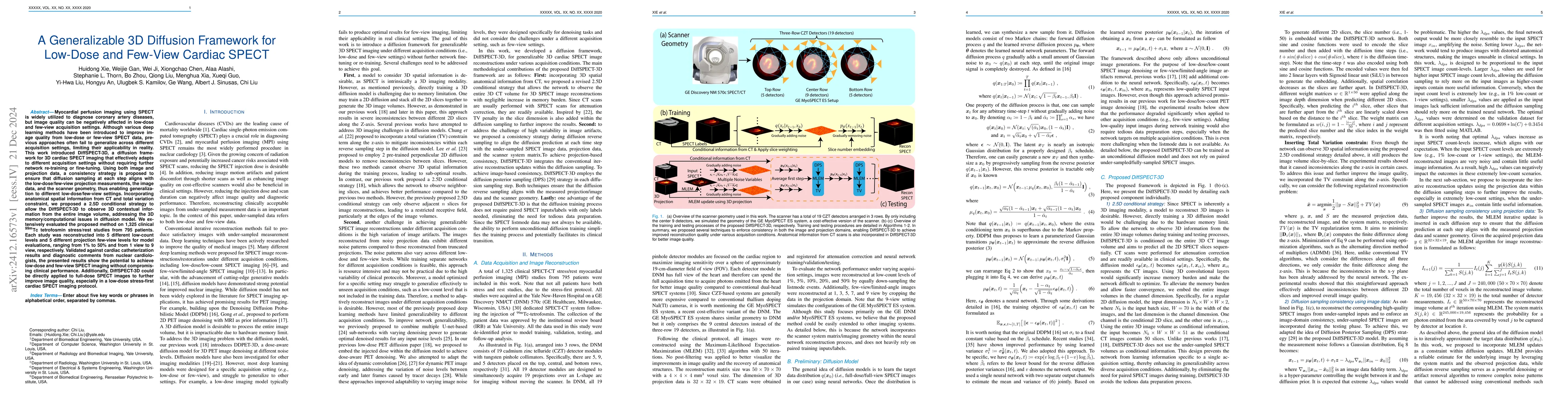
Myocardial perfusion imaging using SPECT is widely utilized to diagnose coronary artery diseases, but image quality can be negatively affected in low-dose and few-view acquisition settings. Although v...
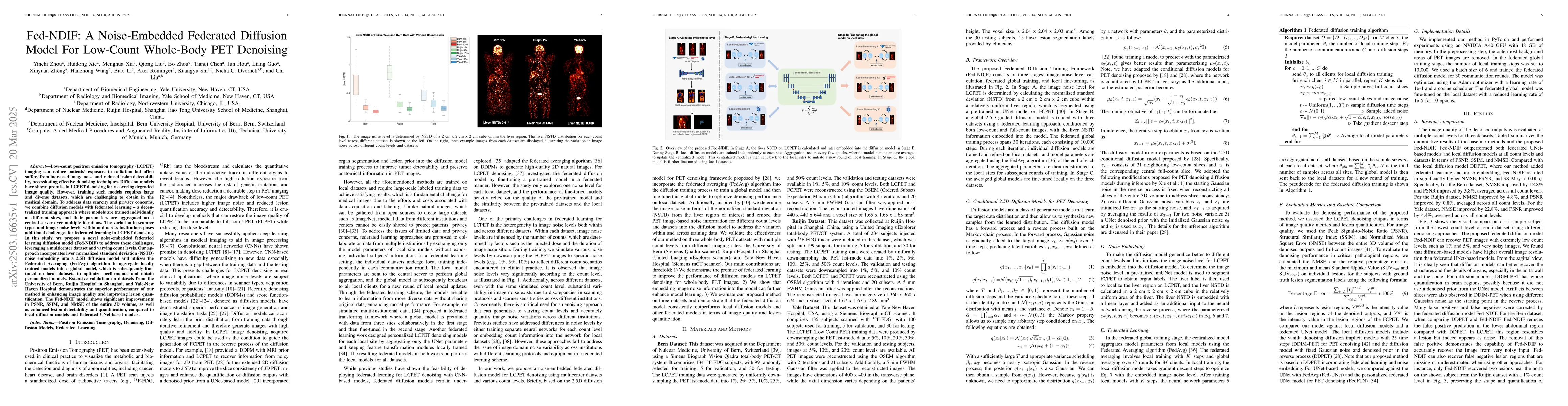
Low-count positron emission tomography (LCPET) imaging can reduce patients' exposure to radiation but often suffers from increased image noise and reduced lesion detectability, necessitating effective...

Positron emission tomography (PET) image denoising, along with lesion and organ segmentation, are critical steps in PET-aided diagnosis. However, existing methods typically treat these tasks independe...
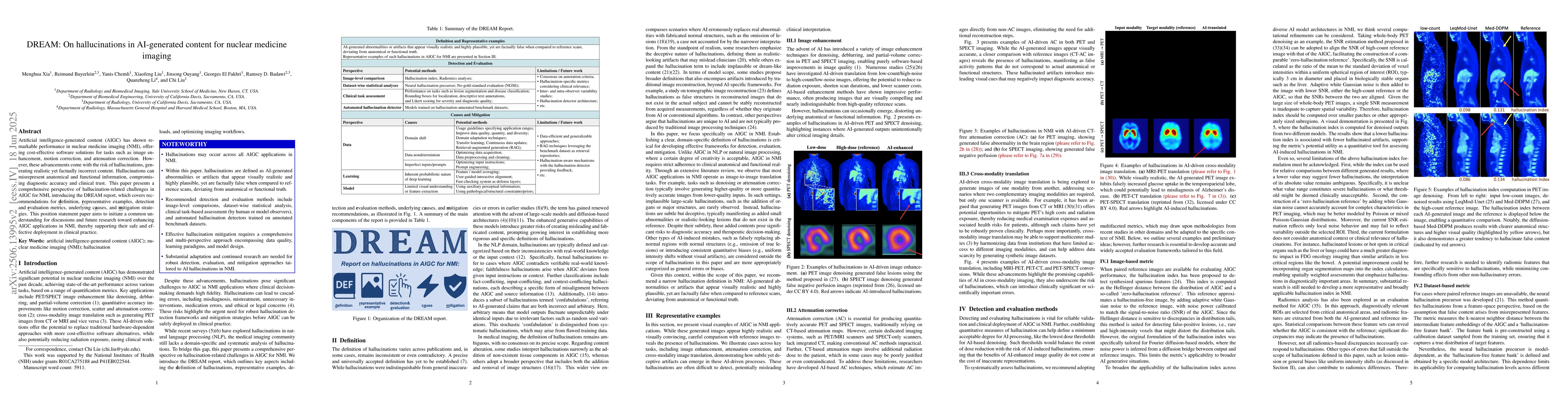
Artificial intelligence-generated content (AIGC) has shown remarkable performance in nuclear medicine imaging (NMI), offering cost-effective software solutions for tasks such as image enhancement, mot...
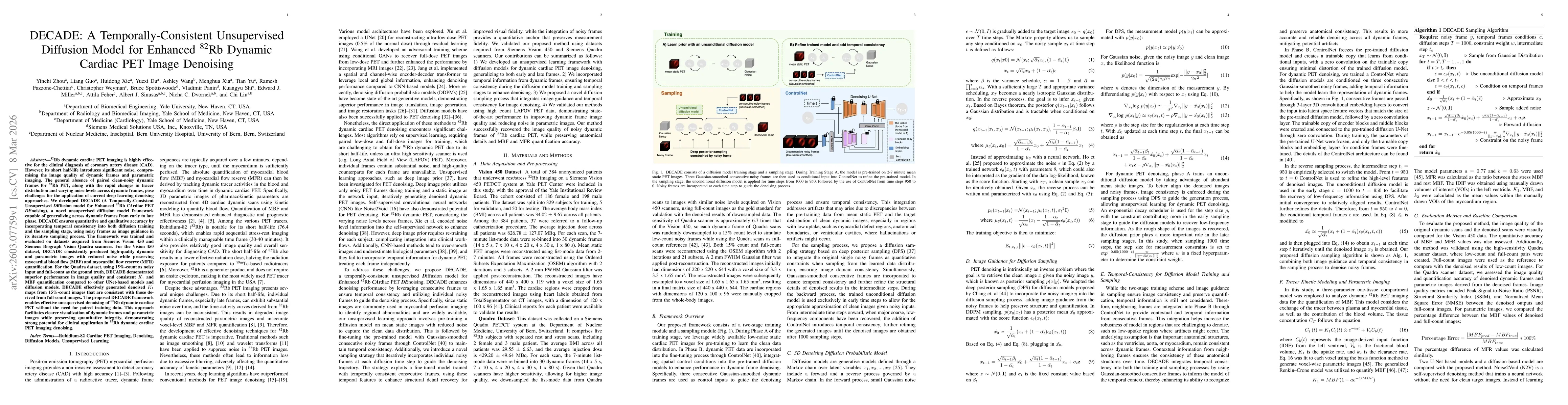
Rb-82 dynamic cardiac PET imaging is widely used for the clinical diagnosis of coronary artery disease (CAD), but its short half-life results in high noise levels that degrade dynamic frame quality an...
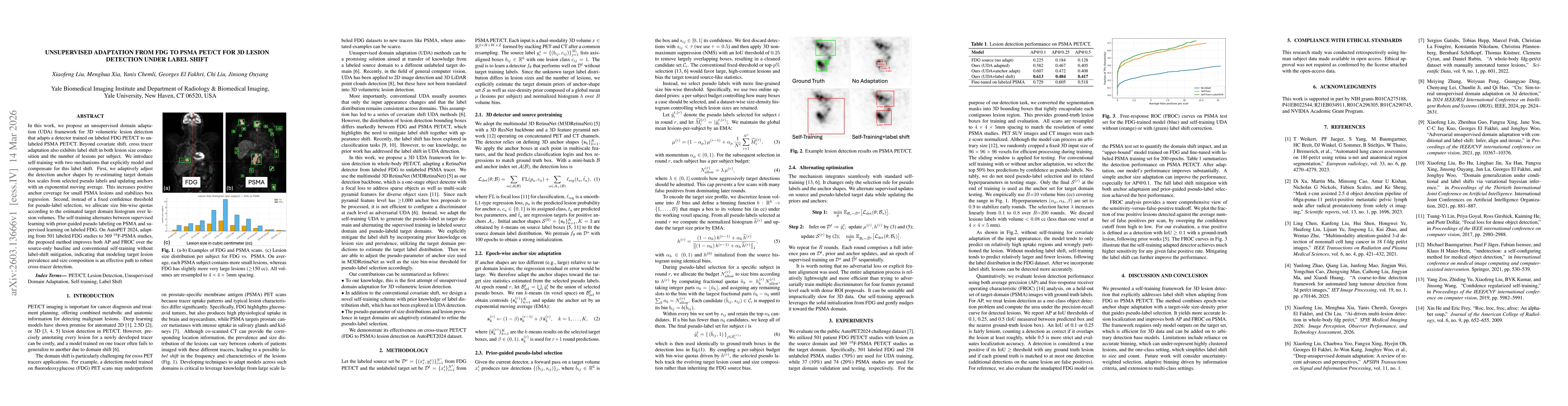
In this work, we propose an unsupervised domain adaptation (UDA) framework for 3D volumetric lesion detection that adapts a detector trained on labeled FDG PET/CT to unlabeled PSMA PET/CT. Beyond cova...
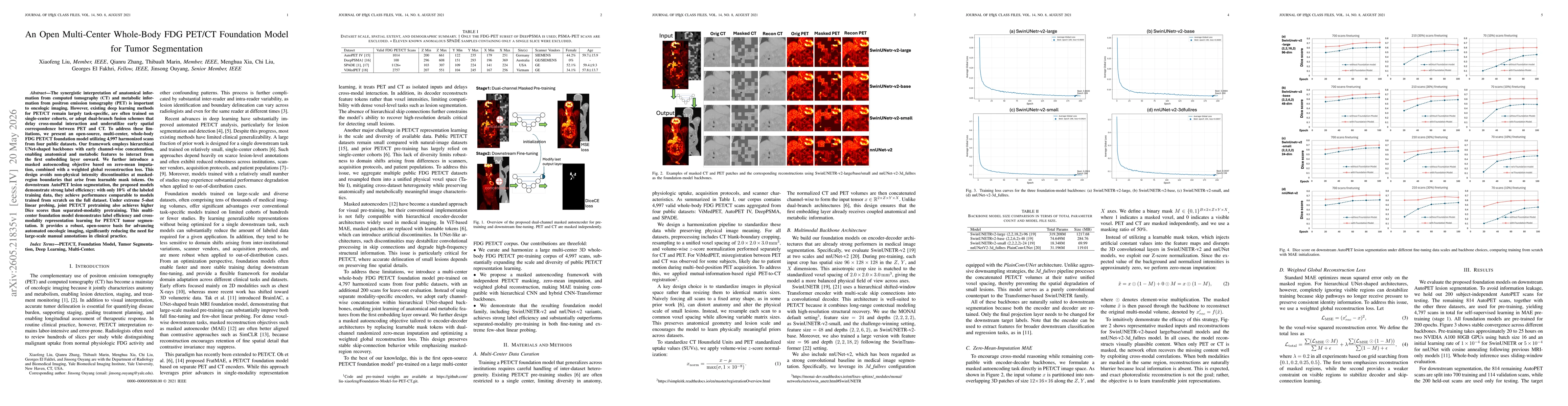
The synergistic interpretation of anatomical information from computed tomography (CT) and metabolic information from positron emission tomography (PET) is important to oncologic imaging. However, exi...