Academic Profile
Statistics
Similar Authors
Papers on arXiv
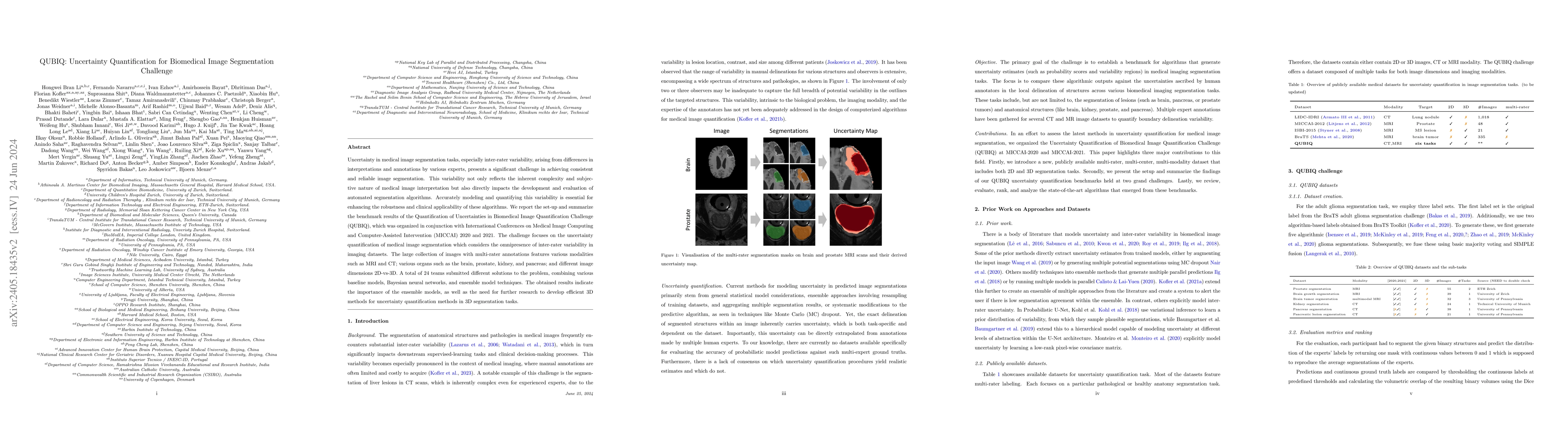
Uncertainty in medical image segmentation tasks, especially inter-rater variability, arising from differences in interpretations and annotations by various experts, presents a significant challenge in...
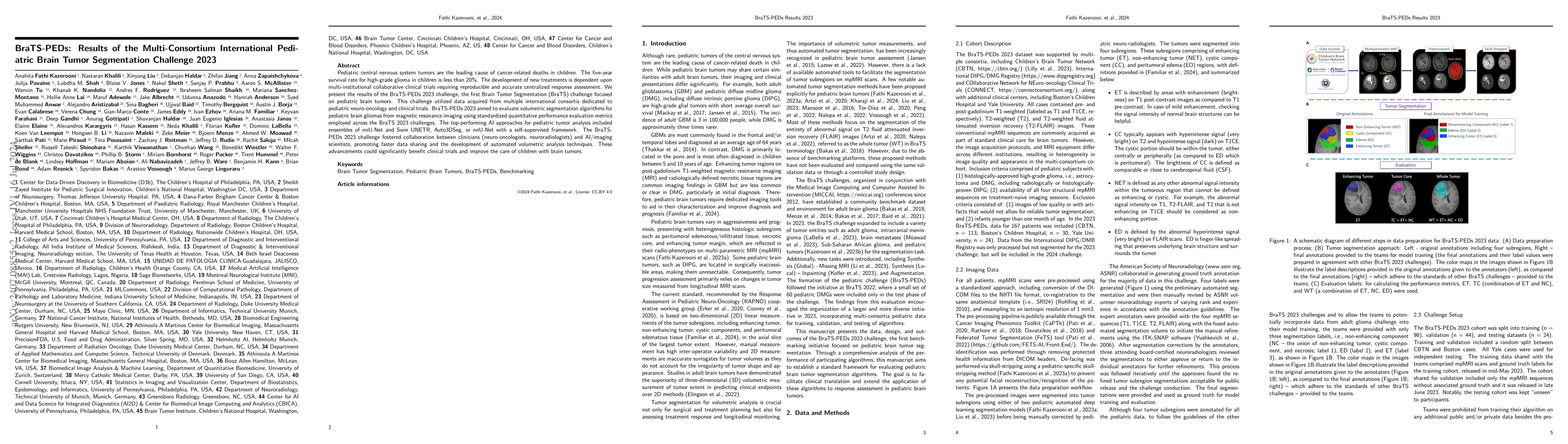
Pediatric central nervous system tumors are the leading cause of cancer-related deaths in children. The five-year survival rate for high-grade glioma in children is less than 20%. The development of n...
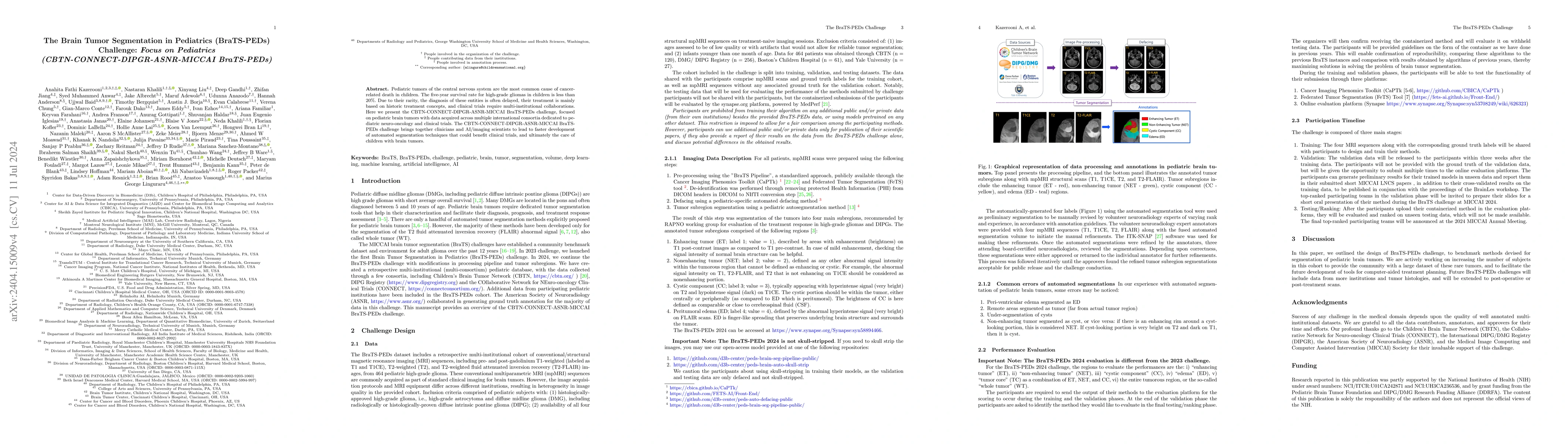
Pediatric tumors of the central nervous system are the most common cause of cancer-related death in children. The five-year survival rate for high-grade gliomas in children is less than 20%. Due to th...
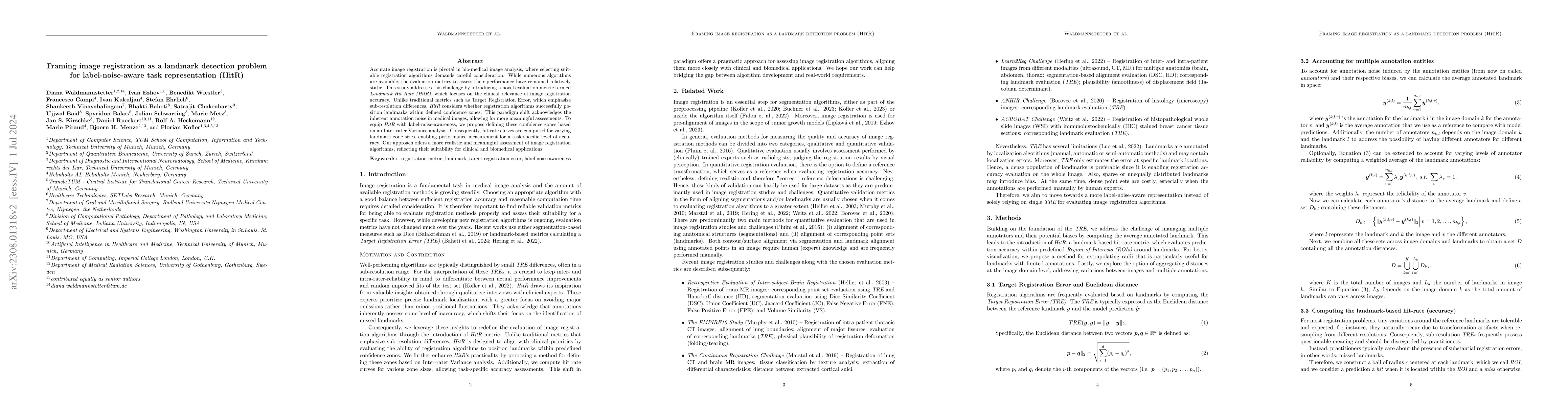
Accurate image registration is pivotal in biomedical image analysis, where selecting suitable registration algorithms demands careful consideration. While numerous algorithms are available, the evalua...
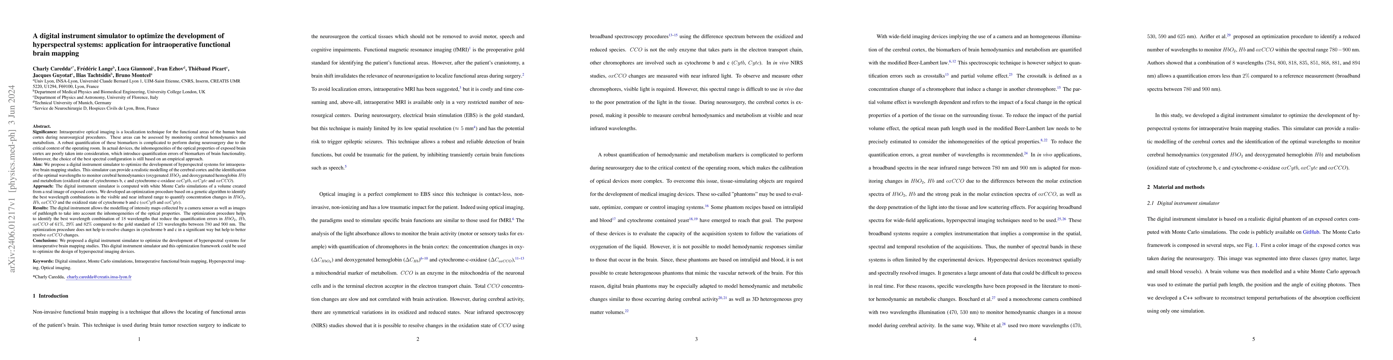
Intraoperative optical imaging is a localization technique for the functional areas of the human brain cortex during neurosurgical procedures. These areas can be assessed by monitoring cerebral hemo...
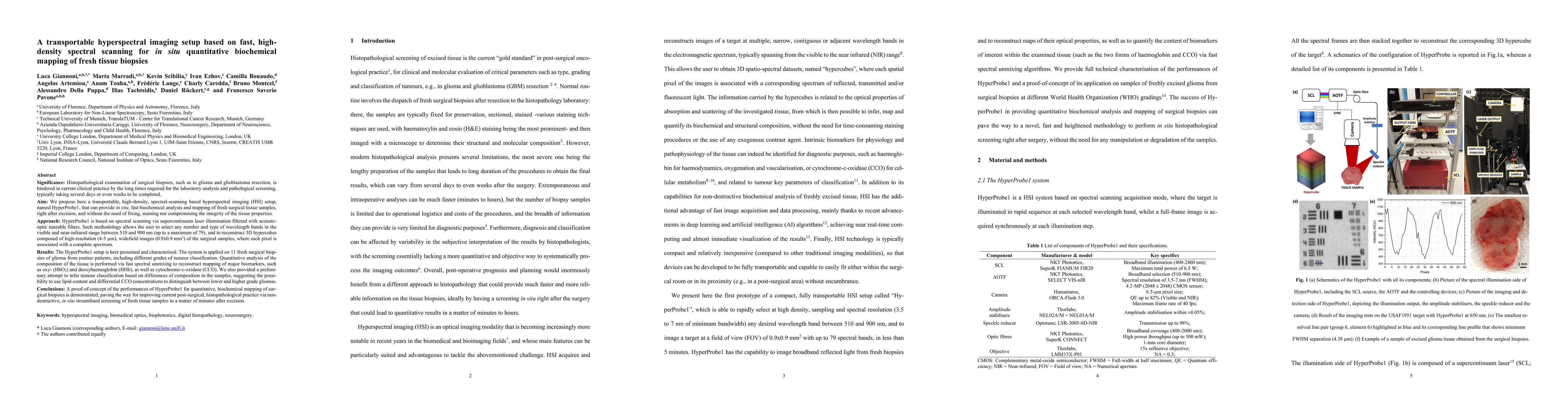
Histopathological examination of surgical biopsies, such as in glioma and glioblastoma resection, is hindered in current clinical practice by the long times required for the laboratory analysis and ...
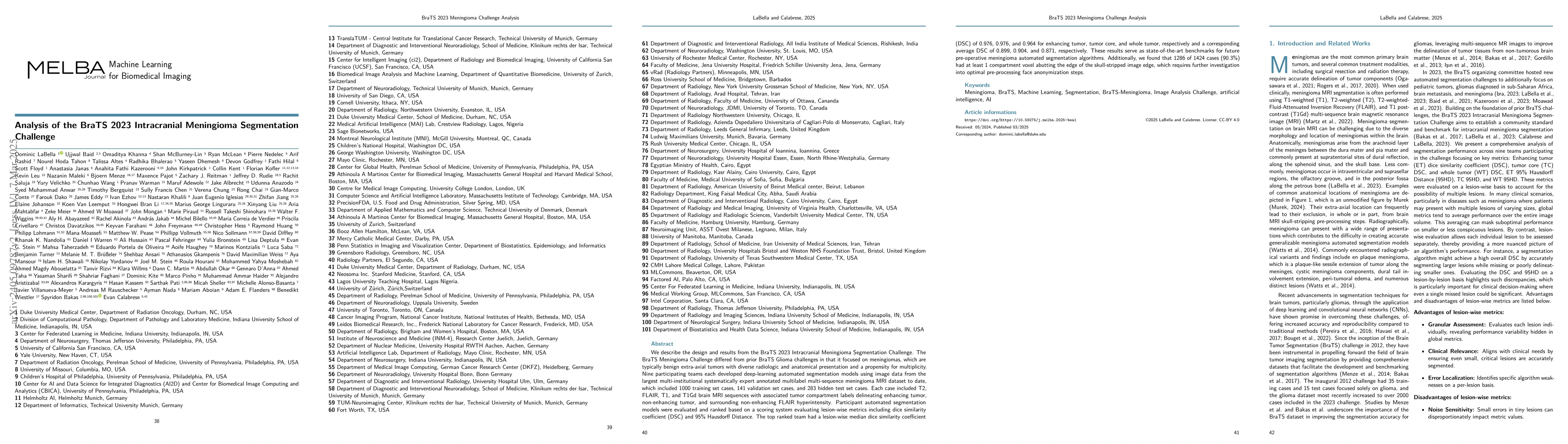
We describe the design and results from the BraTS 2023 Intracranial Meningioma Segmentation Challenge. The BraTS Meningioma Challenge differed from prior BraTS Glioma challenges in that it focused o...
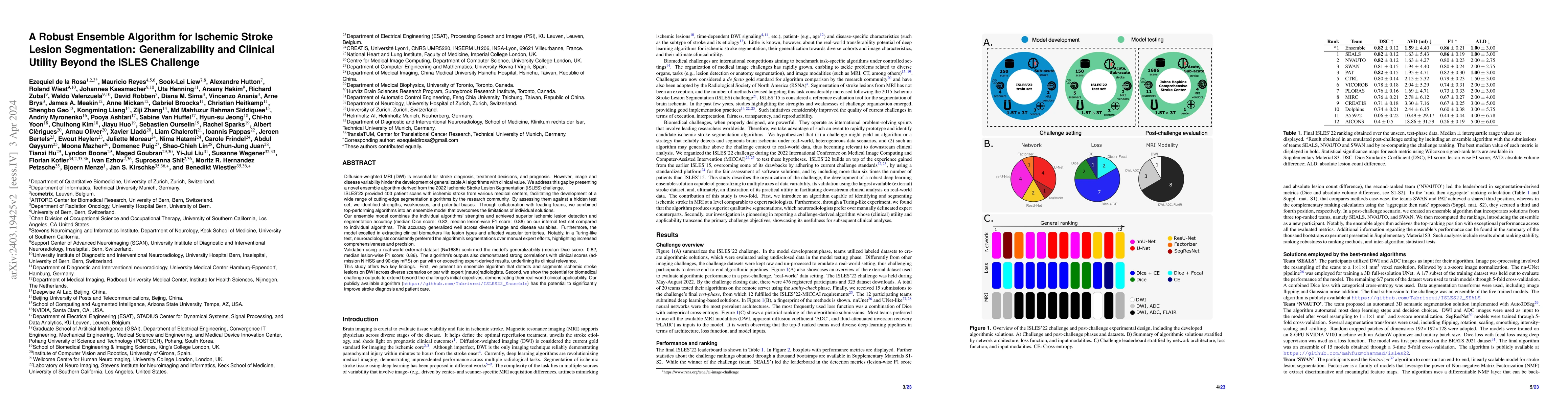
Diffusion-weighted MRI (DWI) is essential for stroke diagnosis, treatment decisions, and prognosis. However, image and disease variability hinder the development of generalizable AI algorithms with ...

Direct image-to-graph transformation is a challenging task that solves object detection and relationship prediction in a single model. Due to the complexity of this task, large training datasets are...
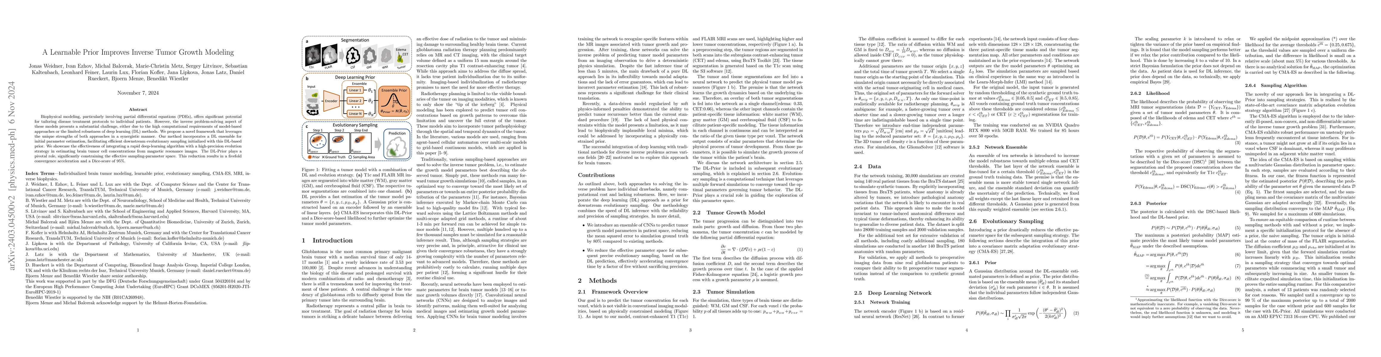
Biophysical modeling, particularly involving partial differential equations (PDEs), offers significant potential for tailoring disease treatment protocols to individual patients. However, the invers...
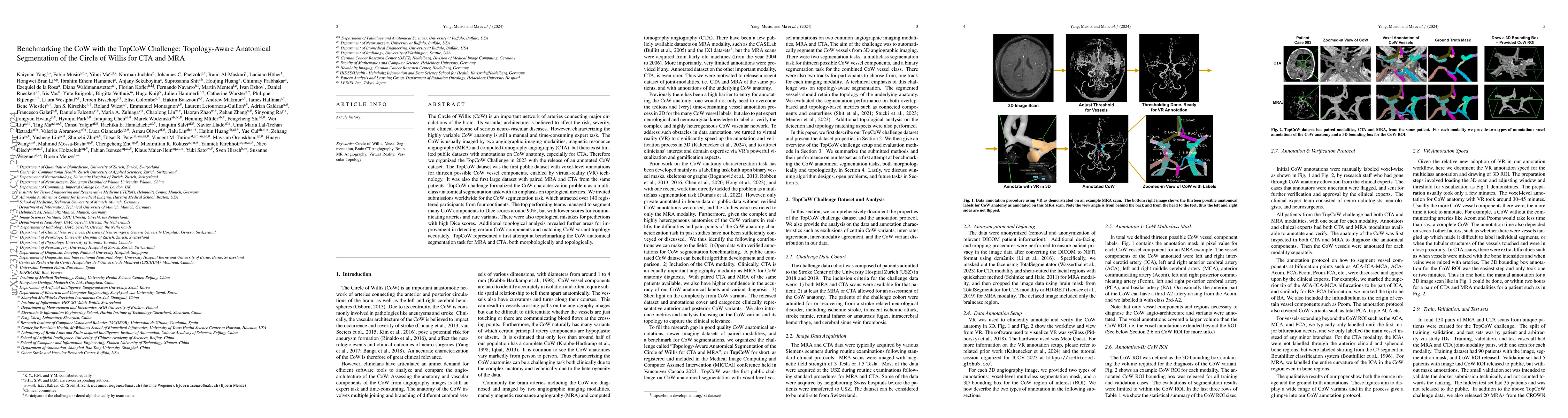
The Circle of Willis (CoW) is an important network of arteries connecting major circulations of the brain. Its vascular architecture is believed to affect the risk, severity, and clinical outcome of...
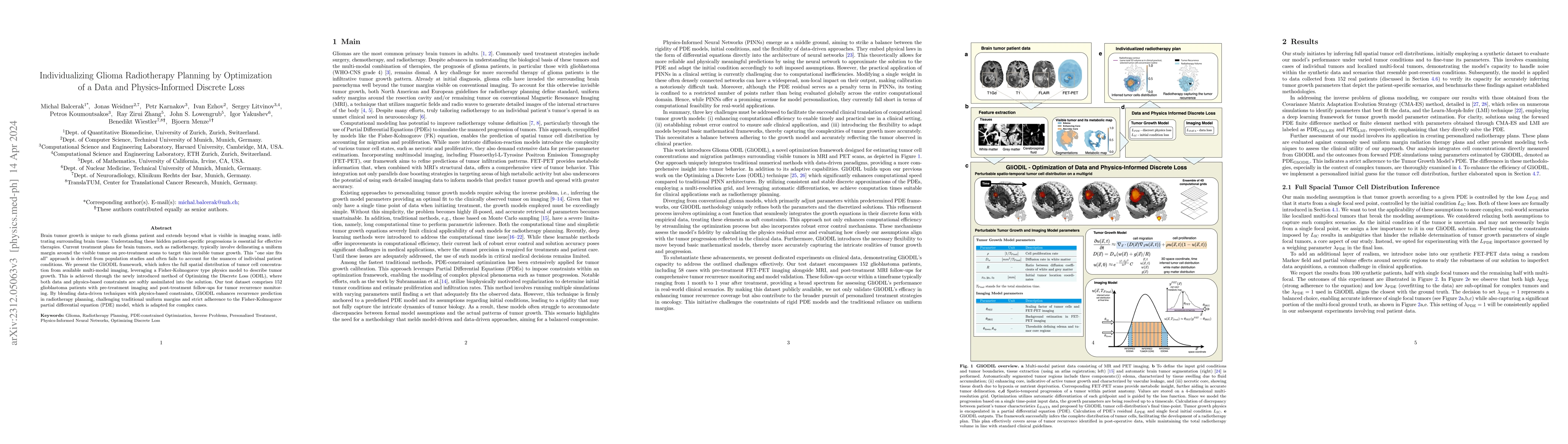
Brain tumor growth is unique to each glioma patient and extends beyond what is visible in imaging scans, infiltrating surrounding brain tissue. Understanding these hidden patient-specific progressio...
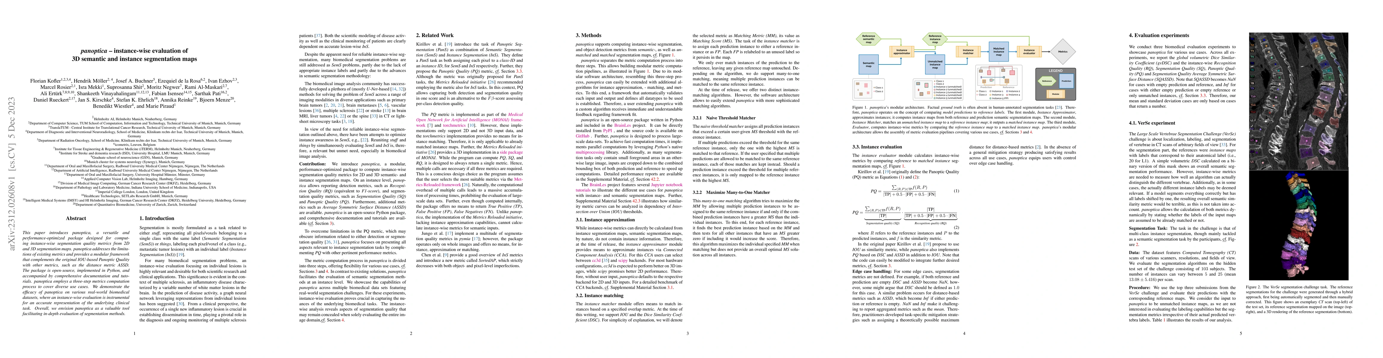
This paper introduces panoptica, a versatile and performance-optimized package designed for computing instance-wise segmentation quality metrics from 2D and 3D segmentation maps. panoptica addresses...
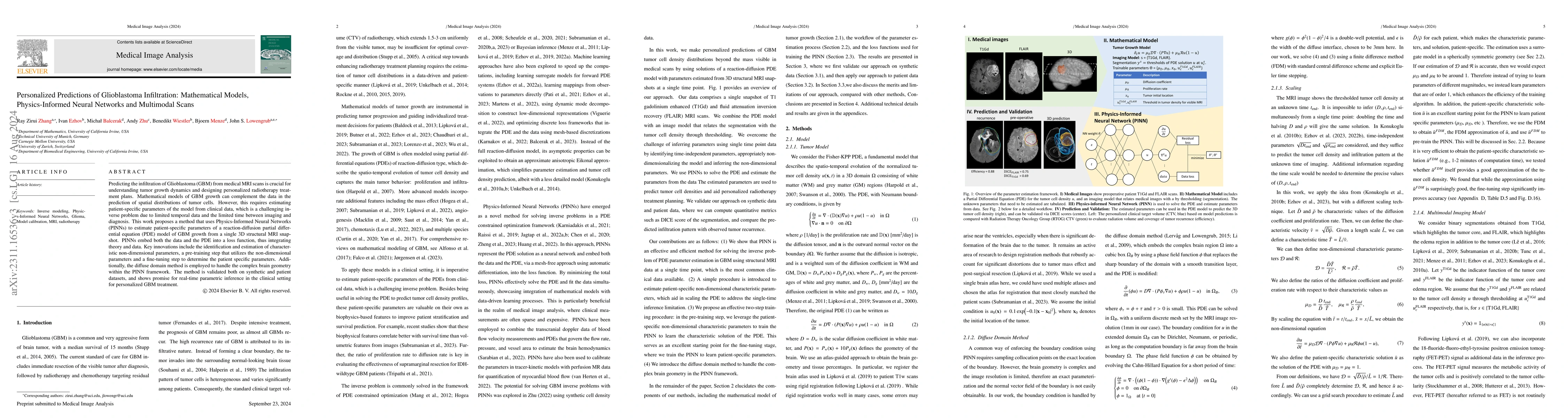
Predicting the infiltration of Glioblastoma (GBM) from medical MRI scans is crucial for understanding tumor growth dynamics and designing personalized radiotherapy treatment plans.Mathematical model...
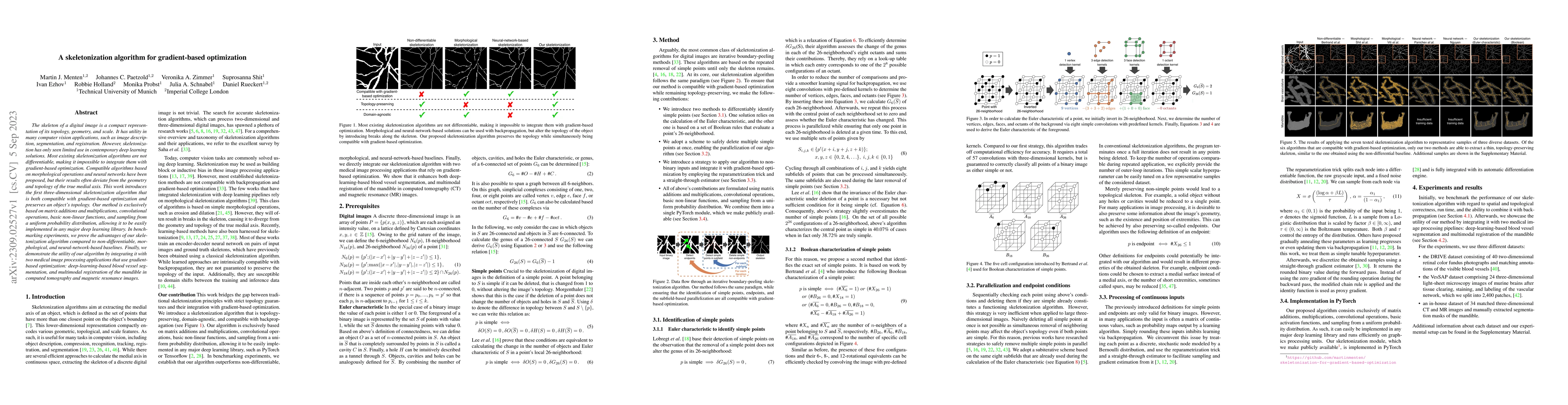
The skeleton of a digital image is a compact representation of its topology, geometry, and scale. It has utility in many computer vision applications, such as image description, segmentation, and re...
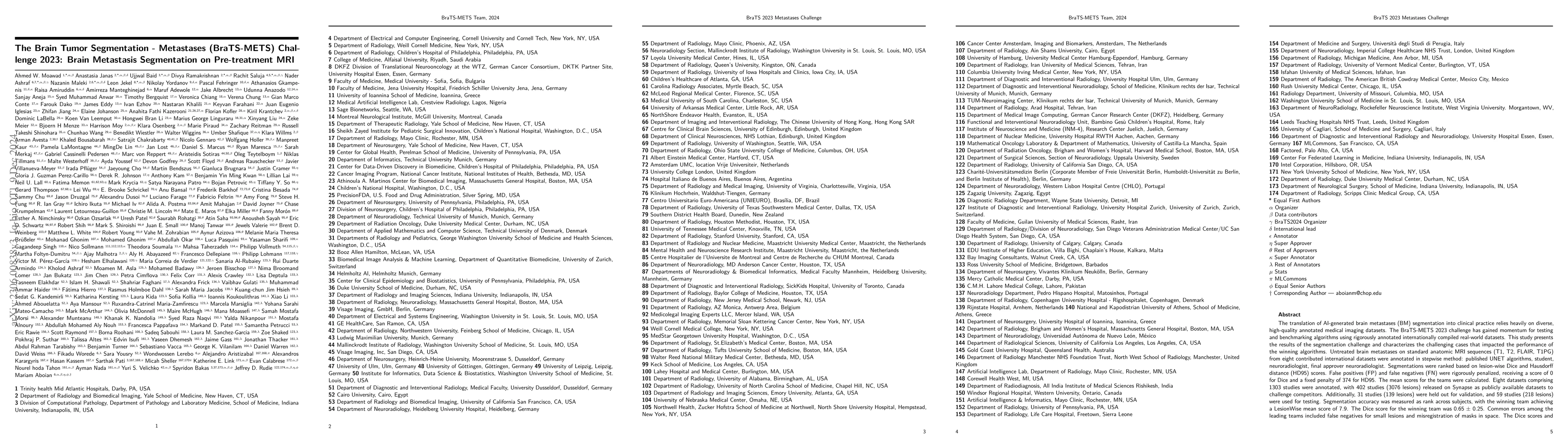
The translation of AI-generated brain metastases (BM) segmentation into clinical practice relies heavily on diverse, high-quality annotated medical imaging datasets. The BraTS-METS 2023 challenge ha...
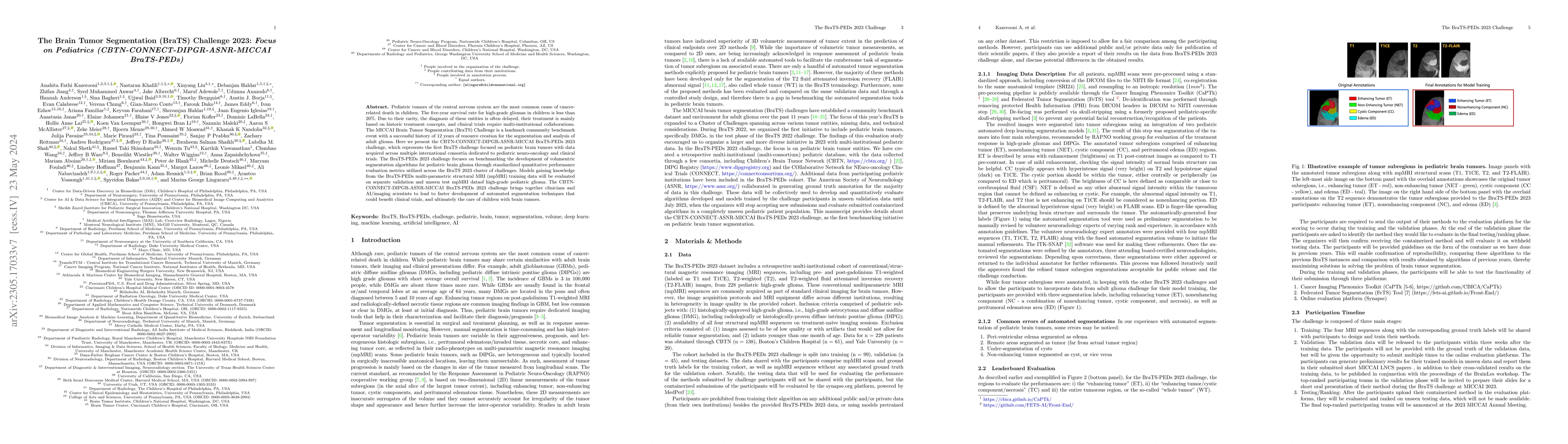
Pediatric tumors of the central nervous system are the most common cause of cancer-related death in children. The five-year survival rate for high-grade gliomas in children is less than 20\%. Due to...
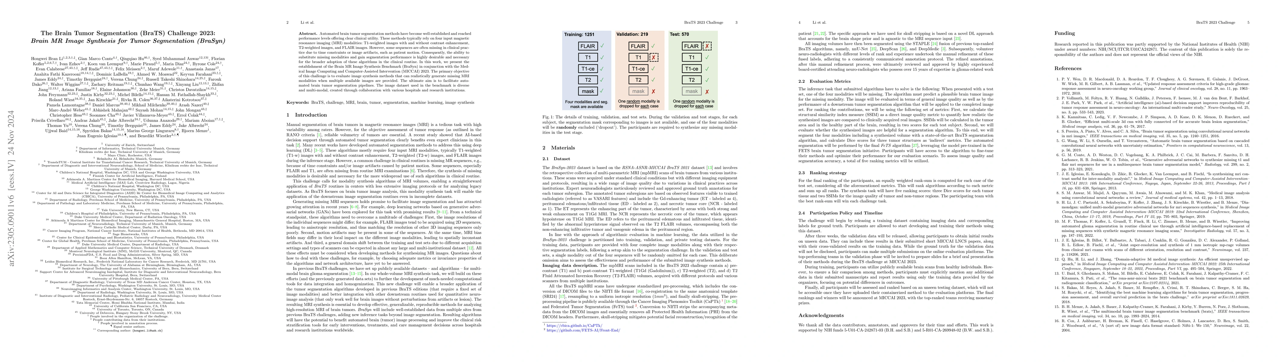
Automated brain tumor segmentation methods have become well-established and reached performance levels offering clear clinical utility. These methods typically rely on four input magnetic resonance ...
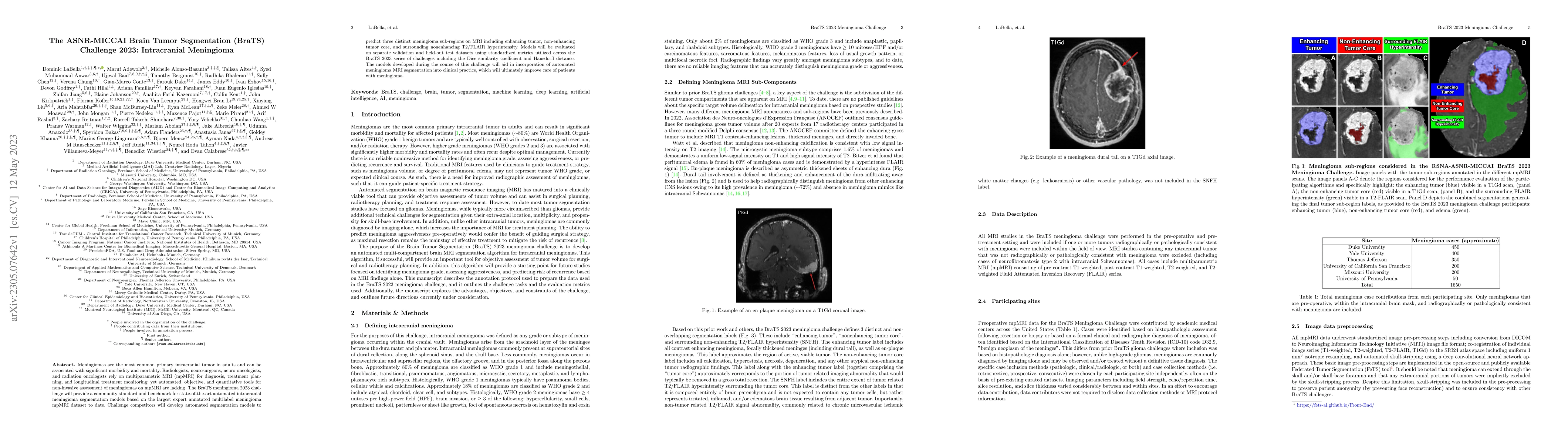
Meningiomas are the most common primary intracranial tumor in adults and can be associated with significant morbidity and mortality. Radiologists, neurosurgeons, neuro-oncologists, and radiation onc...
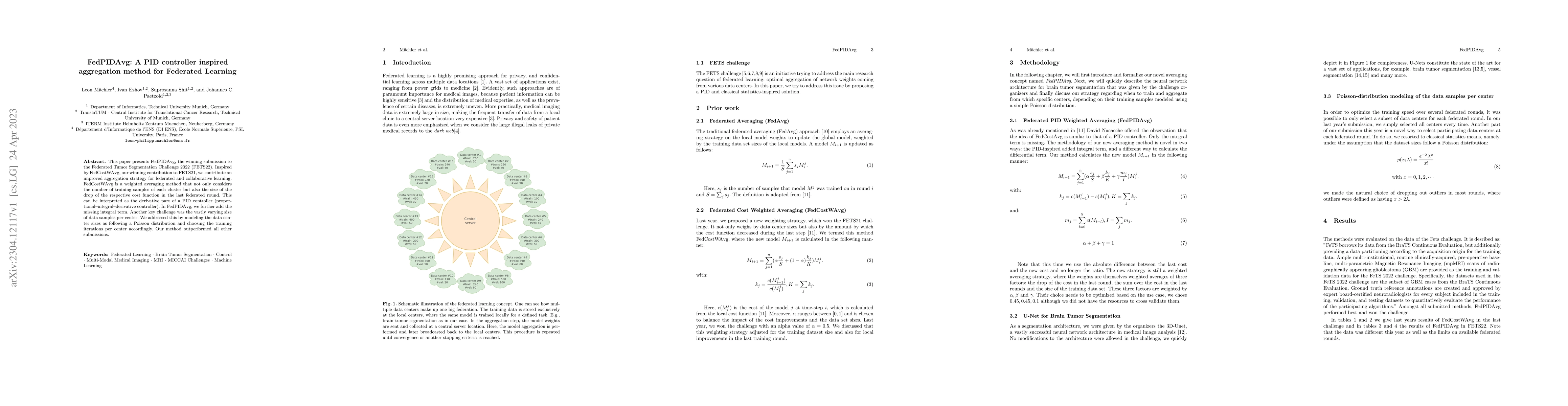
This paper presents FedPIDAvg, the winning submission to the Federated Tumor Segmentation Challenge 2022 (FETS22). Inspired by FedCostWAvg, our winning contribution to FETS21, we contribute an impro...
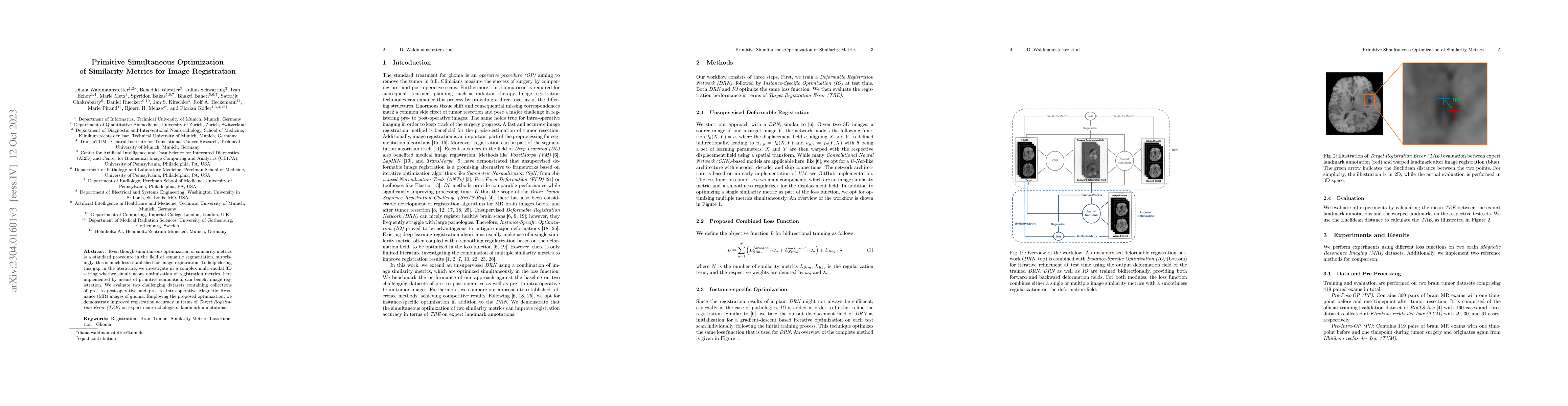
Even though simultaneous optimization of similarity metrics is a standard procedure in the field of semantic segmentation, surprisingly, this is much less established for image registration. To help...
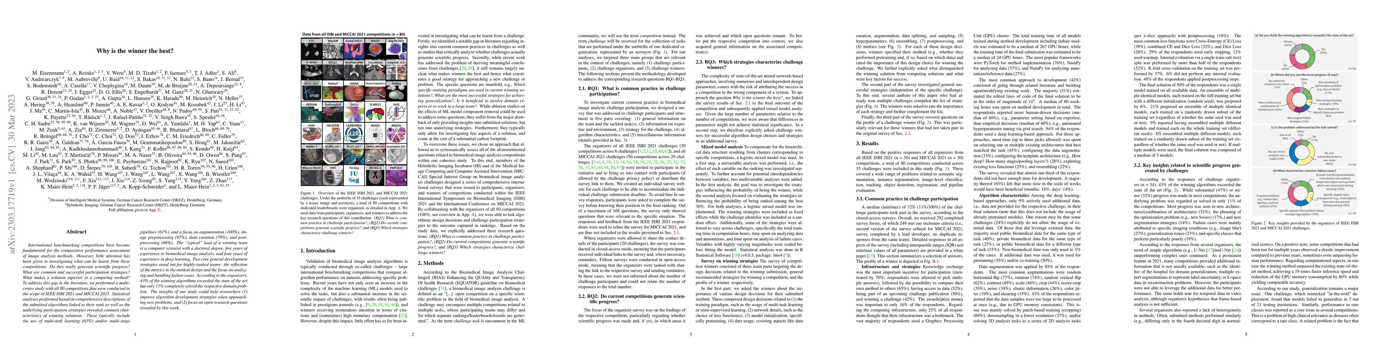
International benchmarking competitions have become fundamental for the comparative performance assessment of image analysis methods. However, little attention has been given to investigating what c...
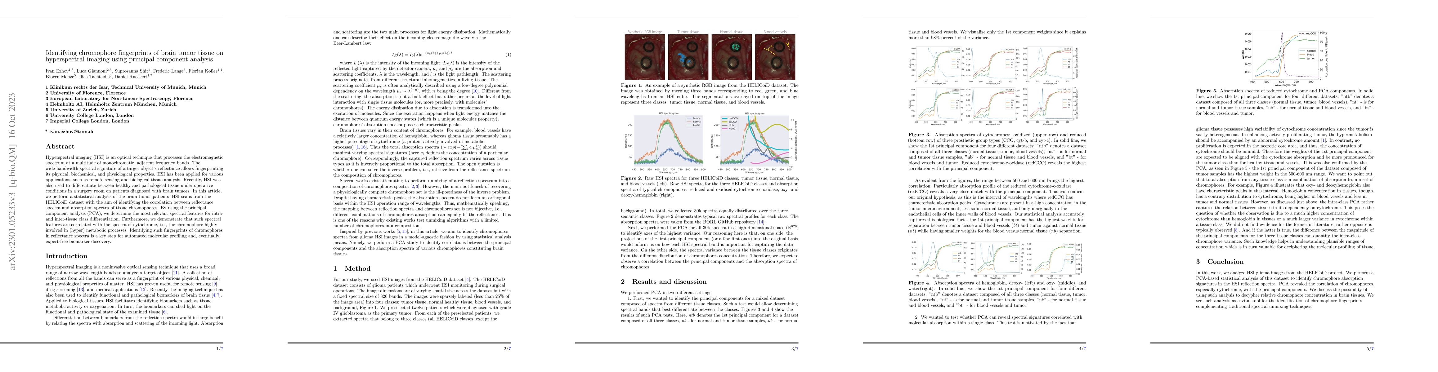
Hyperspectral imaging (HSI) is an optical technique that processes the electromagnetic spectrum at a multitude of monochromatic, adjacent frequency bands. The wide-bandwidth spectral signature of a ...
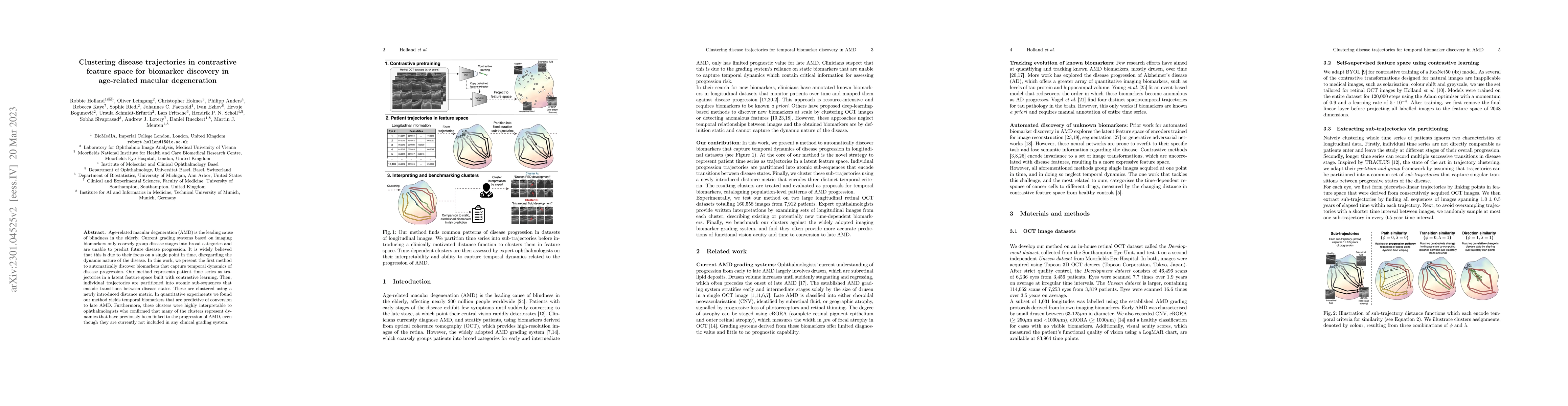
Age-related macular degeneration (AMD) is the leading cause of blindness in the elderly. Current grading systems based on imaging biomarkers only coarsely group disease stages into broad categories ...
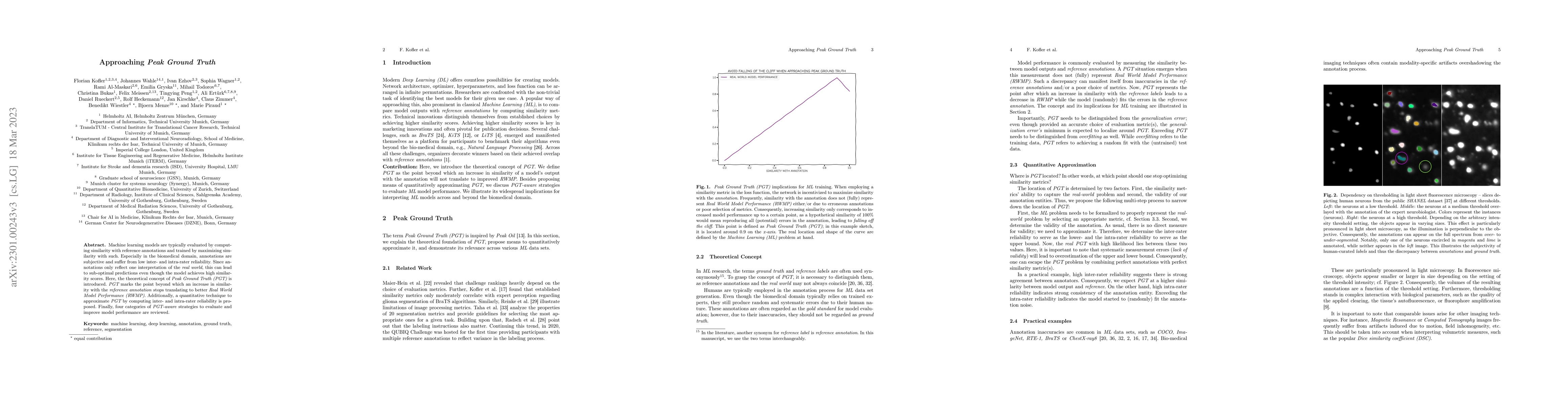
Machine learning models are typically evaluated by computing similarity with reference annotations and trained by maximizing similarity with such. Especially in the biomedical domain, annotations ar...
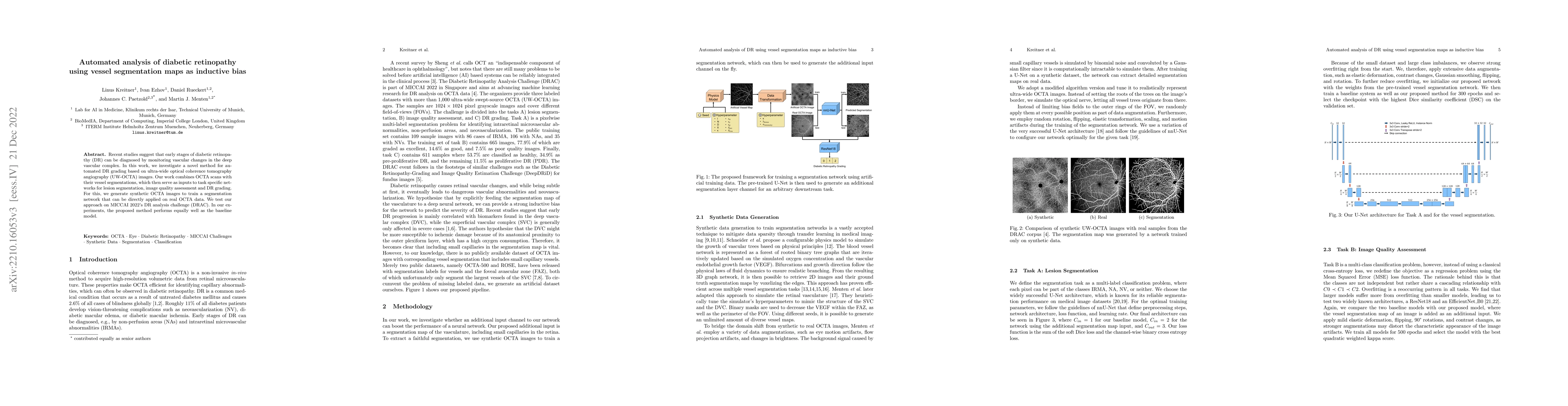
Recent studies suggest that early stages of diabetic retinopathy (DR) can be diagnosed by monitoring vascular changes in the deep vascular complex. In this work, we investigate a novel method for au...
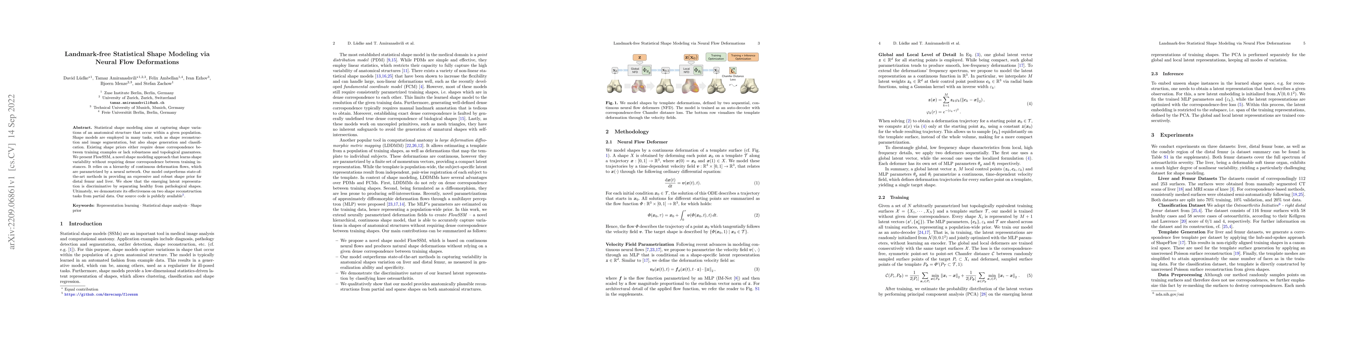
Statistical shape modeling aims at capturing shape variations of an anatomical structure that occur within a given population. Shape models are employed in many tasks, such as shape reconstruction a...
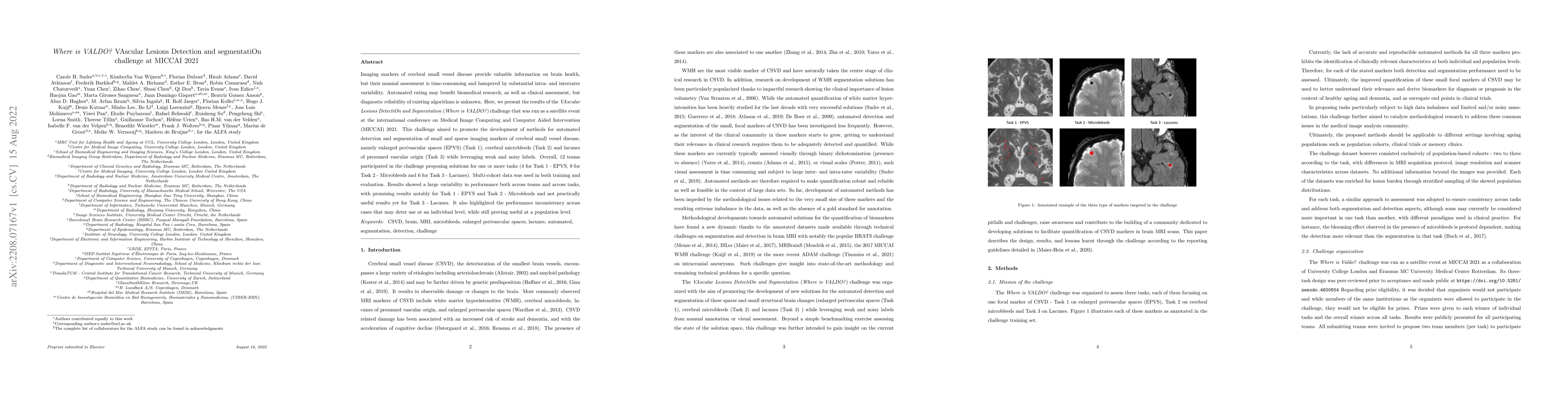
Imaging markers of cerebral small vessel disease provide valuable information on brain health, but their manual assessment is time-consuming and hampered by substantial intra- and interrater variabi...
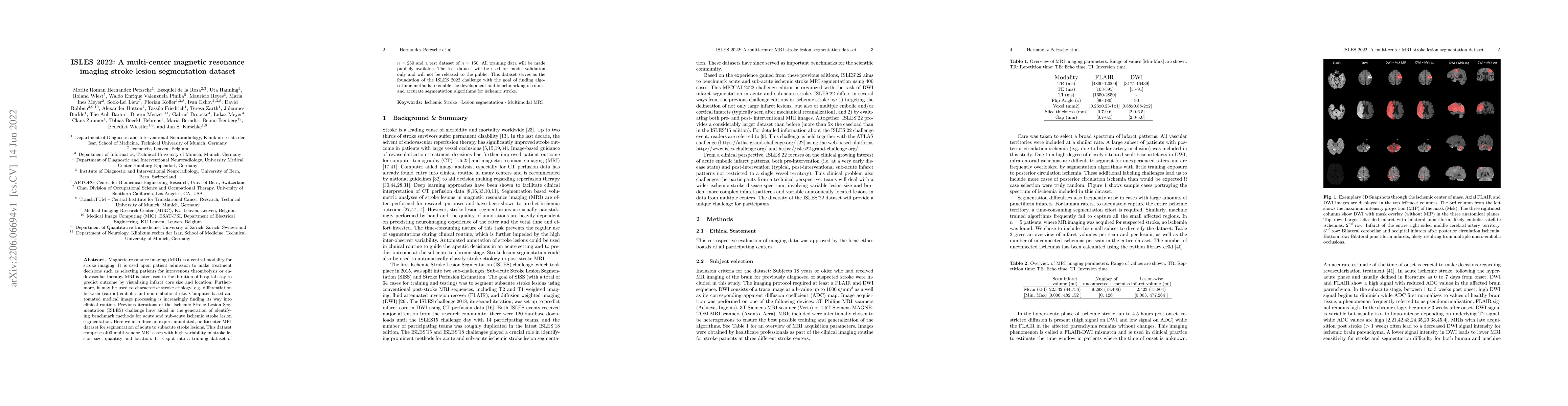
Magnetic resonance imaging (MRI) is a central modality for stroke imaging. It is used upon patient admission to make treatment decisions such as selecting patients for intravenous thrombolysis or en...
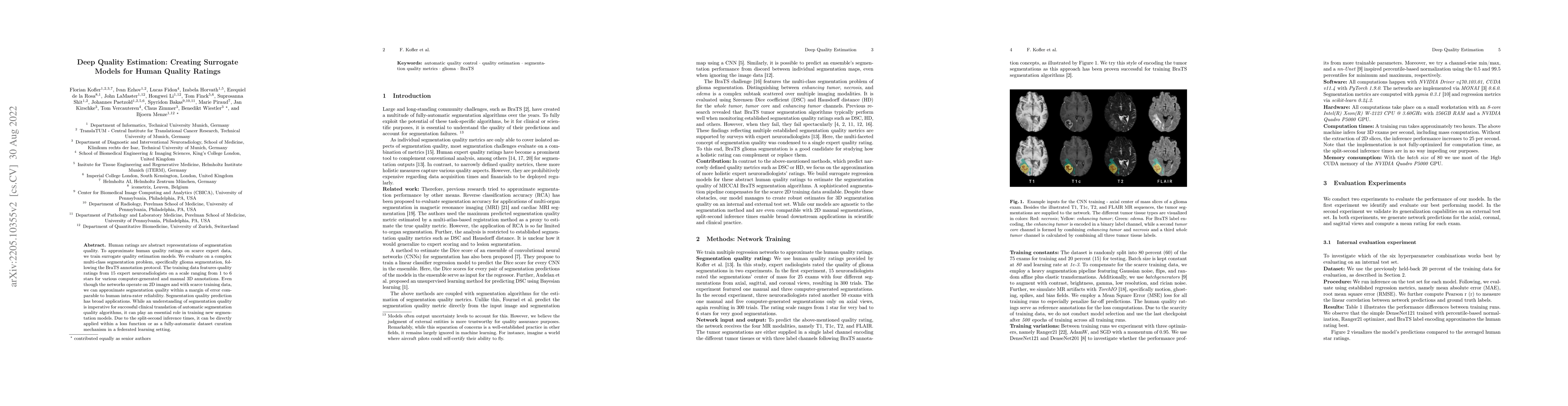
Human ratings are abstract representations of segmentation quality. To approximate human quality ratings on scarce expert data, we train surrogate quality estimation models. We evaluate on a complex...
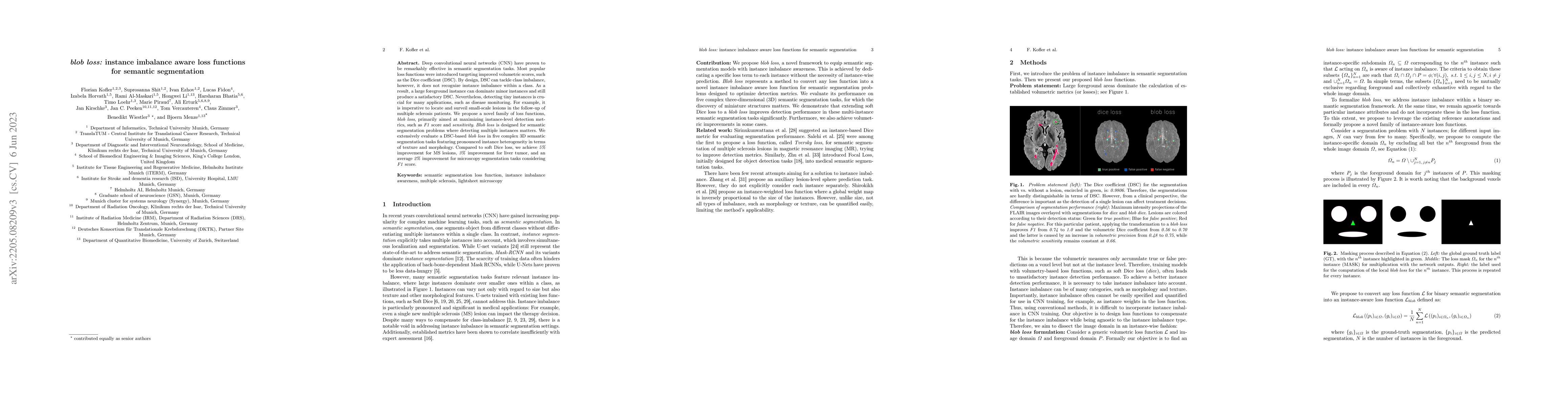
Deep convolutional neural networks (CNN) have proven to be remarkably effective in semantic segmentation tasks. Most popular loss functions were introduced targeting improved volumetric scores, such...
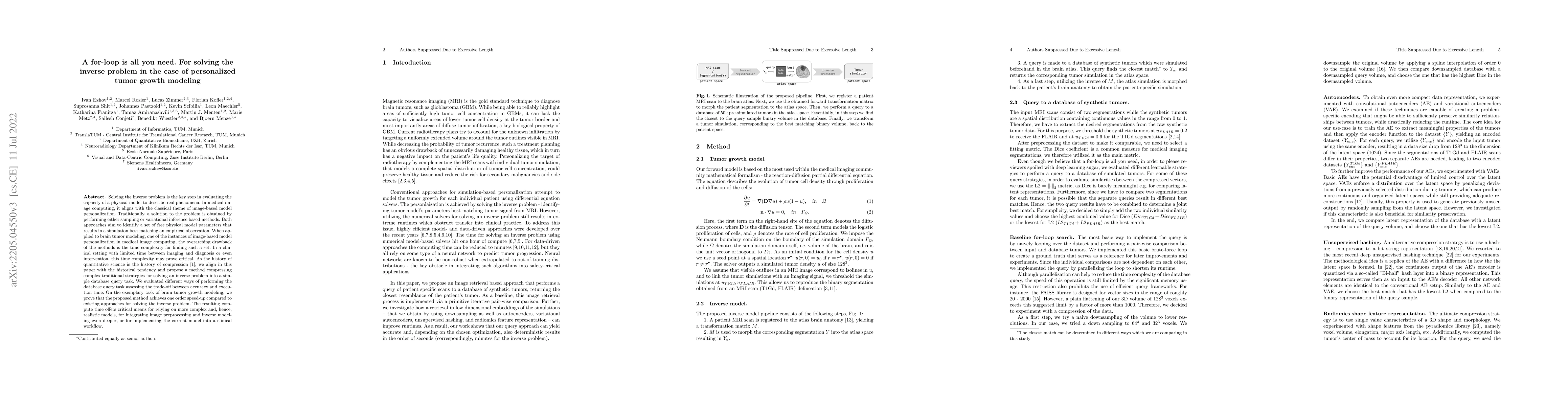
Solving the inverse problem is the key step in evaluating the capacity of a physical model to describe real phenomena. In medical image computing, it aligns with the classical theme of image-based m...
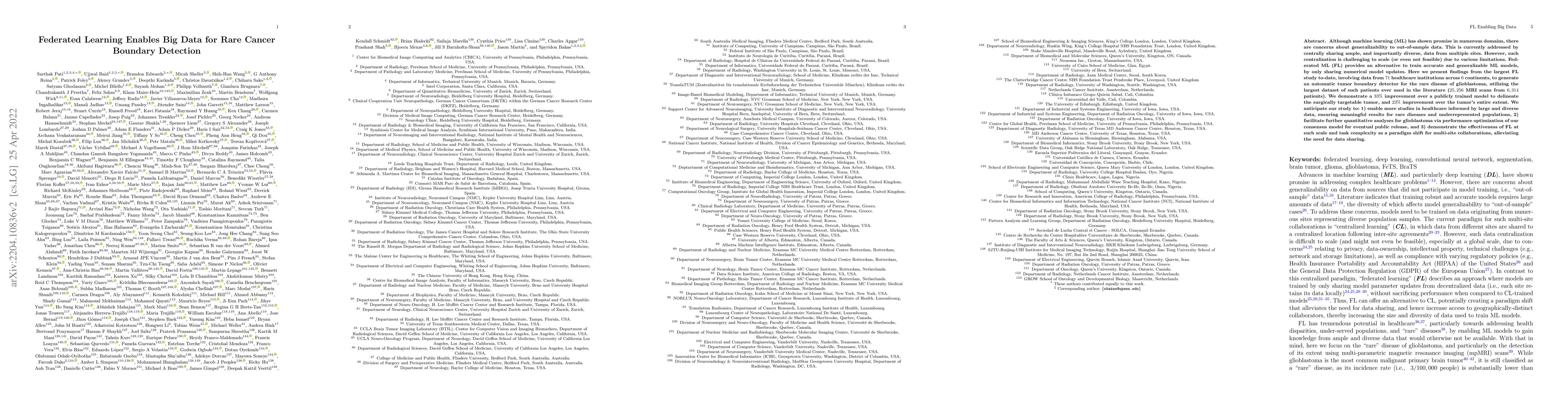
Although machine learning (ML) has shown promise in numerous domains, there are concerns about generalizability to out-of-sample data. This is currently addressed by centrally sharing ample, and imp...
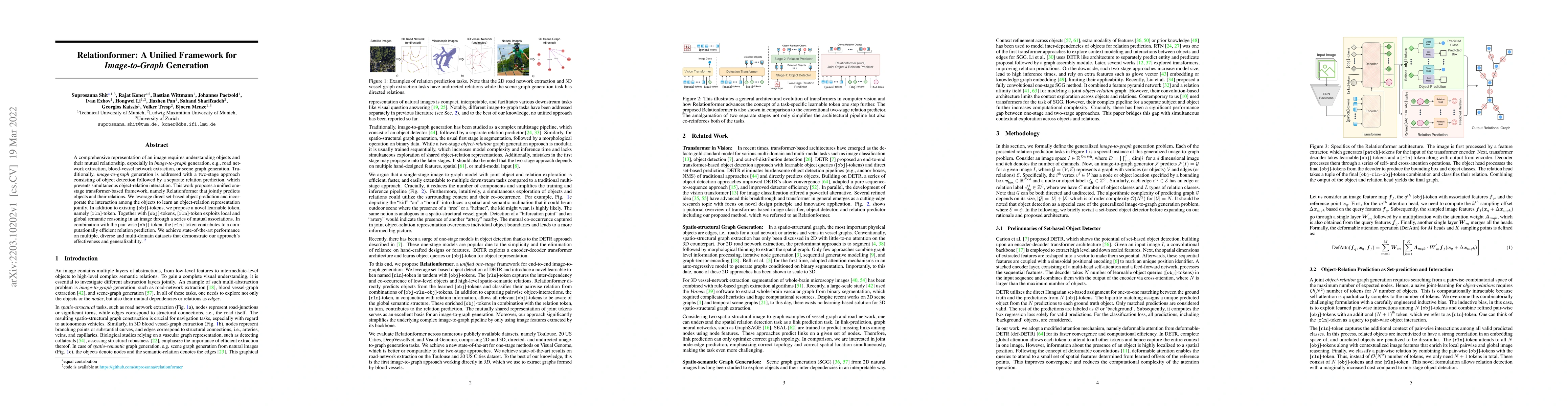
A comprehensive representation of an image requires understanding objects and their mutual relationship, especially in image-to-graph generation, e.g., road network extraction, blood-vessel network ...
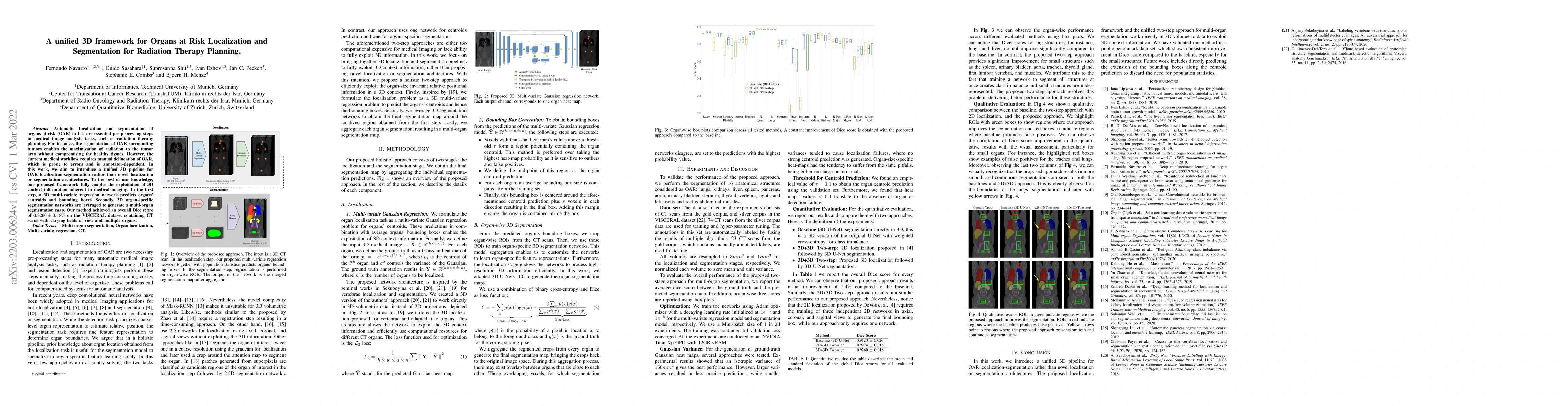
Automatic localization and segmentation of organs-at-risk (OAR) in CT are essential pre-processing steps in medical image analysis tasks, such as radiation therapy planning. For instance, the segmen...
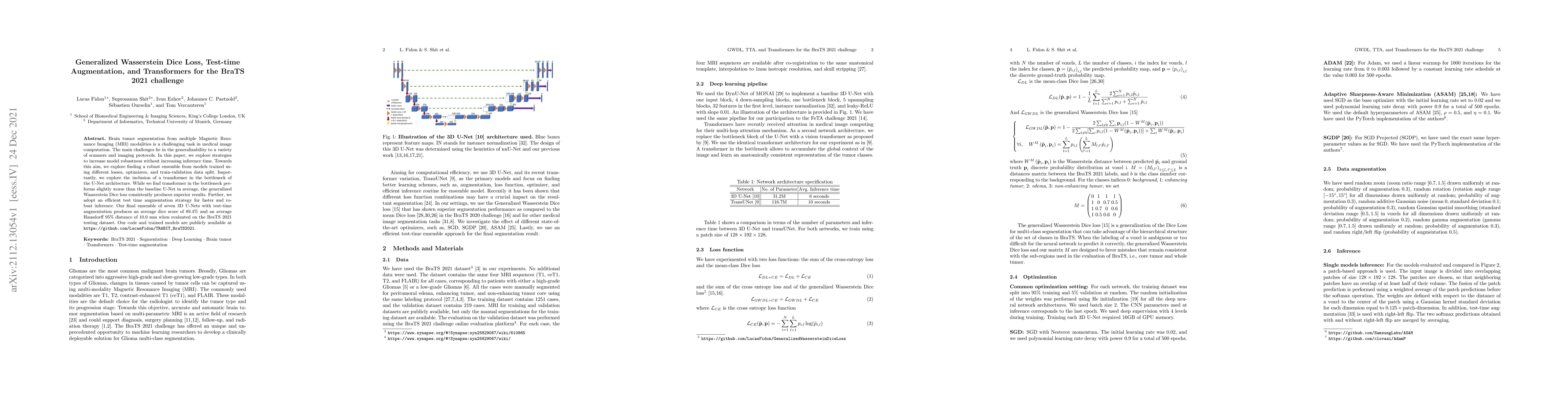
Brain tumor segmentation from multiple Magnetic Resonance Imaging (MRI) modalities is a challenging task in medical image computation. The main challenges lie in the generalizability to a variety of...
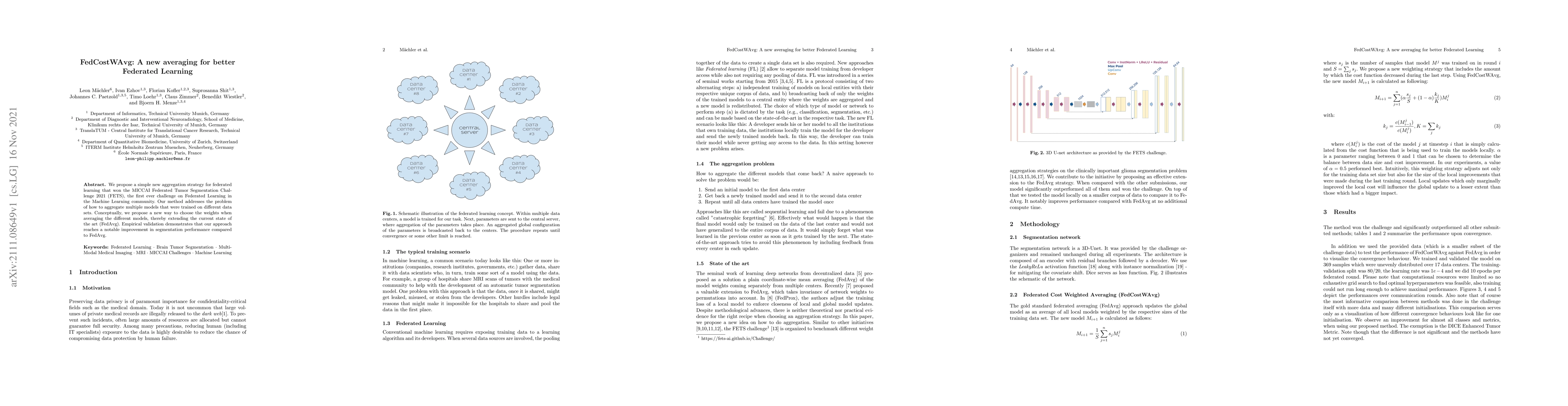
We propose a simple new aggregation strategy for federated learning that won the MICCAI Federated Tumor Segmentation Challenge 2021 (FETS), the first ever challenge on Federated Learning in the Mach...
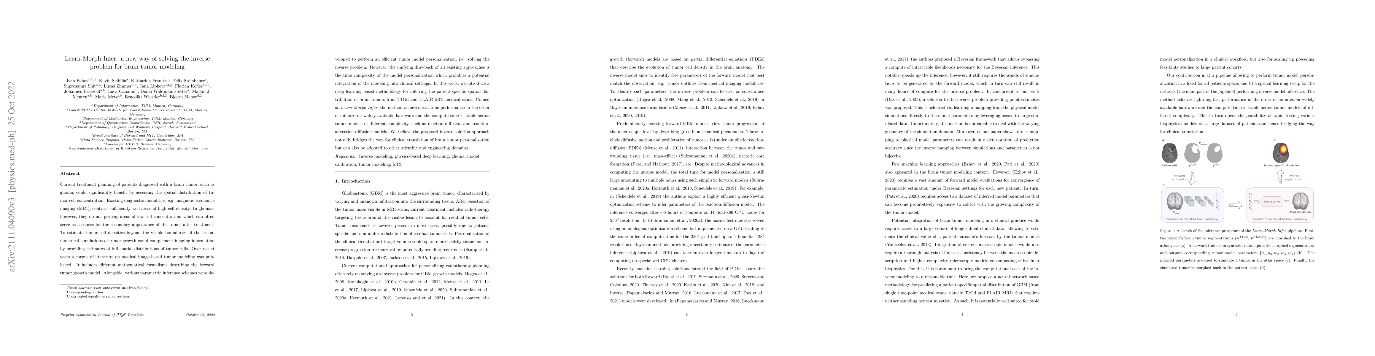
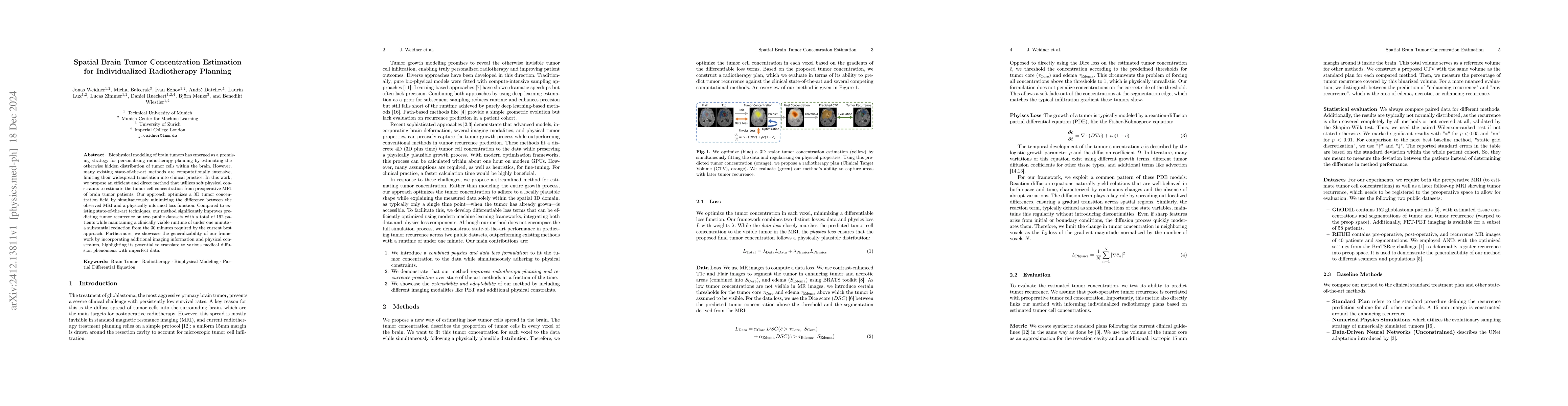
Current treatment planning of patients diagnosed with a brain tumor, such as glioma, could significantly benefit by accessing the spatial distribution of tumor cell concentration. Existing diagnosti...
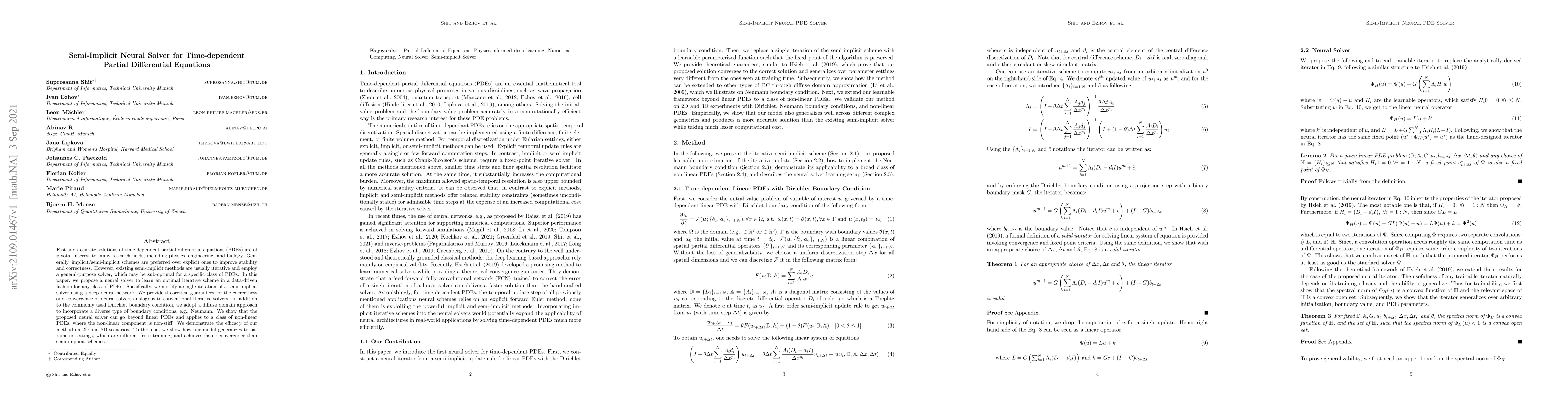
Fast and accurate solutions of time-dependent partial differential equations (PDEs) are of pivotal interest to many research fields, including physics, engineering, and biology. Generally, implicit/...
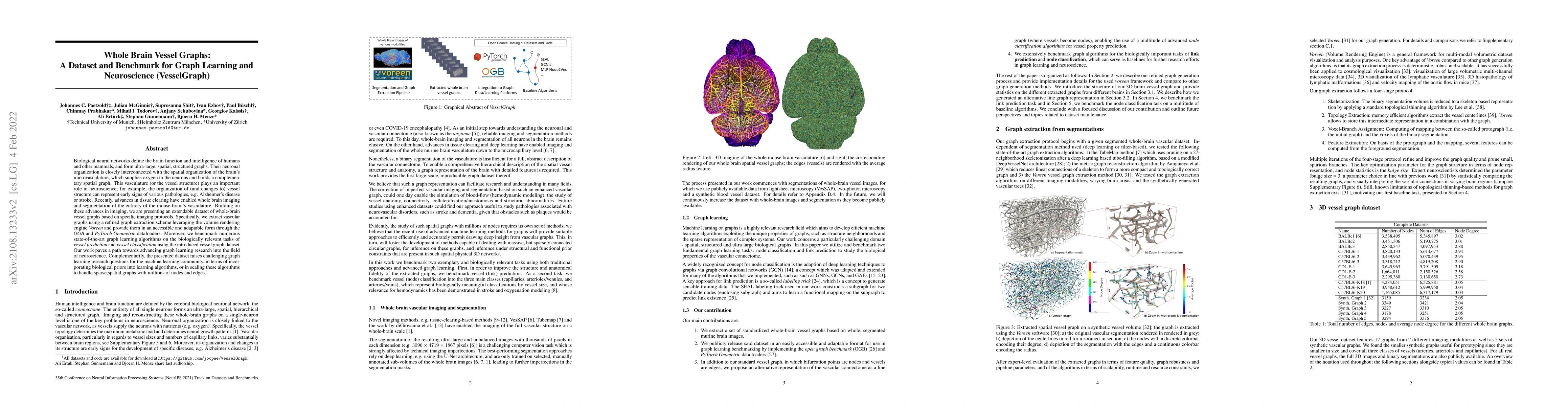
Biological neural networks define the brain function and intelligence of humans and other mammals, and form ultra-large, spatial, structured graphs. Their neuronal organization is closely interconne...
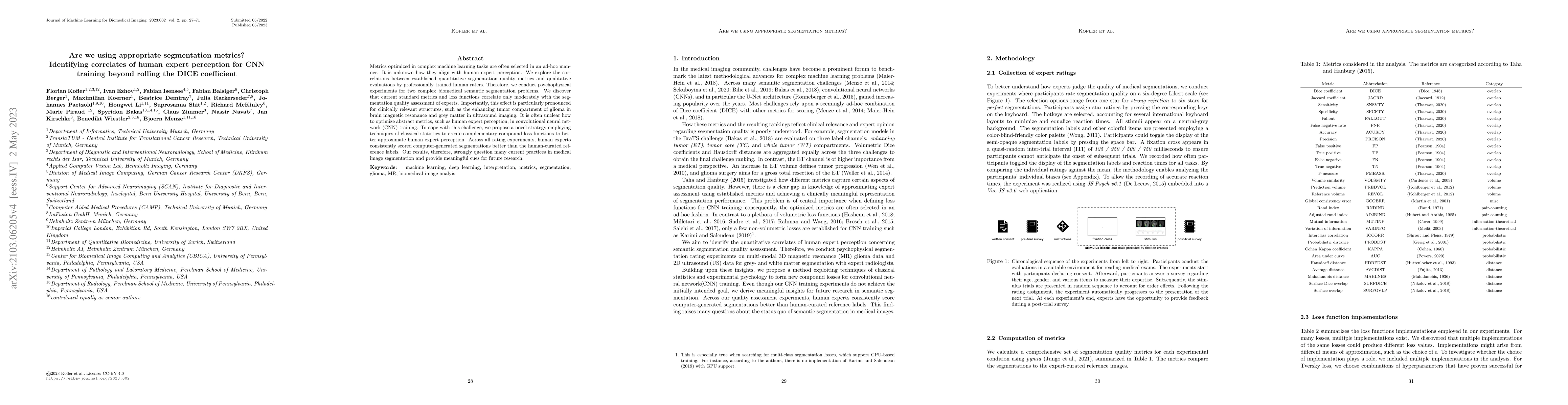
Metrics optimized in complex machine learning tasks are often selected in an ad-hoc manner. It is unknown how they align with human expert perception. We explore the correlations between established...
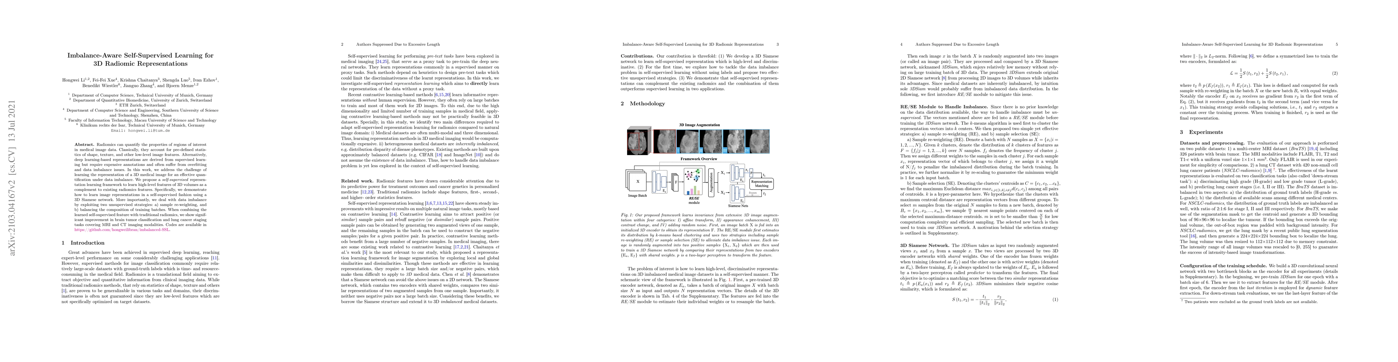
Radiomic representations can quantify properties of regions of interest in medical image data. Classically, they account for pre-defined statistics of shape, texture, and other low-level image featu...
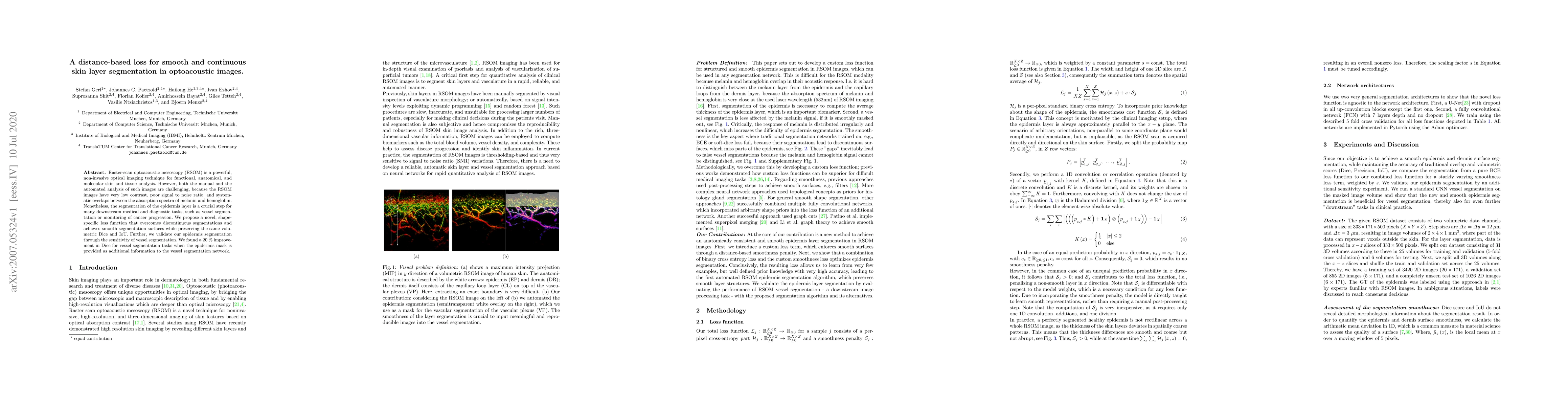
Raster-scan optoacoustic mesoscopy (RSOM) is a powerful, non-invasive optical imaging technique for functional, anatomical, and molecular skin and tissue analysis. However, both the manual and the a...
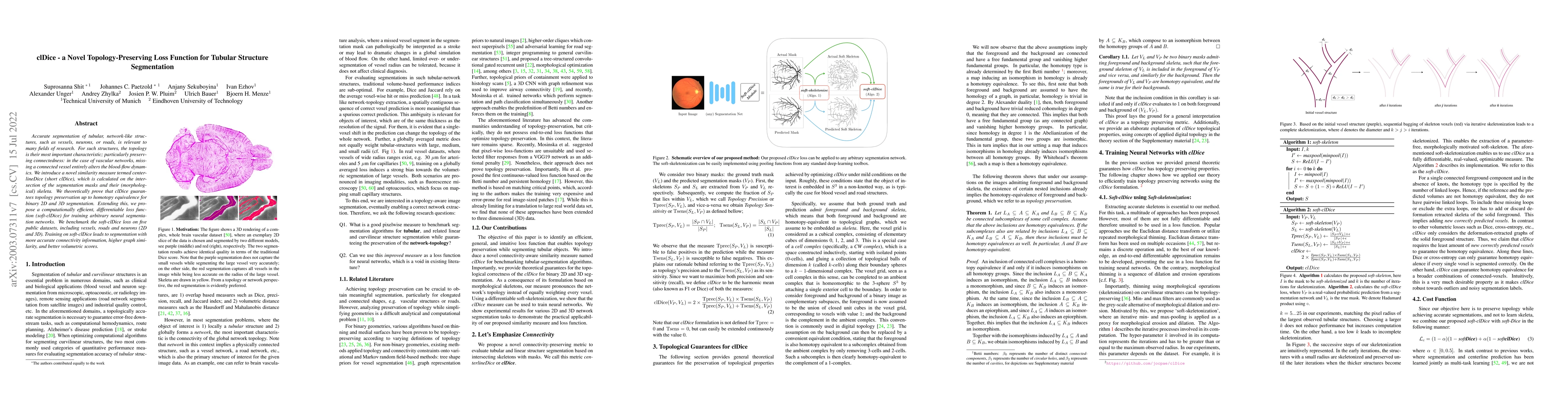
Accurate segmentation of tubular, network-like structures, such as vessels, neurons, or roads, is relevant to many fields of research. For such structures, the topology is their most important chara...
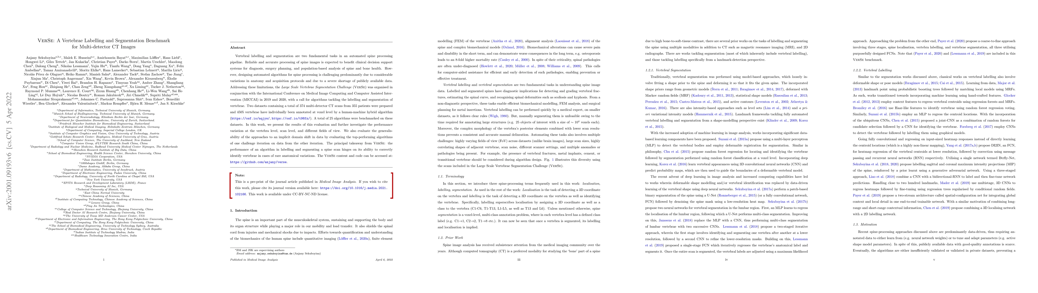
Vertebral labelling and segmentation are two fundamental tasks in an automated spine processing pipeline. Reliable and accurate processing of spine images is expected to benefit clinical decision-su...
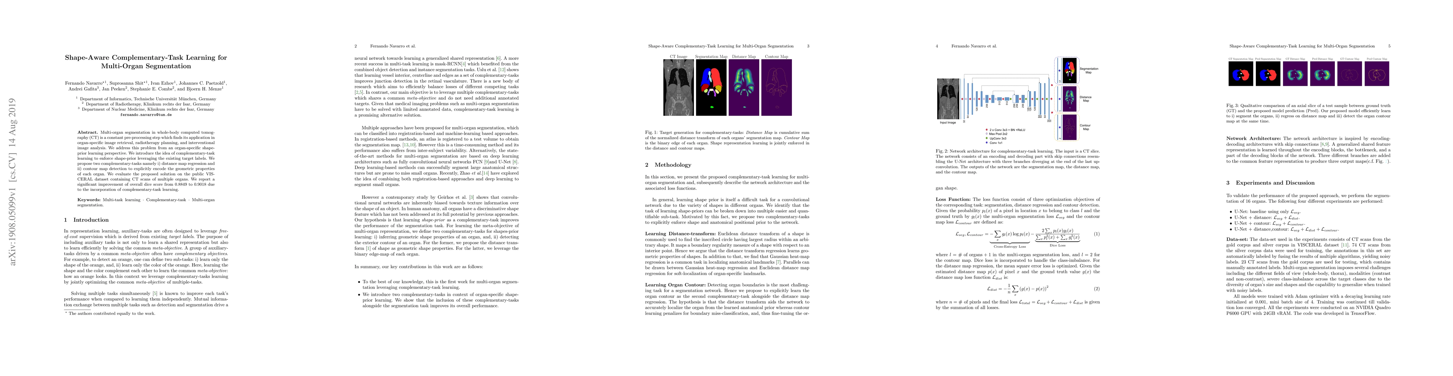
Multi-organ segmentation in whole-body computed tomography (CT) is a constant pre-processing step which finds its application in organ-specific image retrieval, radiotherapy planning, and interventi...
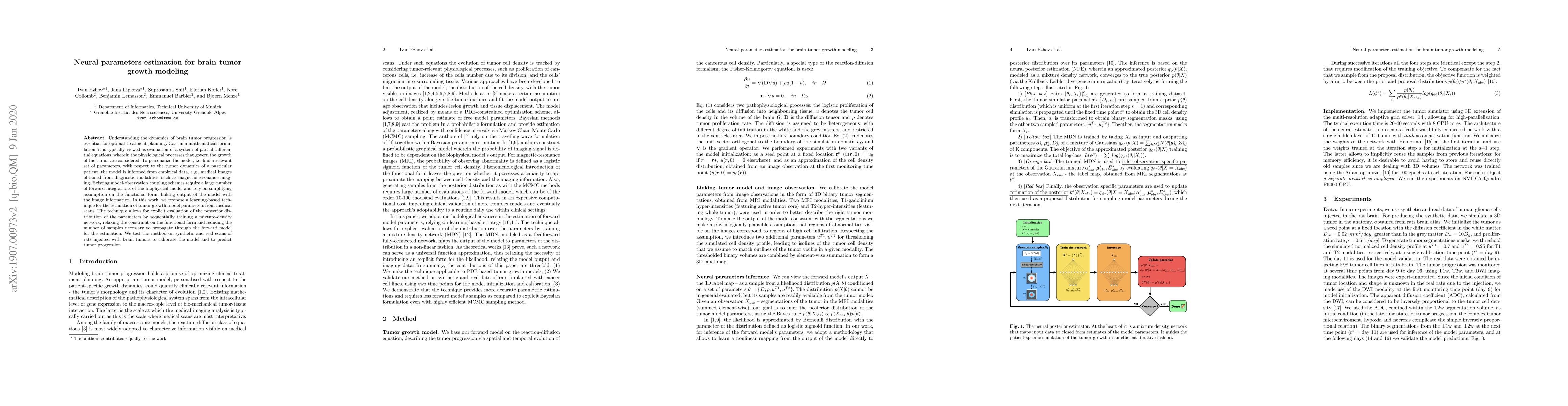
Understanding the dynamics of brain tumor progression is essential for optimal treatment planning. Cast in a mathematical formulation, it is typically viewed as evaluation of a system of partial dif...
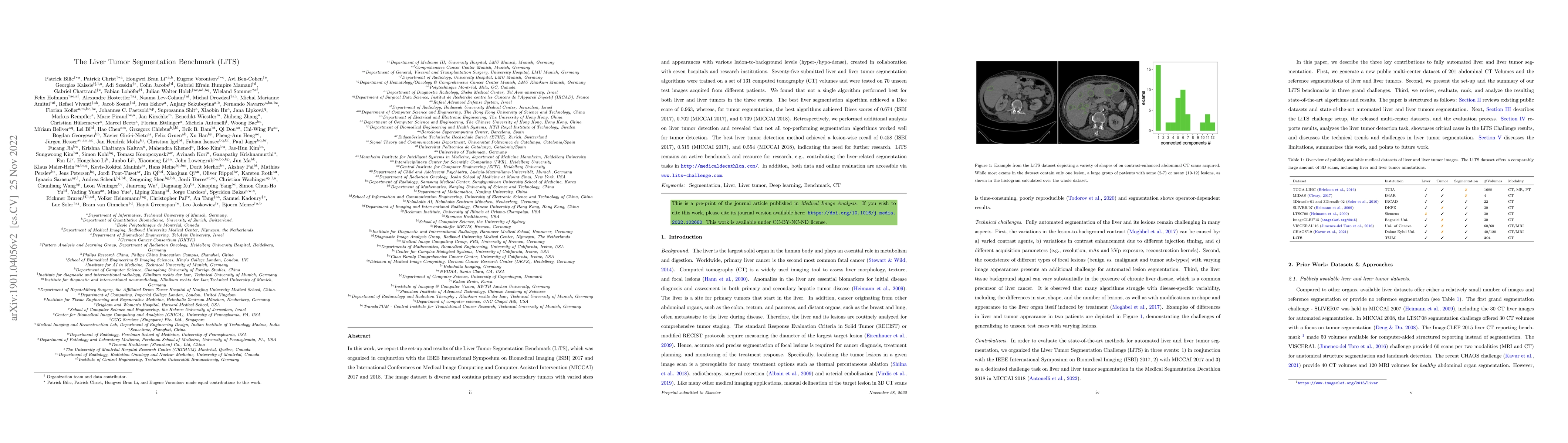
In this work, we report the set-up and results of the Liver Tumor Segmentation Benchmark (LiTS), which was organized in conjunction with the IEEE International Symposium on Biomedical Imaging (ISBI)...
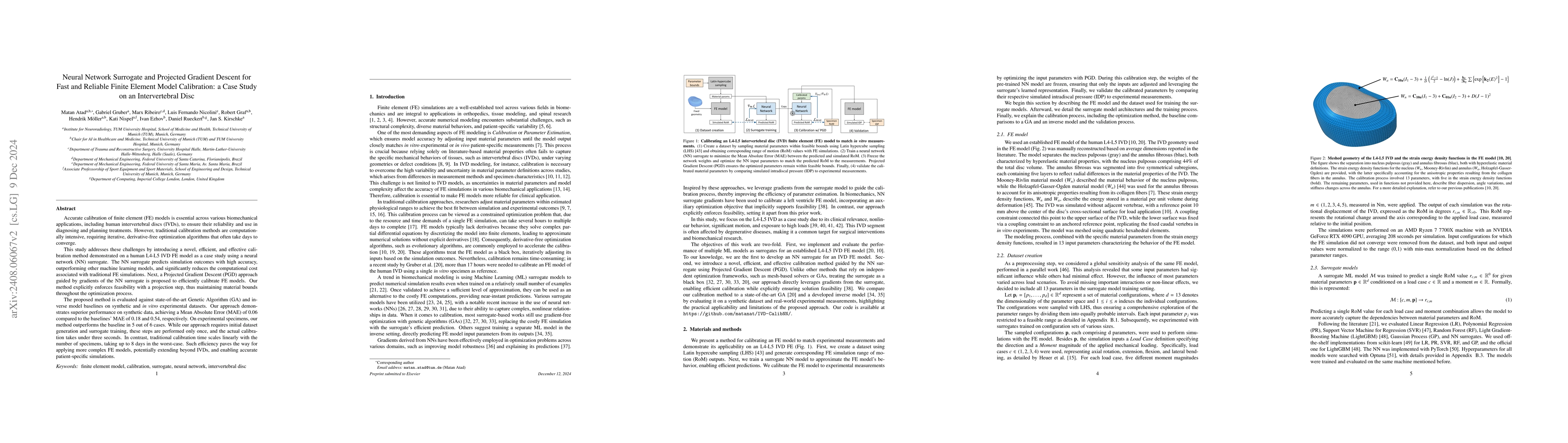
Accurate calibration of finite element (FE) models of human intervertebral discs (IVDs) is essential for their reliability and application in diagnosing and planning treatments for spinal conditions. ...
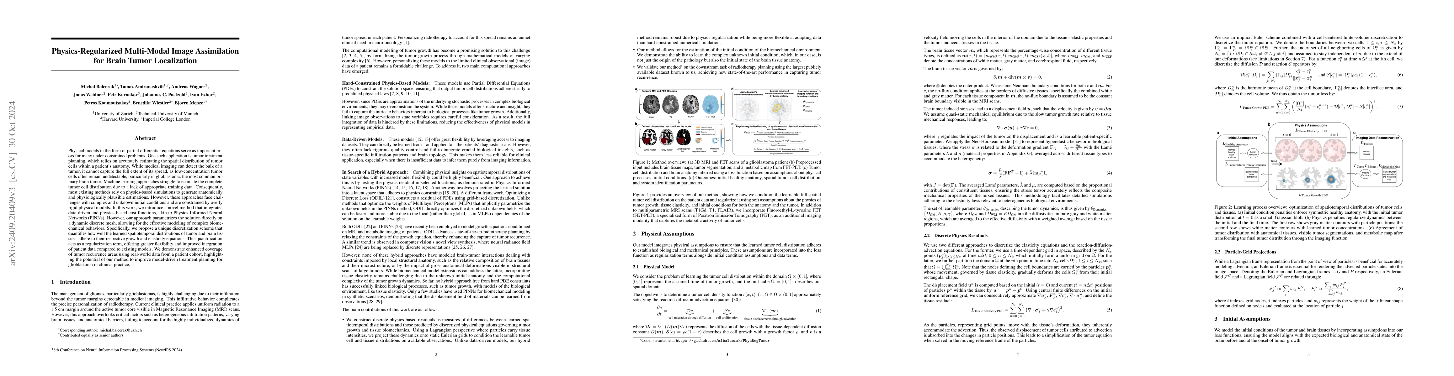
Physical models in the form of partial differential equations represent an important prior for many under-constrained problems. One example is tumor treatment planning, which heavily depends on accura...
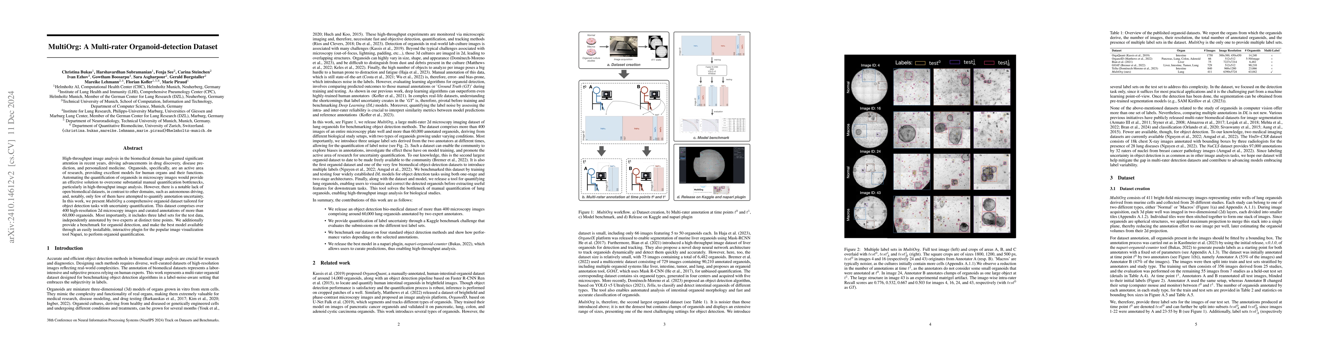
High-throughput image analysis in the biomedical domain has gained significant attention in recent years, driving advancements in drug discovery, disease prediction, and personalized medicine. Organoi...

This paper presents FedPID, our submission to the Federated Tumor Segmentation Challenge 2024 (FETS24). Inspired by FedCostWAvg and FedPIDAvg, our winning contributions to FETS21 and FETS2022, we prop...
Biophysical modeling of brain tumors has emerged as a promising strategy for personalizing radiotherapy planning by estimating the otherwise hidden distribution of tumor cells within the brain. Howeve...
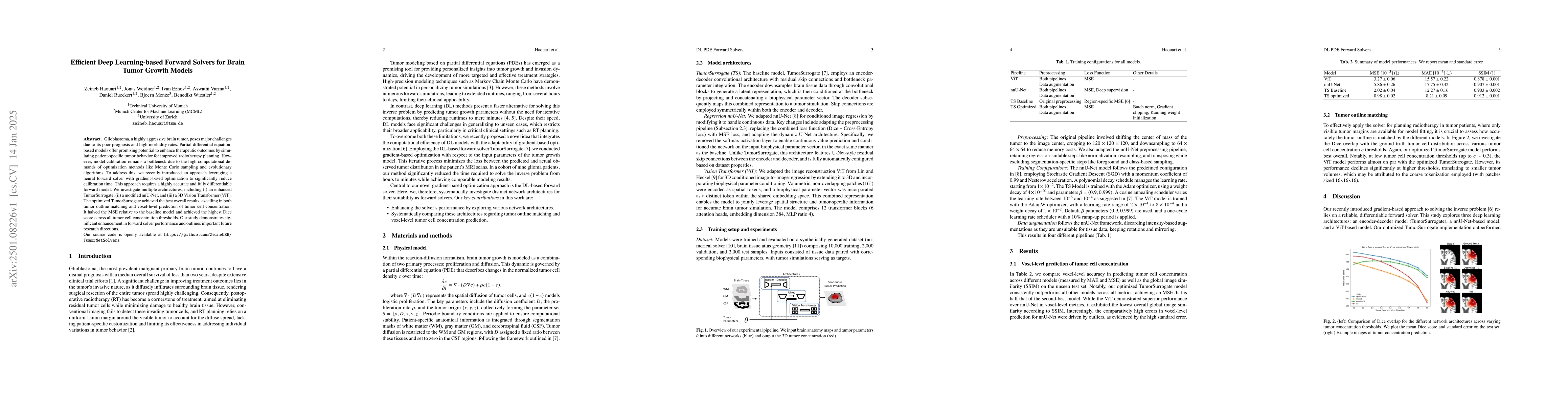
Glioblastoma, a highly aggressive brain tumor, poses major challenges due to its poor prognosis and high morbidity rates. Partial differential equation-based models offer promising potential to enhanc...
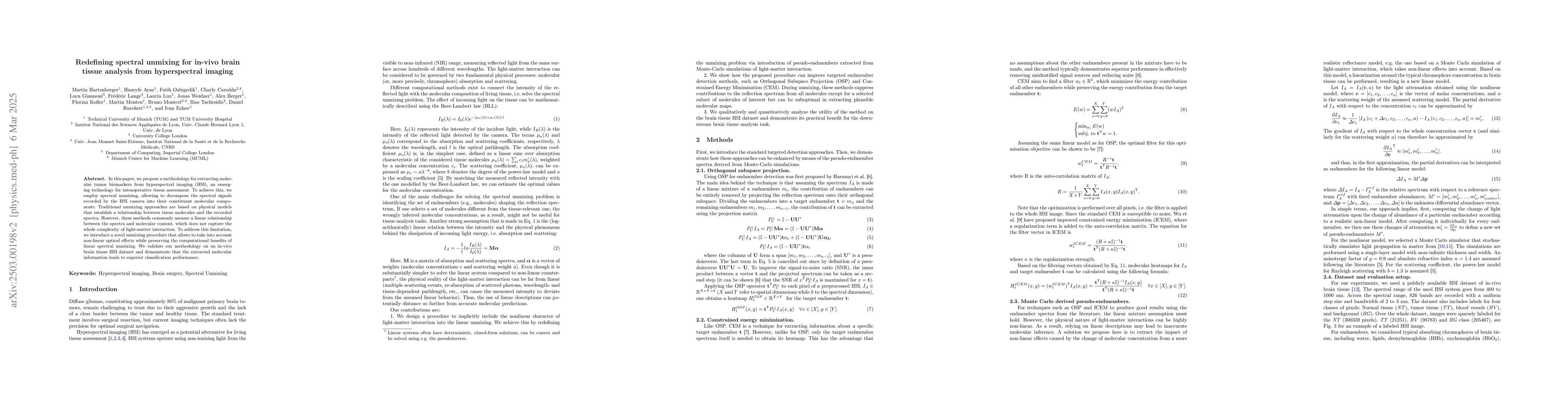
In this paper, we propose a methodology for extracting molecular tumor biomarkers from hyperspectral imaging (HSI), an emerging technology for intraoperative tissue assessment. To achieve this, we emp...
Despite continuous advancements in cancer treatment, brain metastatic disease remains a significant complication of primary cancer and is associated with an unfavorable prognosis. One approach for imp...
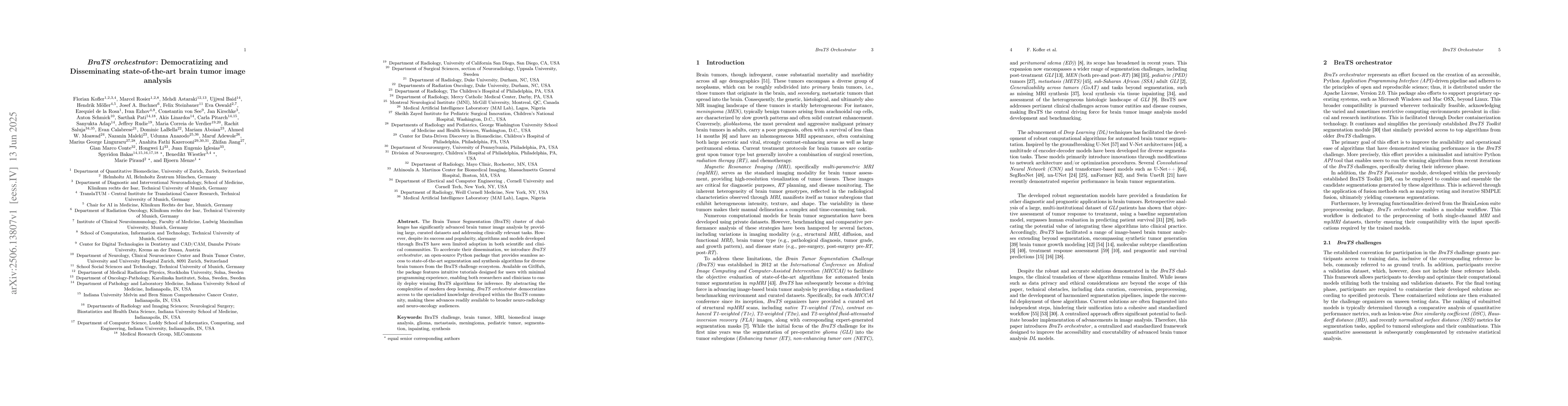
The Brain Tumor Segmentation (BraTS) cluster of challenges has significantly advanced brain tumor image analysis by providing large, curated datasets and addressing clinically relevant tasks. However,...
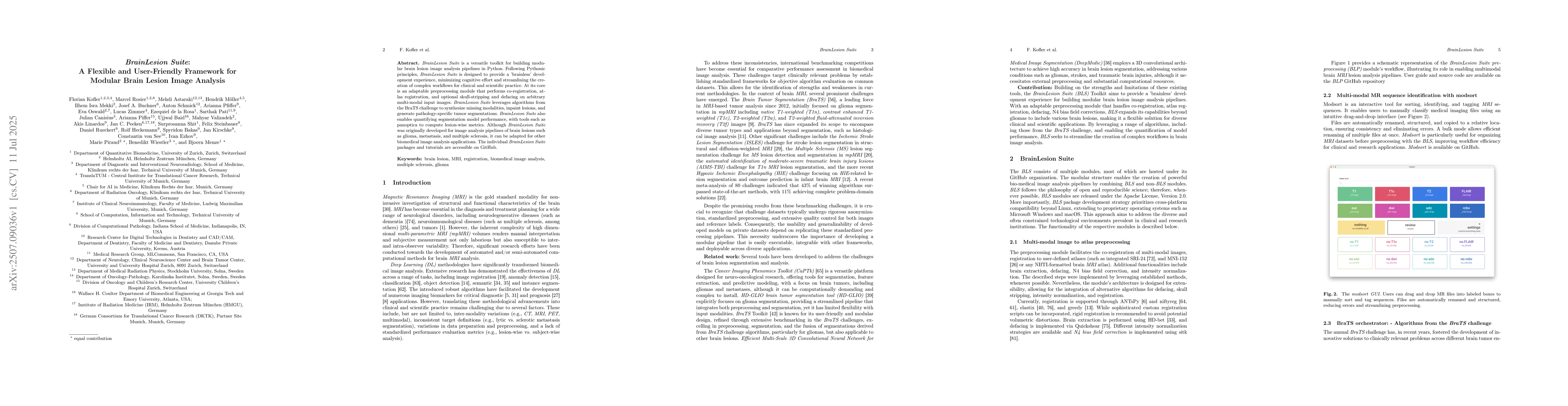
BrainLesion Suite is a versatile toolkit for building modular brain lesion image analysis pipelines in Python. Following Pythonic principles, BrainLesion Suite is designed to provide a 'brainless' dev...
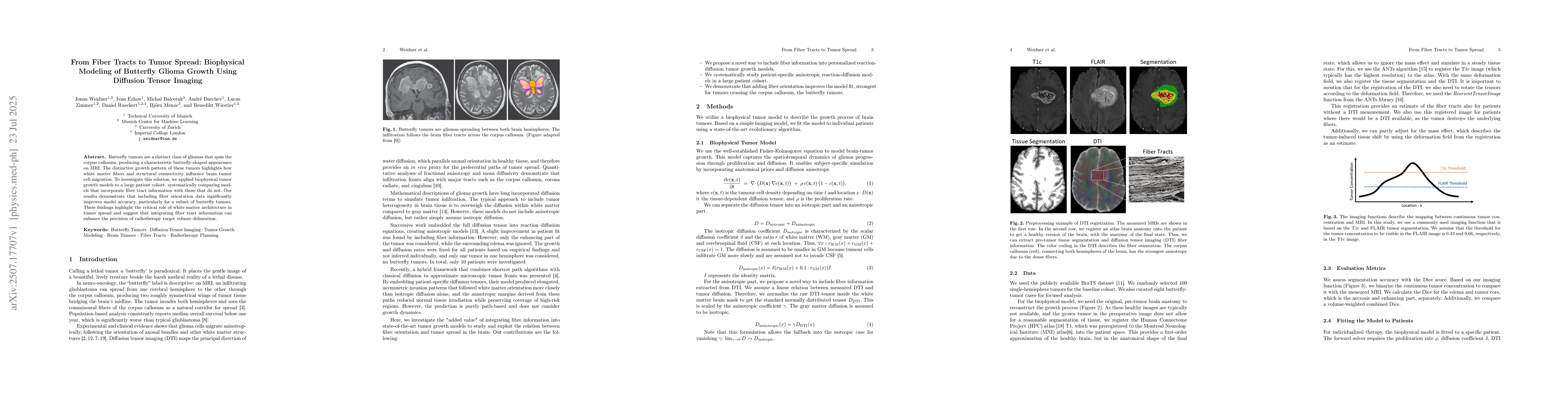
Butterfly tumors are a distinct class of gliomas that span the corpus callosum, producing a characteristic butterfly-shaped appearance on MRI. The distinctive growth pattern of these tumors highlights...
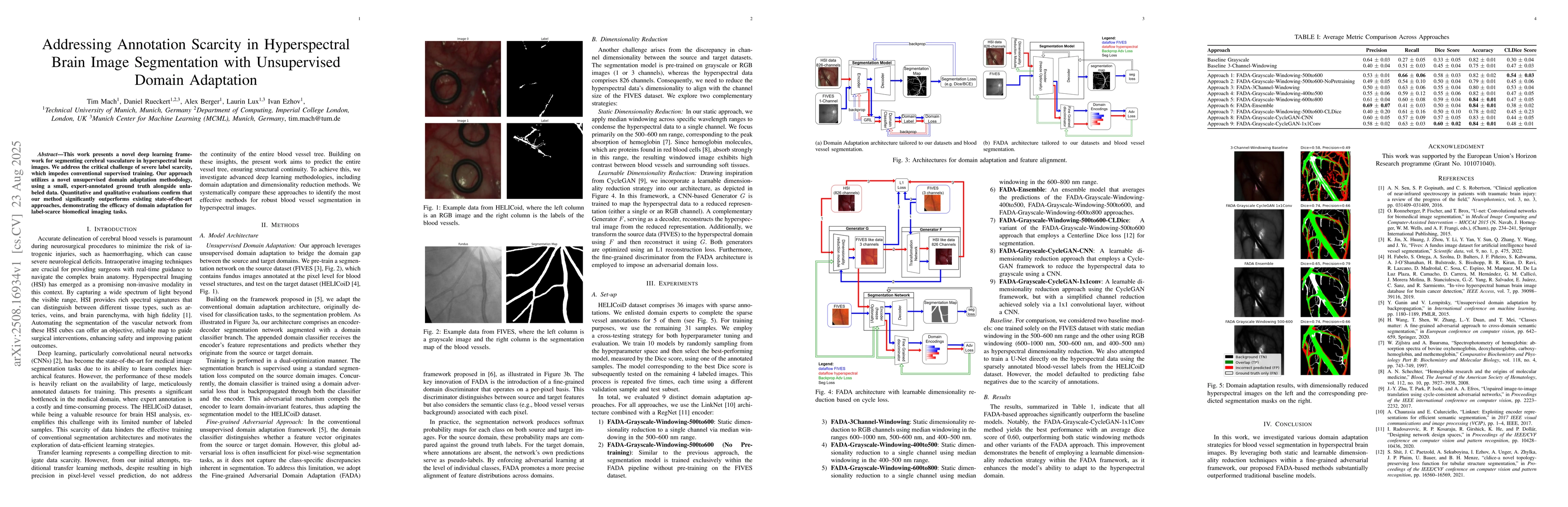
This work presents a novel deep learning framework for segmenting cerebral vasculature in hyperspectral brain images. We address the critical challenge of severe label scarcity, which impedes conventi...
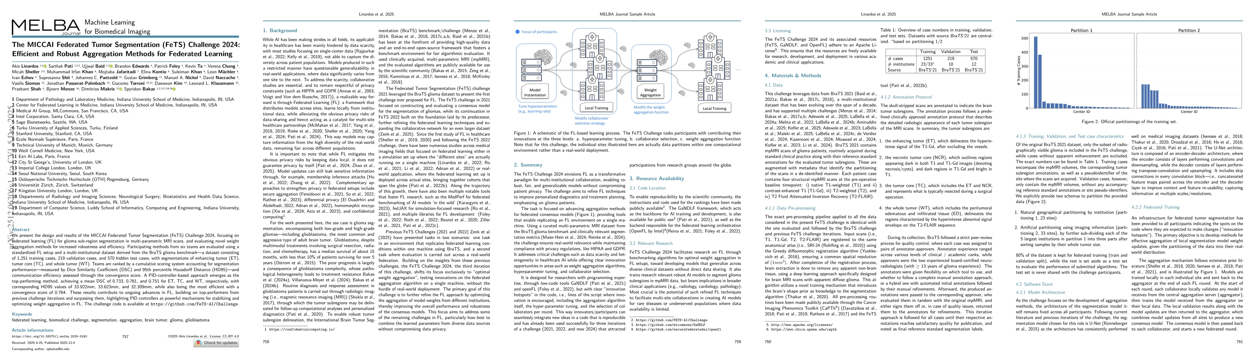
We present the design and results of the MICCAI Federated Tumor Segmentation (FeTS) Challenge 2024, which focuses on federated learning (FL) for glioma sub-region segmentation in multi-parametric MRI ...
Critical breakthroughs in the area of biomedicine and materials science increasingly depend on rapid, non-contact methods for viscoelastic characterization. Laser Speckle Rheology (LSR) is positioned ...
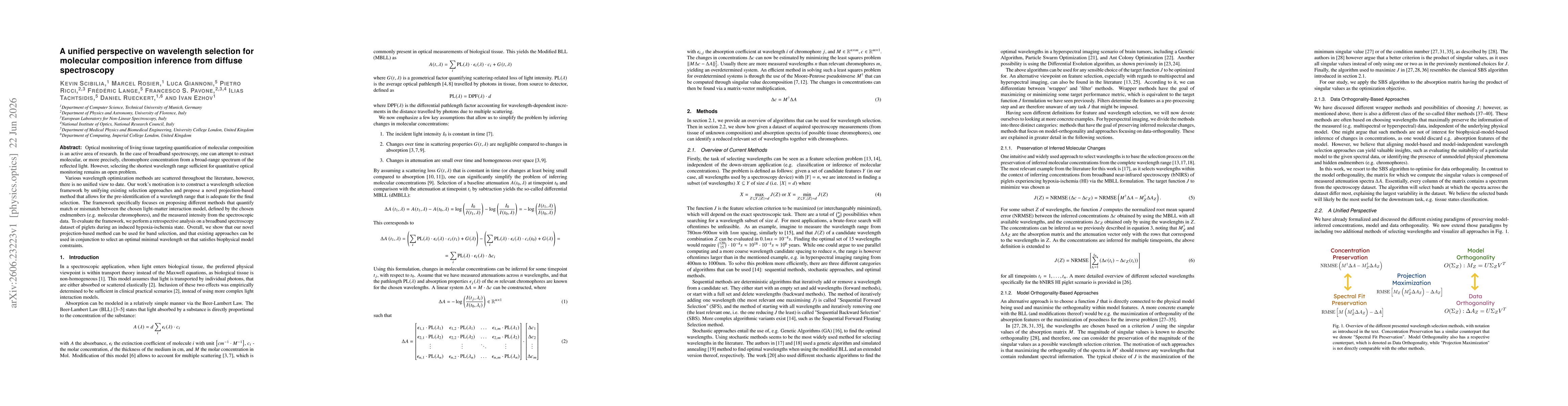
Optical monitoring of living tissue targeting quantification of molecular composition is an active area of research. In the case of broadband spectroscopy, one can attempt to extract molecular, or mor...