Automated analysis of diabetic retinopathy using vessel segmentation maps as inductive bias
Publication
Metrics
AI Quick Summary
This paper presents an automated method for diabetic retinopathy grading using optical coherence tomography angiography (OCTA) images, leveraging vessel segmentation maps as an inductive bias. The approach combines OCTA scans with their segmentations to train networks for lesion segmentation, image quality assessment, and DR grading, achieving performance comparable to the baseline model in the MICCAI 2022 DR analysis challenge.
Paper Preview
Abstract
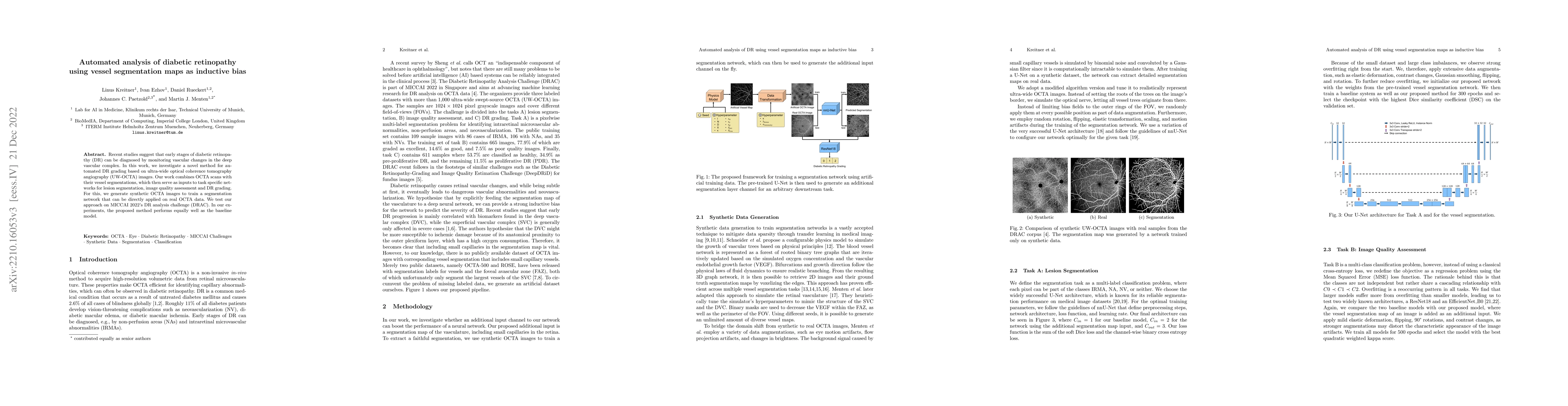
Recent studies suggest that early stages of diabetic retinopathy (DR) can be diagnosed by monitoring vascular changes in the deep vascular complex. In this work, we investigate a novel method for automated DR grading based on optical coherence tomography angiography (OCTA) images. Our work combines OCTA scans with their vessel segmentations, which then serve as inputs to task specific networks for lesion segmentation, image quality assessment and DR grading. For this, we generate synthetic OCTA images to train a segmentation network that can be directly applied on real OCTA data. We test our approach on MICCAI 2022's DR analysis challenge (DRAC). In our experiments, the proposed method performs equally well as the baseline model.
AI Key Findings
Get AI-generated insights about this paper's methodology, results, significance, and more — seven facets brought into focus.
Impact
Paper Details
Authors
PDF Preview
Key Terms
Citation Network
Current paper (gray), citations (green), references (blue)
Display is limited for performance on very large graphs.
Discussion 0