Jean-Philippe Thiran
Centre d'optique, photonique et Laser (COPL), and the Département de génie électrique et génie informatique, Université Laval, Québec, QC, Canada
Academic Profile
Statistics
Similar Authors
Papers on arXiv
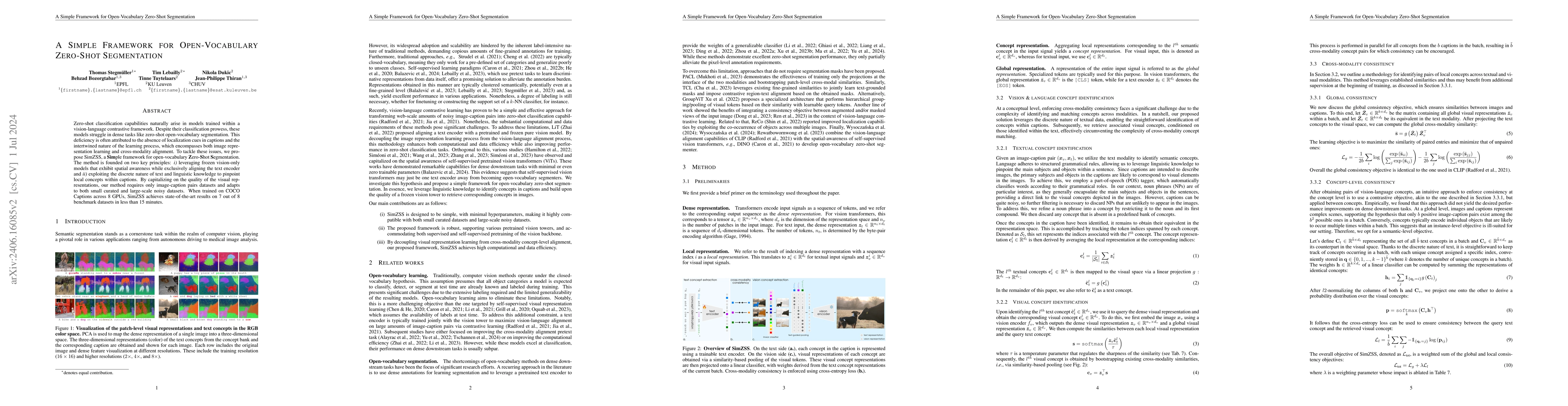
Zero-shot classification capabilities naturally arise in models trained within a vision-language contrastive framework. Despite their classification prowess, these models struggle in dense tasks like ...
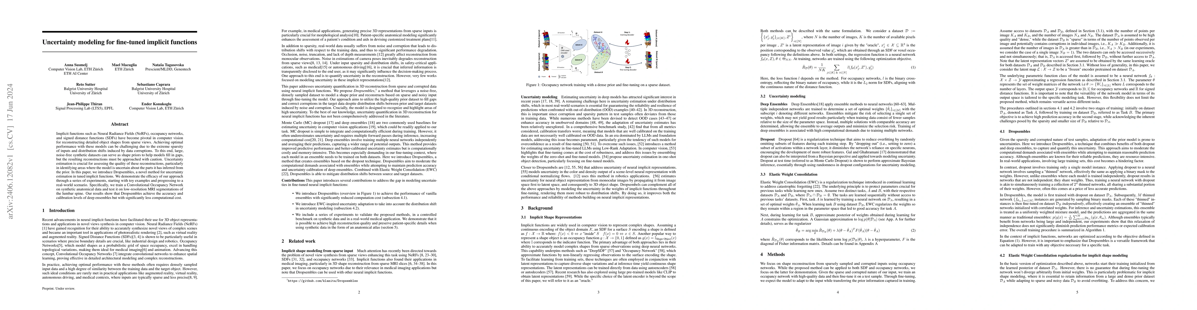
Implicit functions such as Neural Radiance Fields (NeRFs), occupancy networks, and signed distance functions (SDFs) have become pivotal in computer vision for reconstructing detailed object shapes fro...
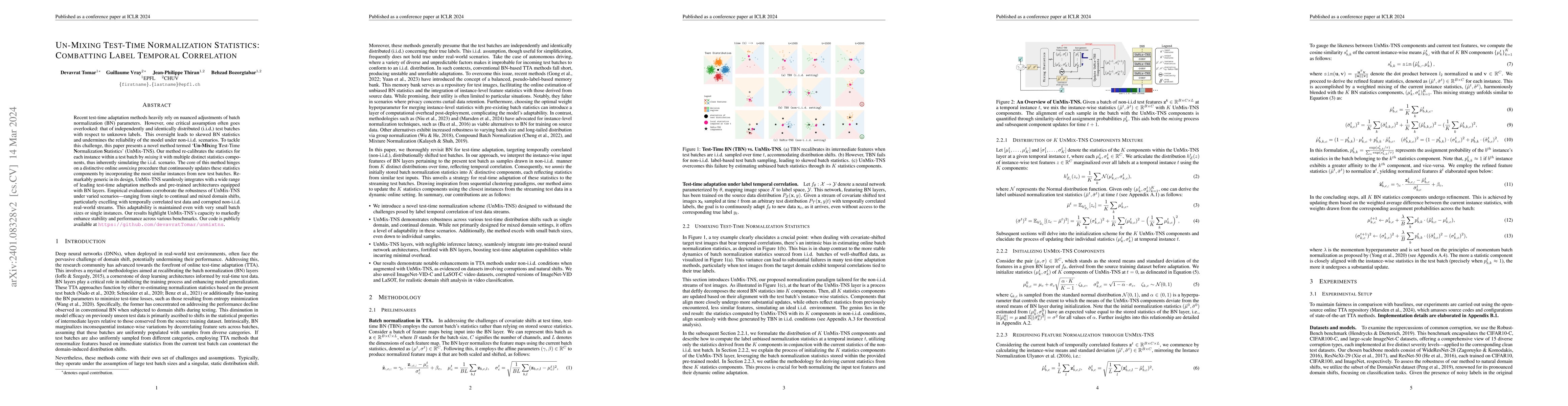
Recent test-time adaptation methods heavily rely on nuanced adjustments of batch normalization (BN) parameters. However, one critical assumption often goes overlooked: that of independently and iden...
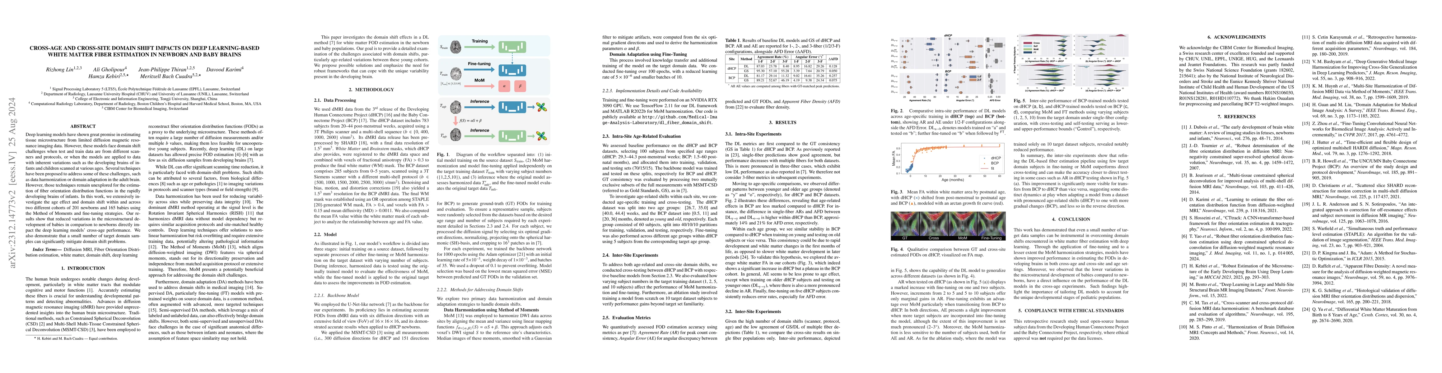
Deep learning models have shown great promise in estimating tissue microstructure from limited diffusion magnetic resonance imaging data. However, these models face domain shift challenges when test...
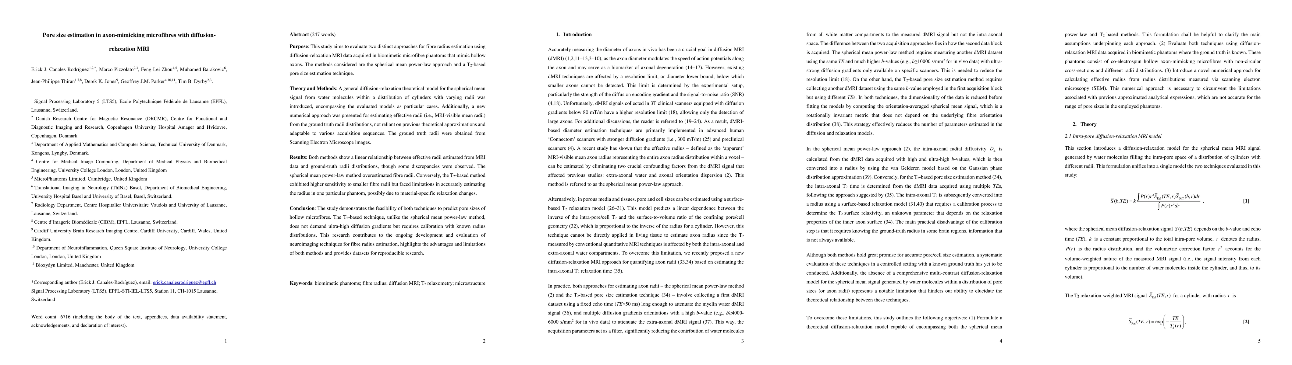
Purpose: This study aims to evaluate two distinct approaches for fibre radius estimation using diffusion-relaxation MRI data acquired in biomimetic microfibre phantoms that mimic hollow axons. The m...
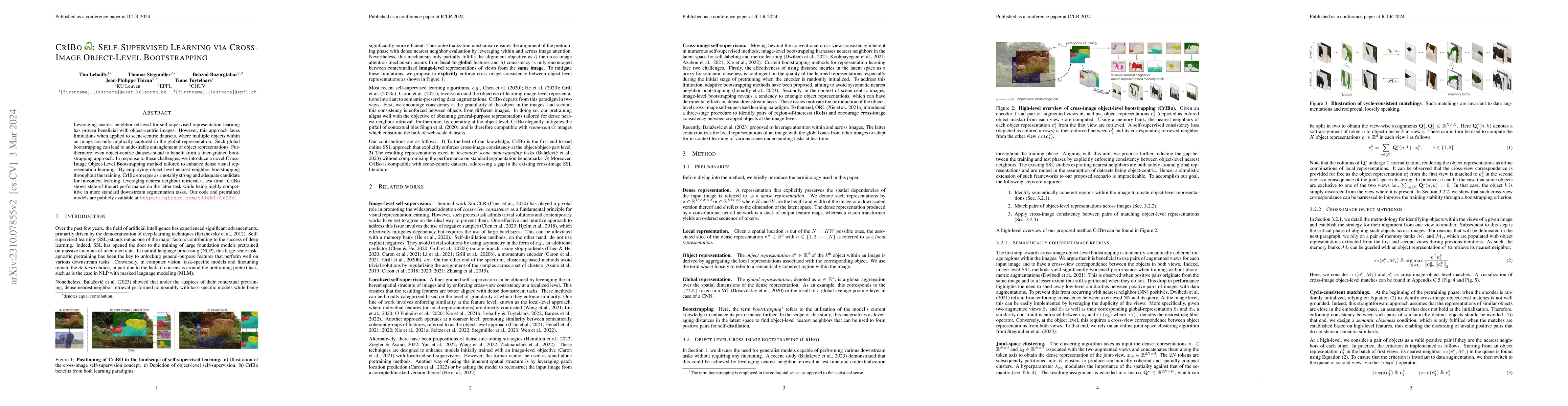
Leveraging nearest neighbor retrieval for self-supervised representation learning has proven beneficial with object-centric images. However, this approach faces limitations when applied to scene-cen...
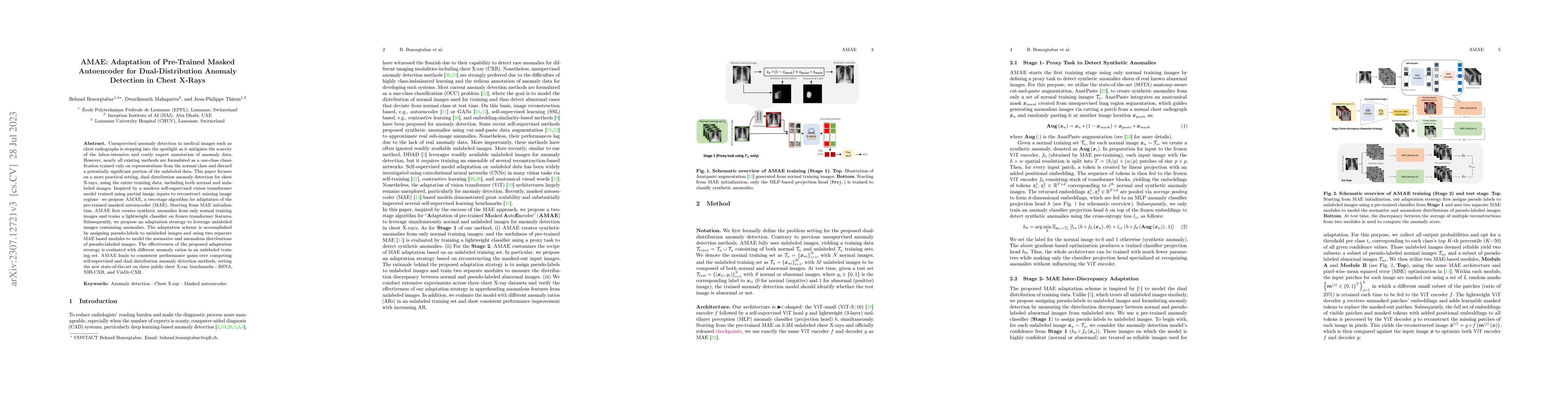
Unsupervised anomaly detection in medical images such as chest radiographs is stepping into the spotlight as it mitigates the scarcity of the labor-intensive and costly expert annotation of anomaly ...
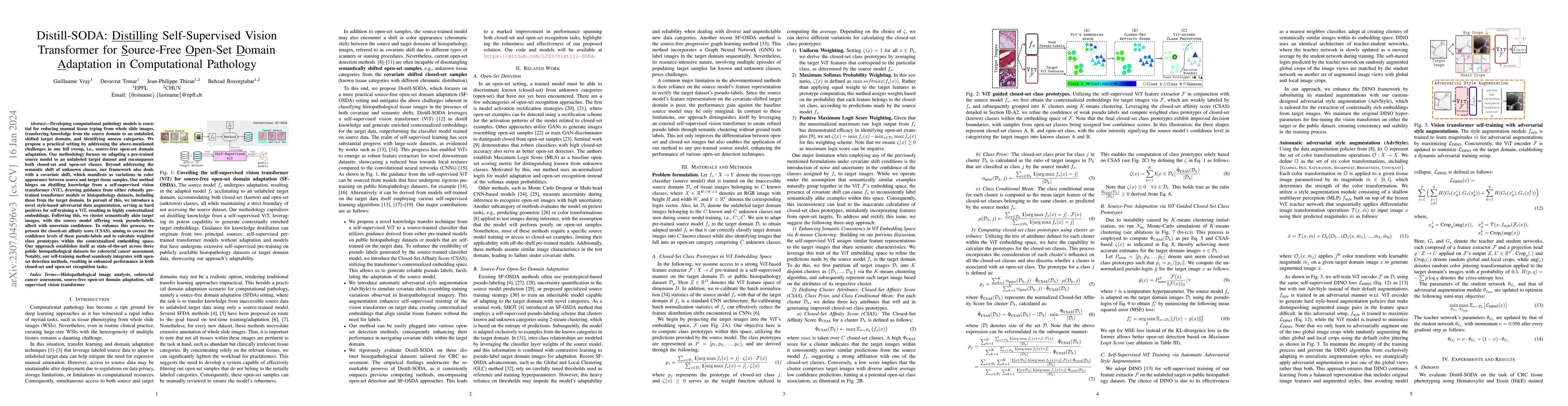
Developing computational pathology models is essential for reducing manual tissue typing from whole slide images, transferring knowledge from the source domain to an unlabeled, shifted target domain...
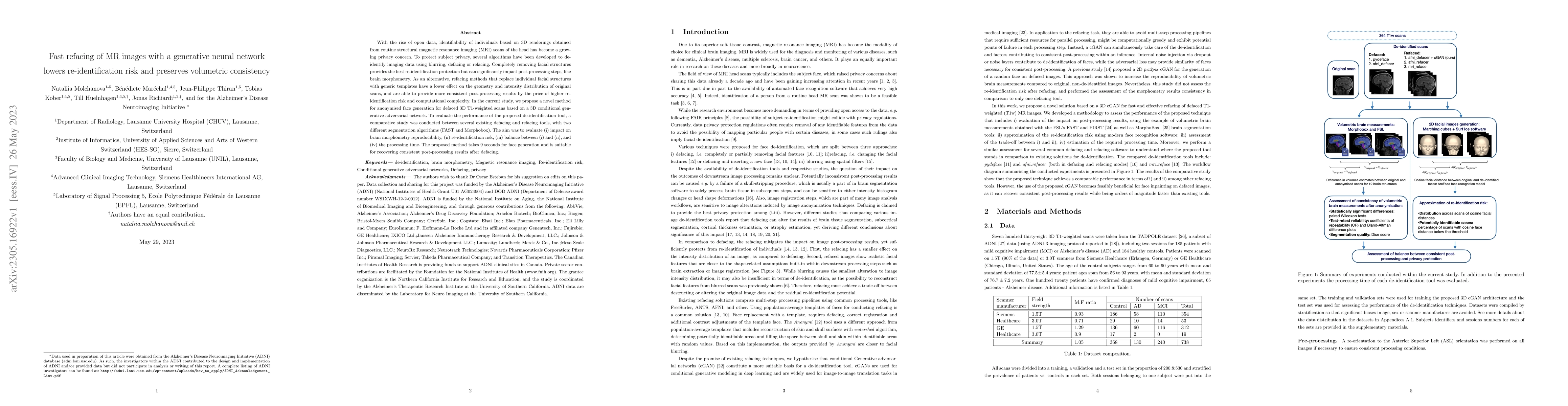
With the rise of open data, identifiability of individuals based on 3D renderings obtained from routine structural magnetic resonance imaging (MRI) scans of the head has become a growing privacy con...
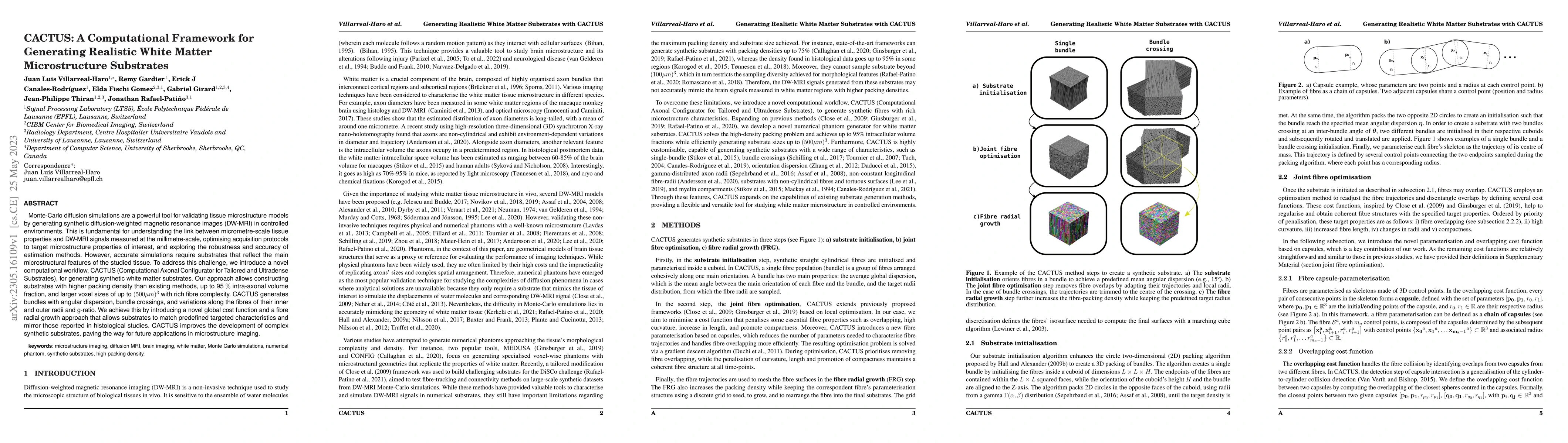
Monte-Carlo diffusion simulations are a powerful tool for validating tissue microstructure models by generating synthetic diffusion-weighted magnetic resonance images (DW-MRI) in controlled environm...
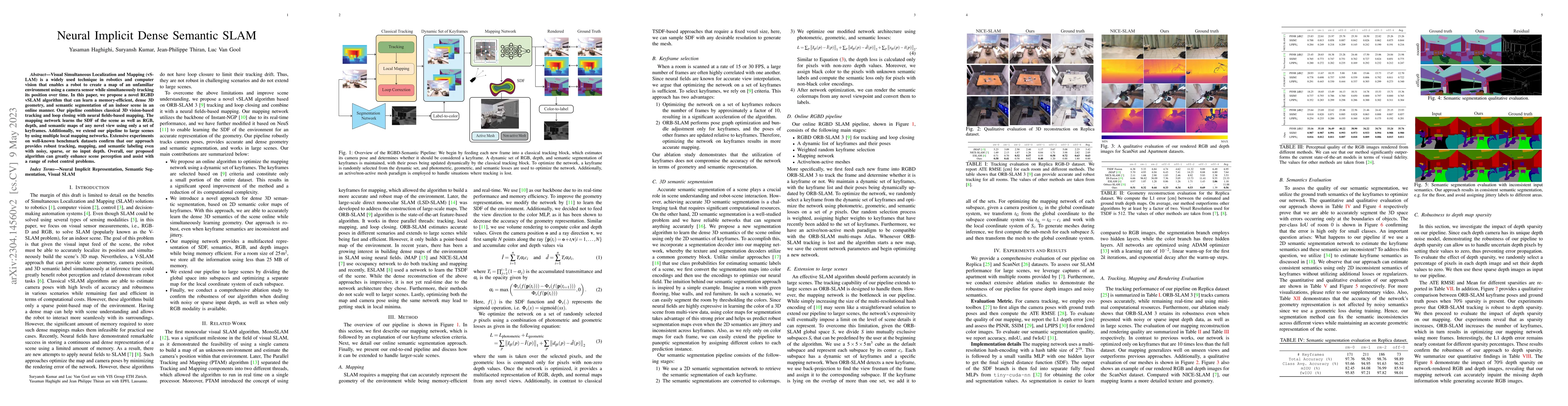
Visual Simultaneous Localization and Mapping (vSLAM) is a widely used technique in robotics and computer vision that enables a robot to create a map of an unfamiliar environment using a camera senso...
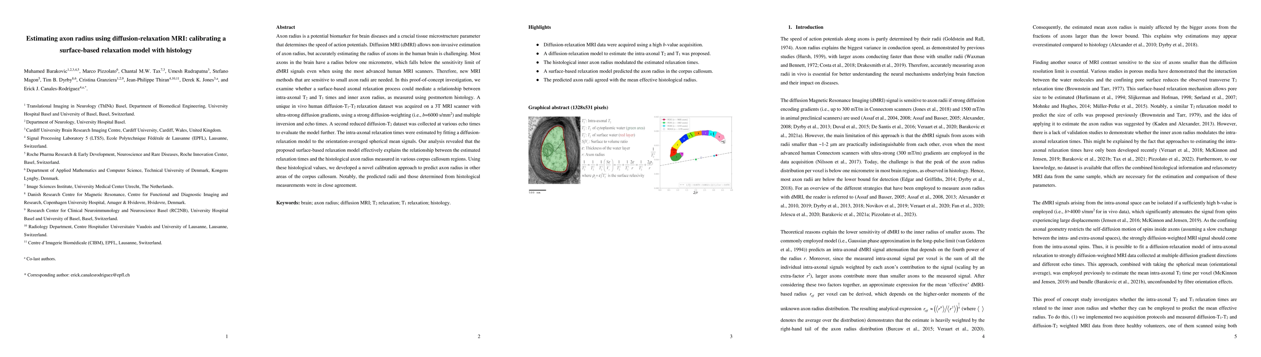
Axon radius is a potential biomarker for brain diseases and a crucial tissue microstructure parameter that determines the speed of action potentials. Diffusion MRI (dMRI) allows non-invasive estimat...
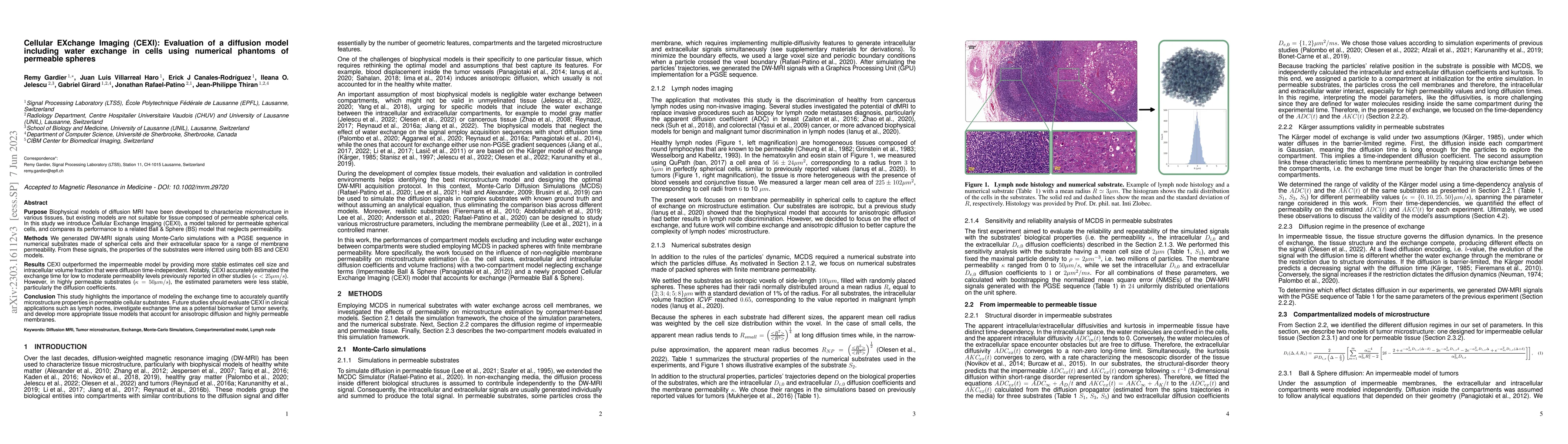
Purpose: Biophysical models of diffusion MRI have been developed to characterize microstructure in various tissues, but existing models are not suitable for tissue composed of permeable spherical ce...
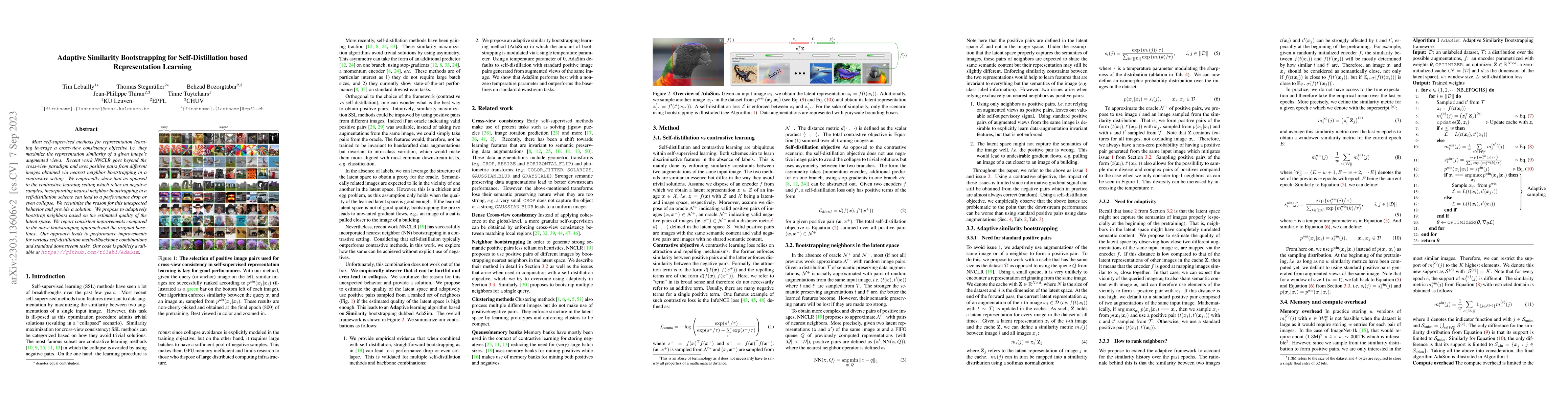
Most self-supervised methods for representation learning leverage a cross-view consistency objective i.e., they maximize the representation similarity of a given image's augmented views. Recent work...
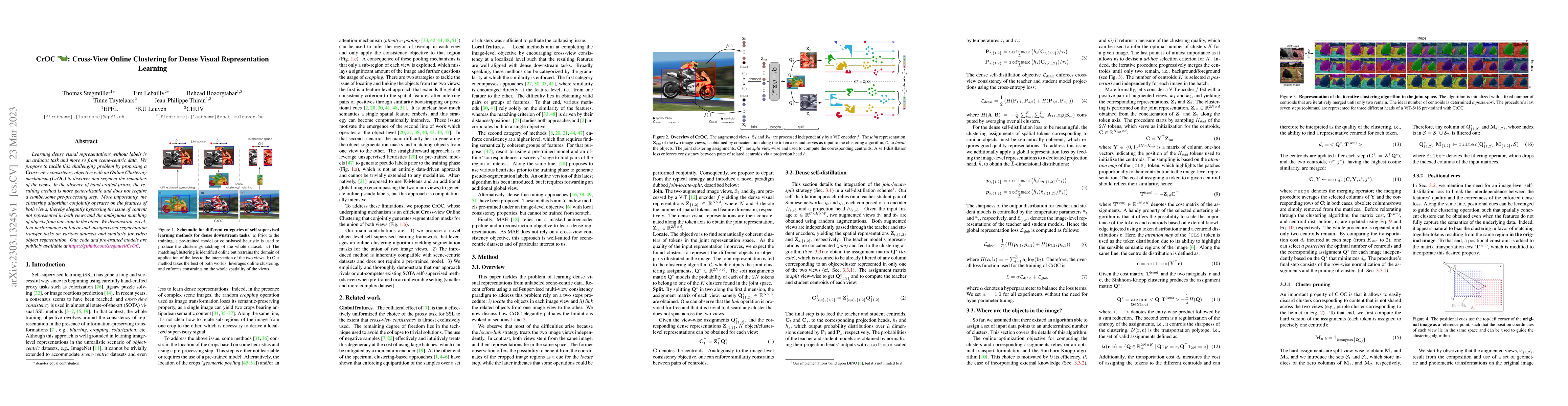
Learning dense visual representations without labels is an arduous task and more so from scene-centric data. We propose to tackle this challenging problem by proposing a Cross-view consistency objec...
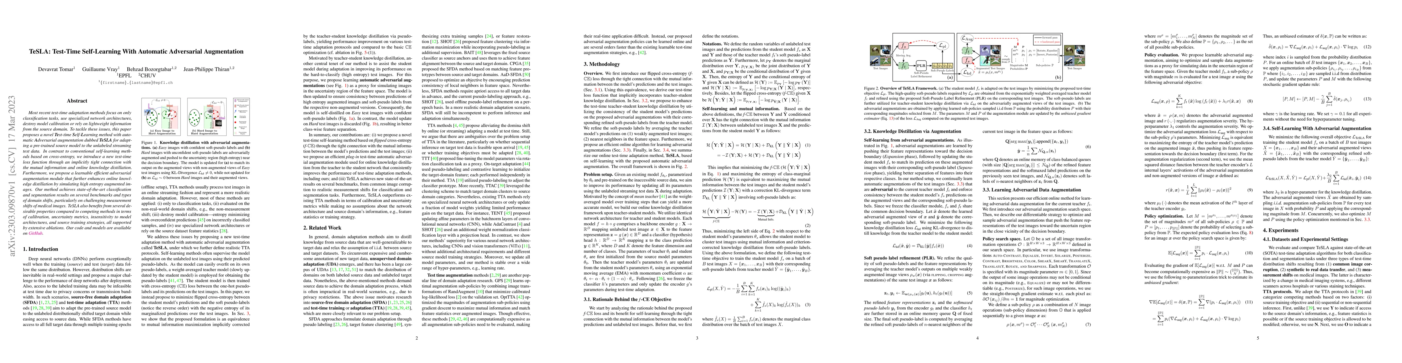
Most recent test-time adaptation methods focus on only classification tasks, use specialized network architectures, destroy model calibration or rely on lightweight information from the source domai...
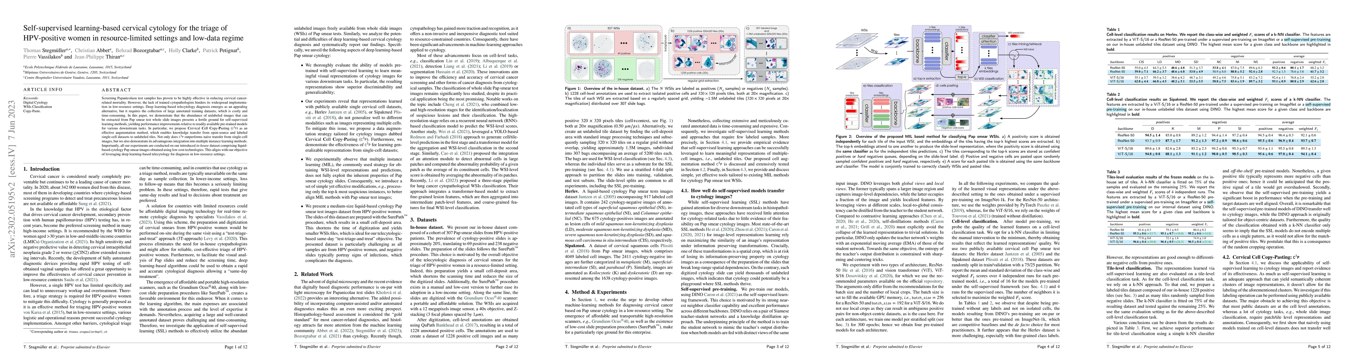
Screening Papanicolaou test samples has proven to be highly effective in reducing cervical cancer-related mortality. However, the lack of trained cytopathologists hinders its widespread implementati...
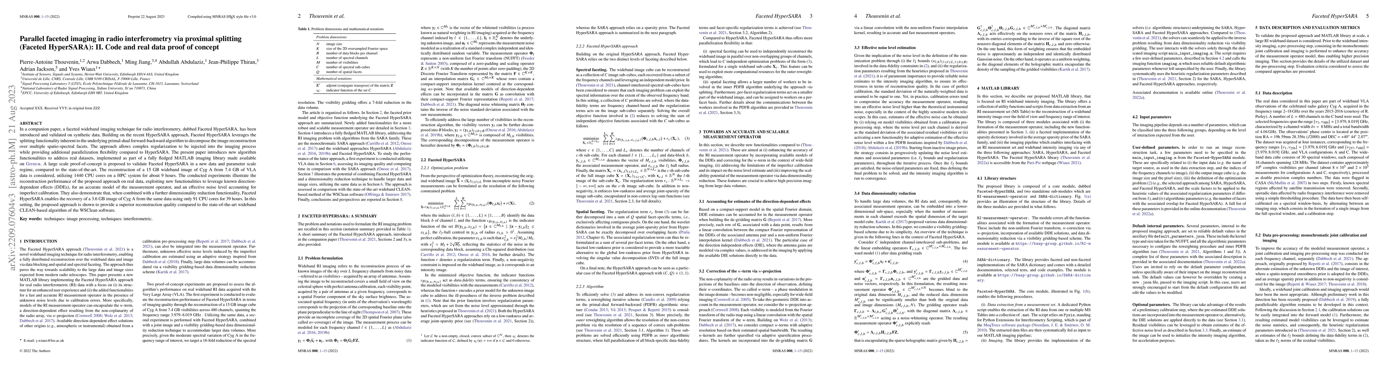
In a companion paper, a faceted wideband imaging technique for radio interferometry, dubbed Faceted HyperSARA, has been introduced and validated on synthetic data. Building on the recent HyperSARA a...
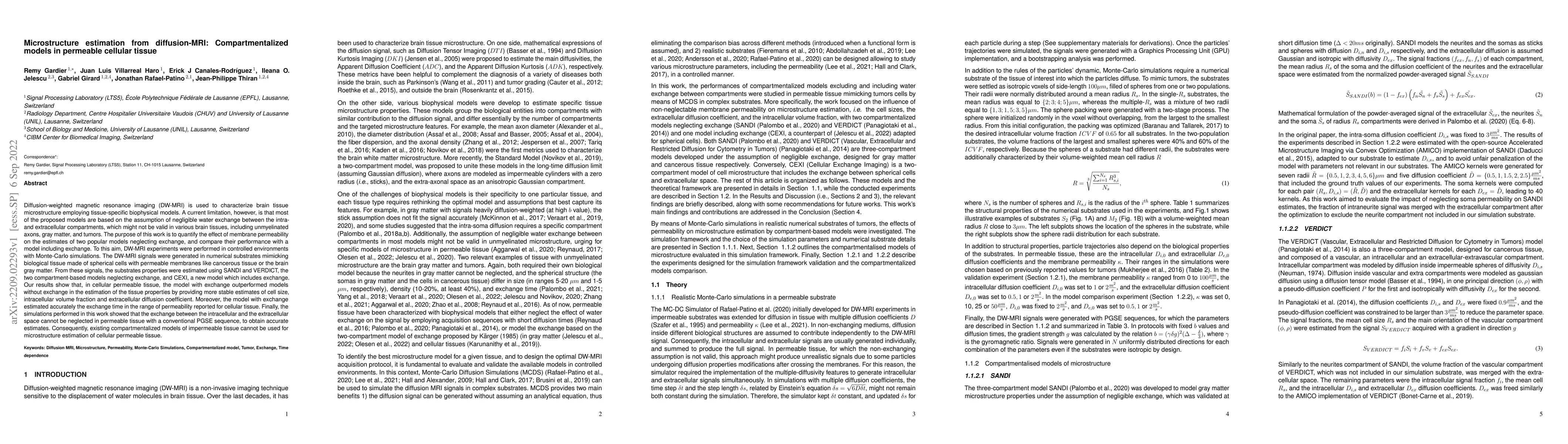
Diffusion-weighted magnetic resonance imaging (DW-MRI) is used to characterize brain tissue microstructure employing tissue-specific biophysical models. A current limitation, however, is that most o...
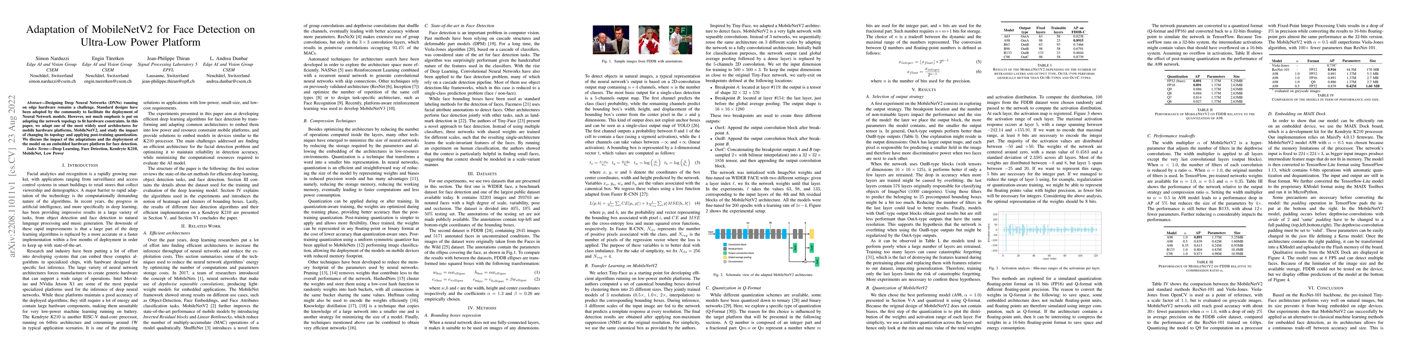
Designing Deep Neural Networks (DNNs) running on edge hardware remains a challenge. Standard designs have been adopted by the community to facilitate the deployment of Neural Network models. However...
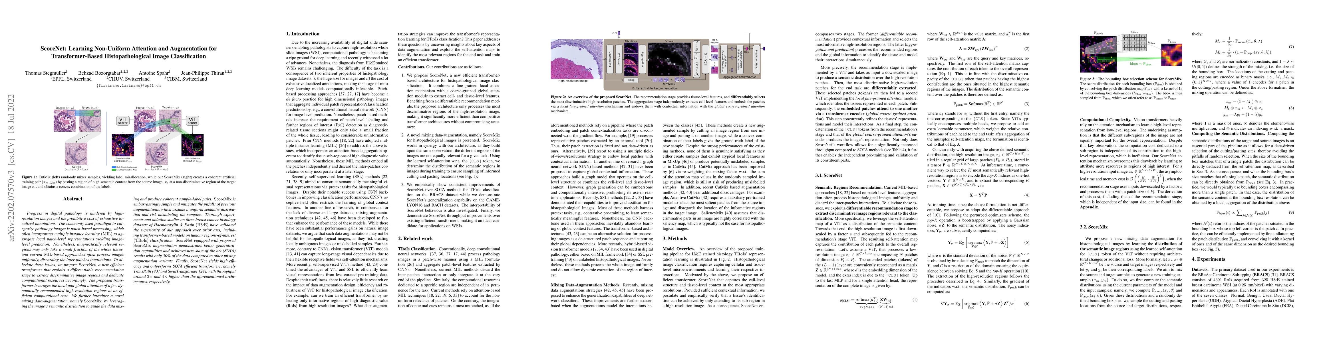
Progress in digital pathology is hindered by high-resolution images and the prohibitive cost of exhaustive localized annotations. The commonly used paradigm to categorize pathology images is patch-b...
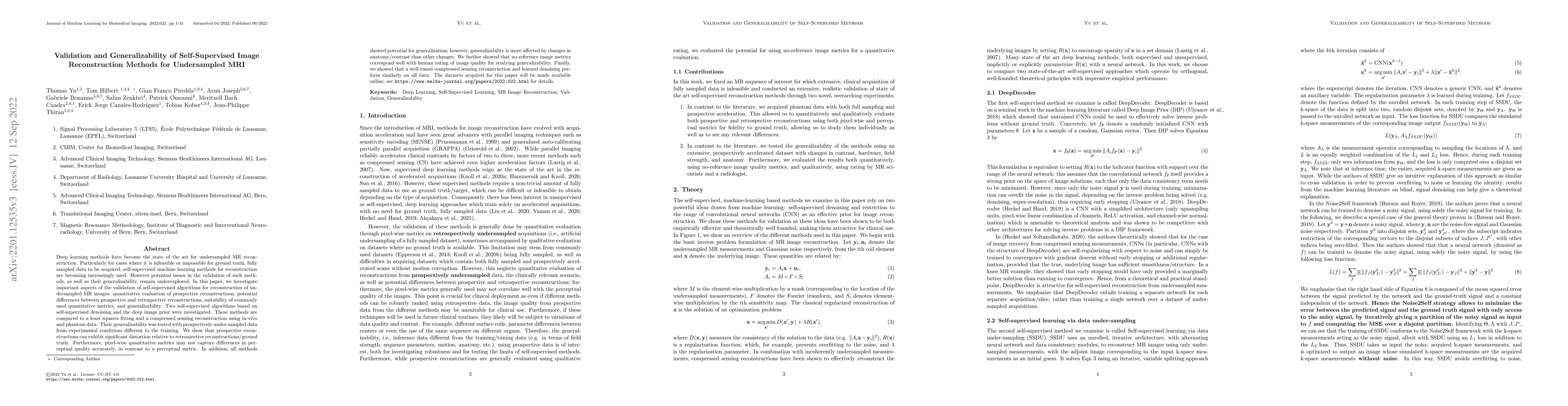
Deep learning methods have become the state of the art for undersampled MR reconstruction. Particularly for cases where it is infeasible or impossible for ground truth, fully sampled data to be acqu...
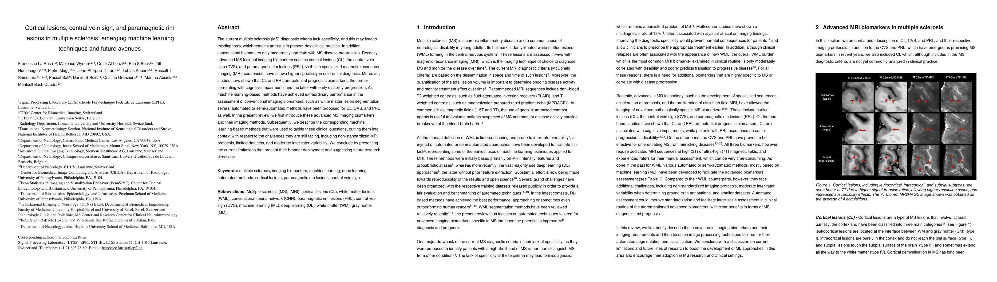
The current multiple sclerosis (MS) diagnostic criteria lack specificity, and this may lead to misdiagnosis, which remains an issue in present-day clinical practice. In addition, conventional biomar...
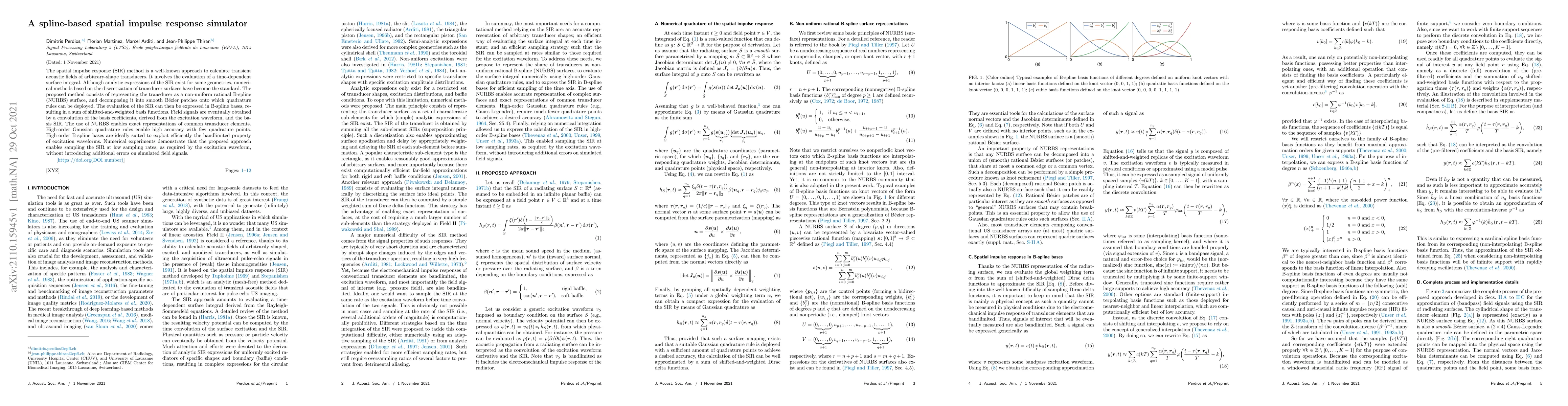
The spatial impulse response (SIR) method is a well-known approach to calculate transient acoustic fields of arbitrary-shape transducers. It involves the evaluation of a time-dependent surface integ...
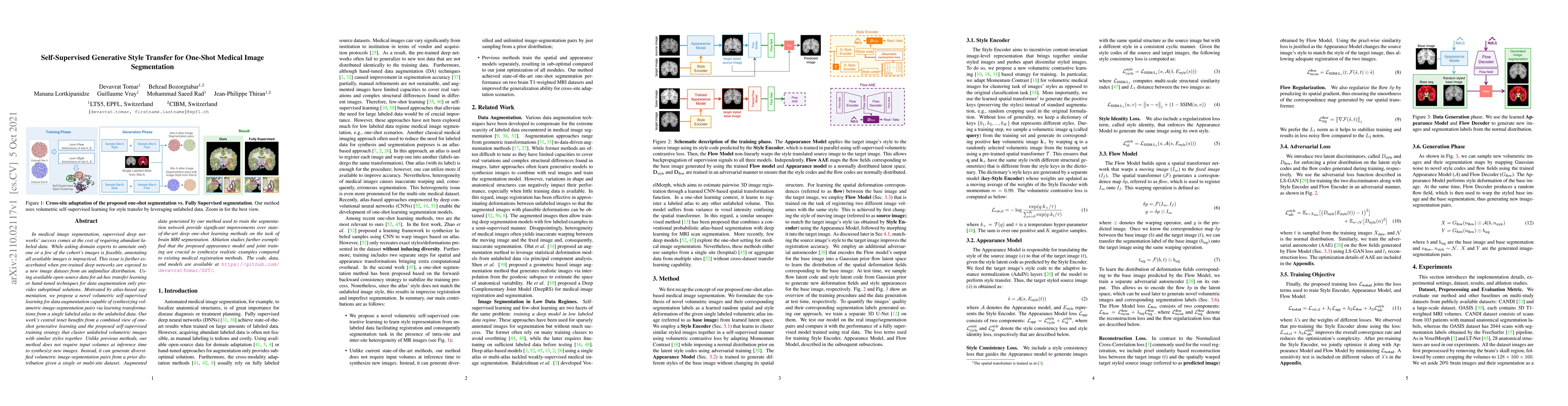
In medical image segmentation, supervised deep networks' success comes at the cost of requiring abundant labeled data. While asking domain experts to annotate only one or a few of the cohort's image...
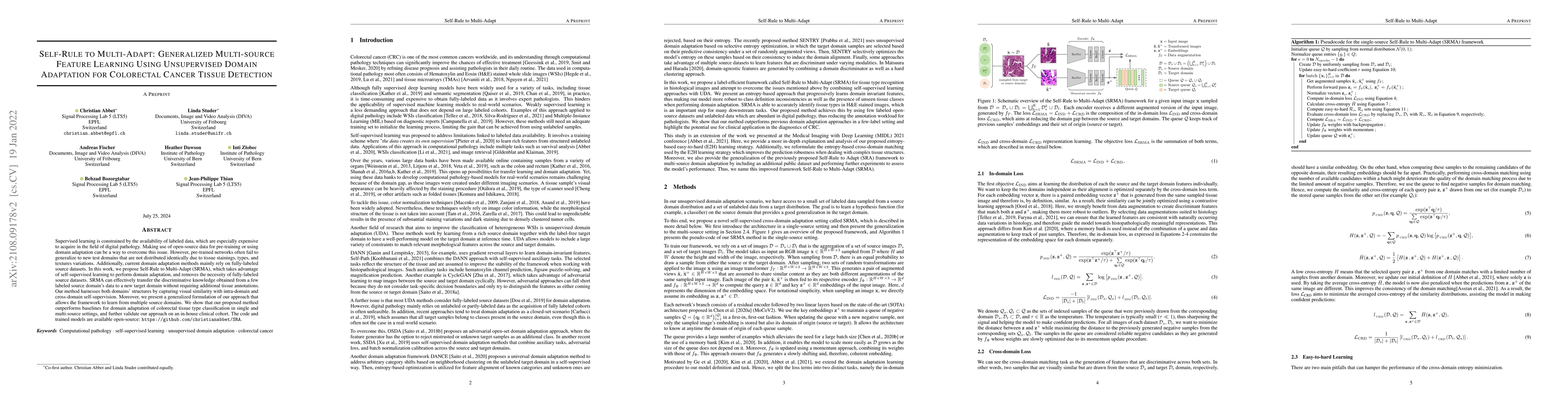
Supervised learning is constrained by the availability of labeled data, which are especially expensive to acquire in the field of digital pathology. Making use of open-source data for pre-training o...
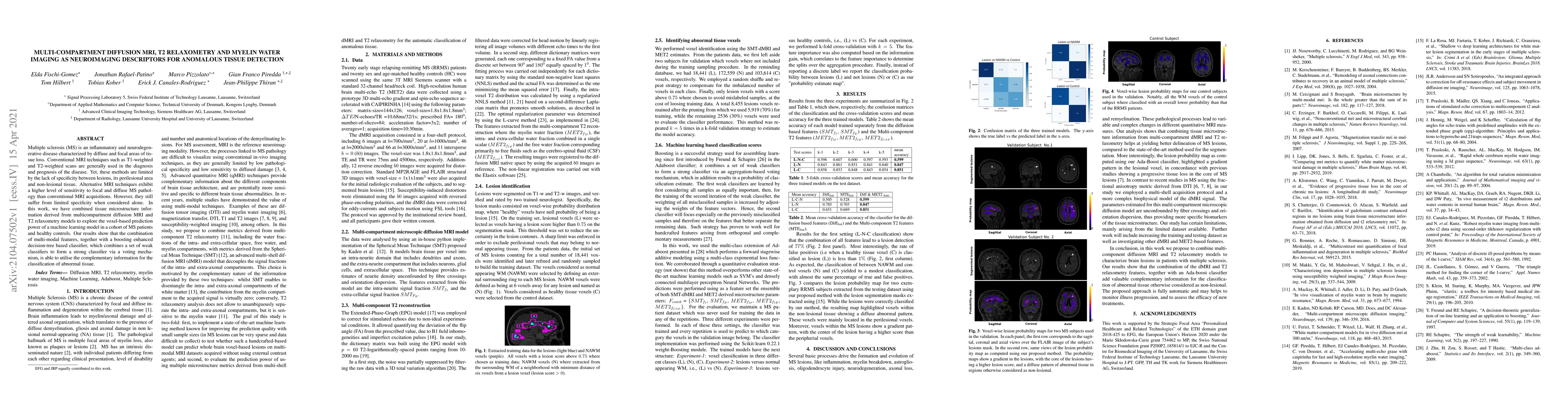
Multiple sclerosis (MS) is an inflammatory and neurodegenerative disease characterized by diffuse and focal areas of tissue loss. Conventional MRI techniques such as T1-weighted and T2-weighted scan...
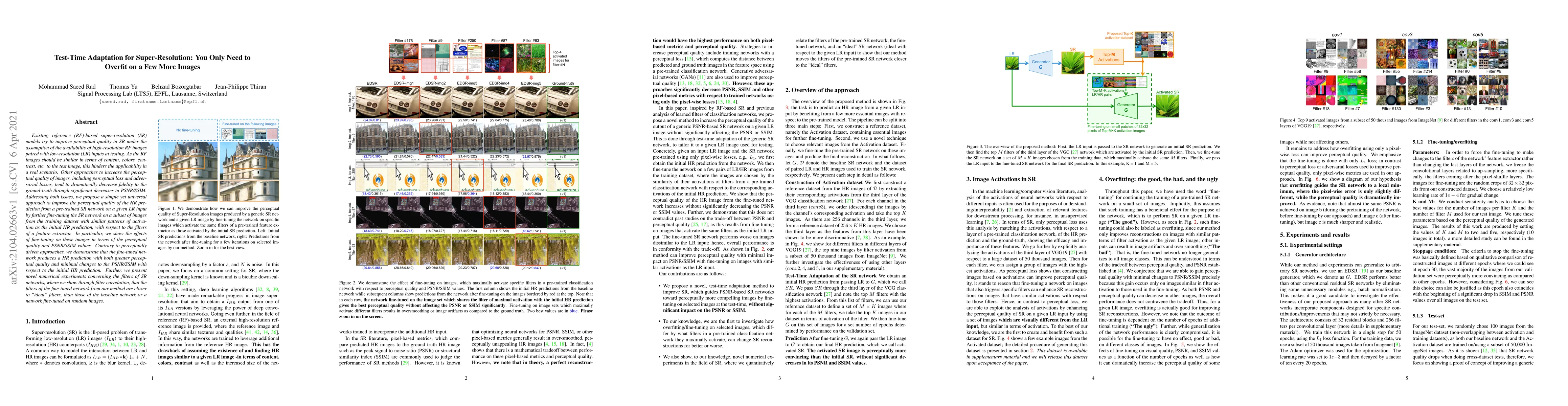
Existing reference (RF)-based super-resolution (SR) models try to improve perceptual quality in SR under the assumption of the availability of high-resolution RF images paired with low-resolution (L...
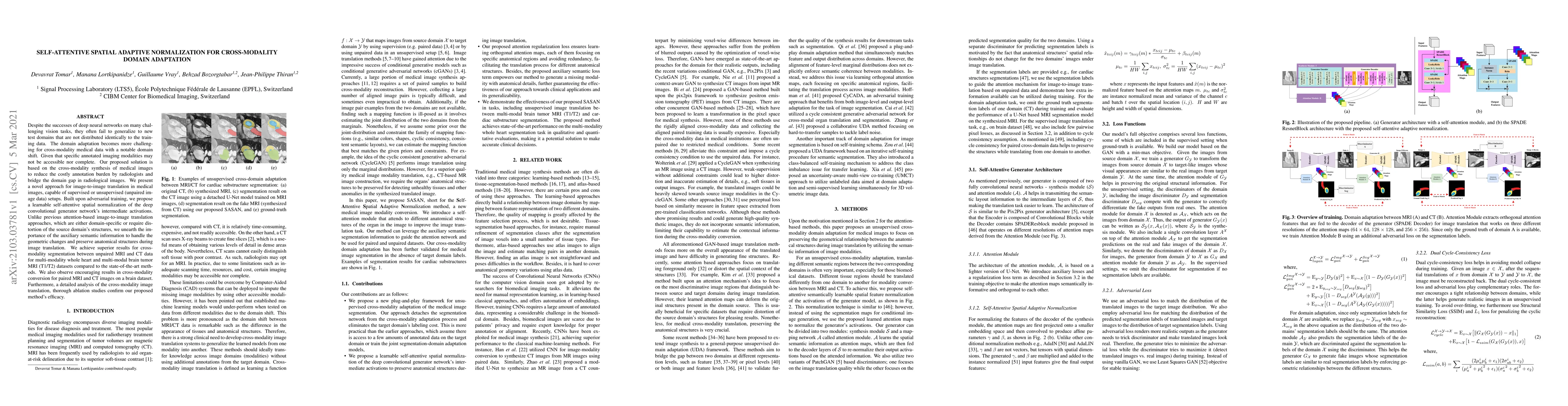
Despite the successes of deep neural networks on many challenging vision tasks, they often fail to generalize to new test domains that are not distributed identically to the training data. The domai...
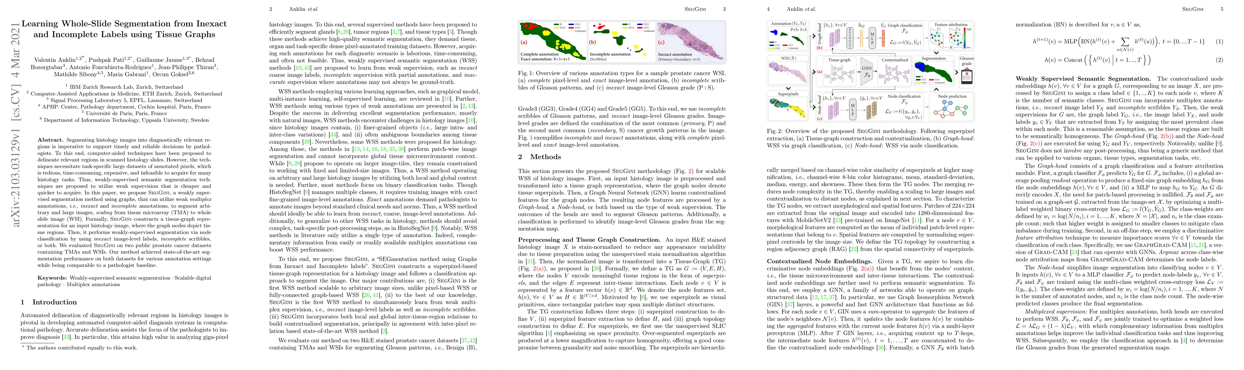
Segmenting histology images into diagnostically relevant regions is imperative to support timely and reliable decisions by pathologists. To this end, computer-aided techniques have been proposed to ...
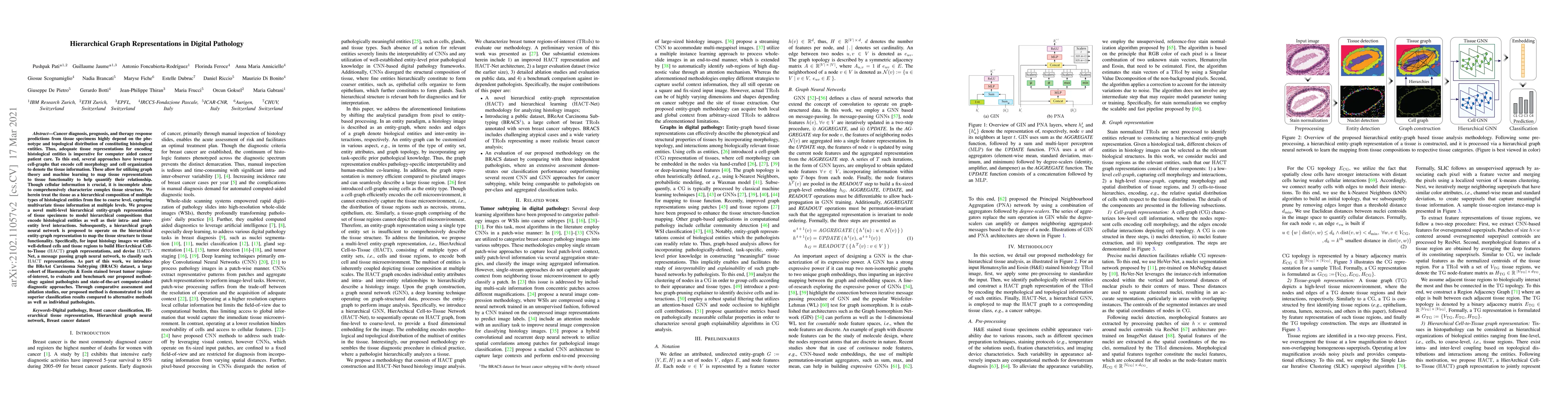
Cancer diagnosis, prognosis, and therapy response predictions from tissue specimens highly depend on the phenotype and topological distribution of constituting histological entities. Thus, adequate ...
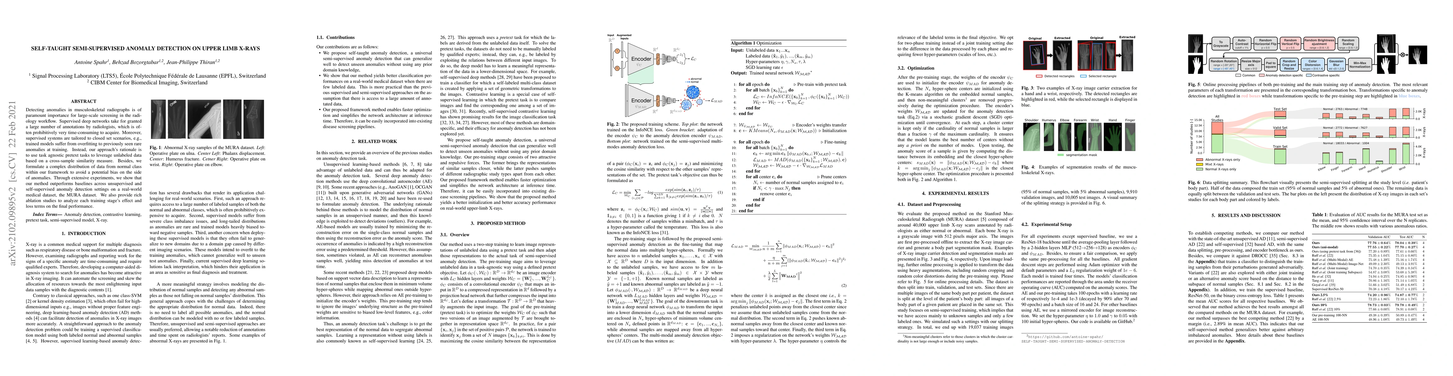
Detecting anomalies in musculoskeletal radiographs is of paramount importance for large-scale screening in the radiology workflow. Supervised deep networks take for granted a large number of annotat...
This paper describes the development of a novel medical Xray imaging system adapted to the needs and constraints of low and middle income countries. The developed system is based on an indirect conv...
Deep anomaly detection models using a supervised mode of learning usually work under a closed set assumption and suffer from overfitting to previously seen rare anomalies at training, which hinders ...
Diffusion-weighted MRI (DW-MRI) has recently seen a rising interest in planar, spherical and general B-tensor encodings. Some of these sequences have aided traditional linear encoding in the estimat...
Ultrafast ultrasound (US) revolutionized biomedical imaging with its capability of acquiring full-view frames at over 1 kHz, unlocking breakthrough modalities such as shear-wave elastography and fun...
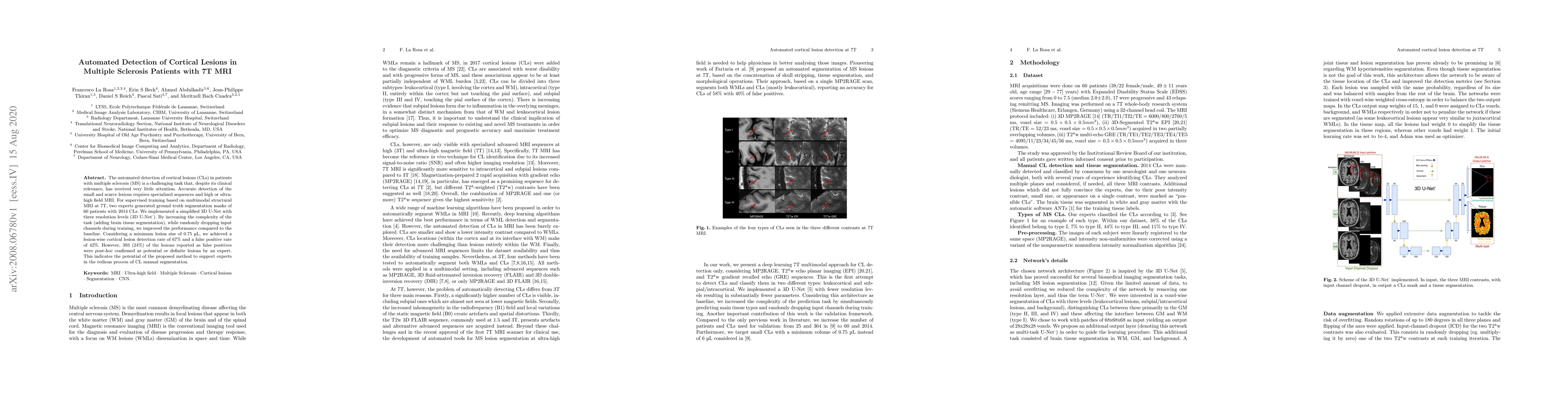
The automated detection of cortical lesions (CLs) in patients with multiple sclerosis (MS) is a challenging task that, despite its clinical relevance, has received very little attention. Accurate de...
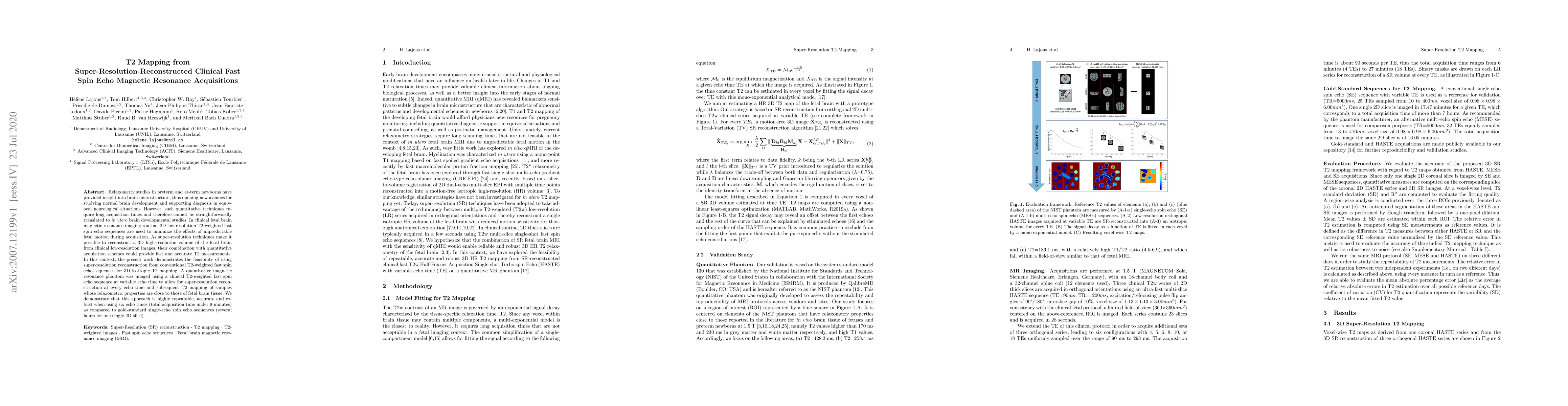
Relaxometry studies in preterm and at-term newborns have provided insight into brain microstructure, thus opening new avenues for studying normal brain development and supporting diagnosis in equivo...
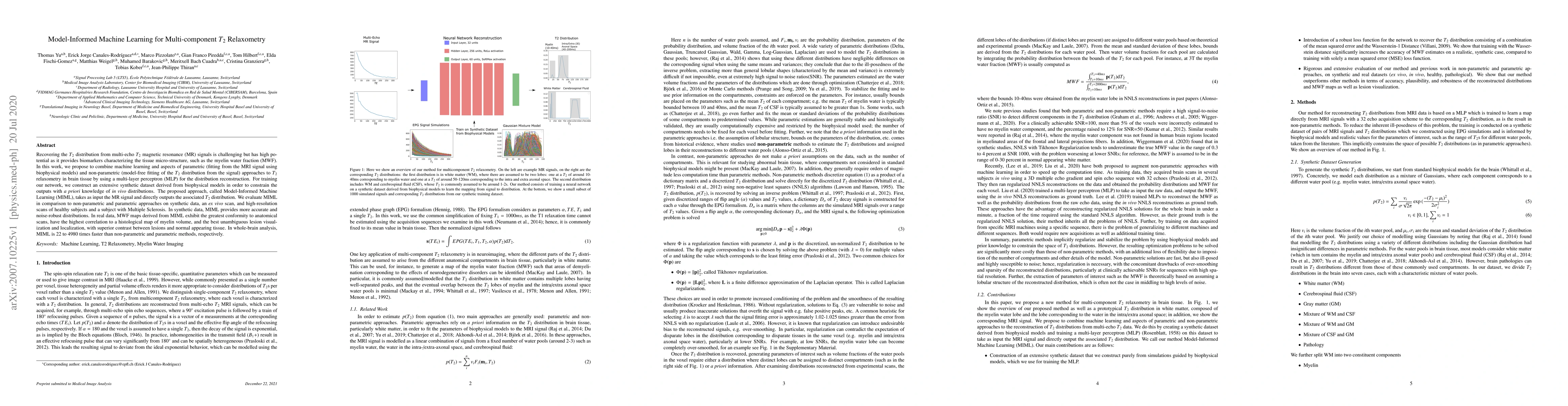
Recovering the T2 distribution from multi-echo T2 magnetic resonance (MR) signals is challenging but has high potential as it provides biomarkers characterizing the tissue micro-structure, such as t...
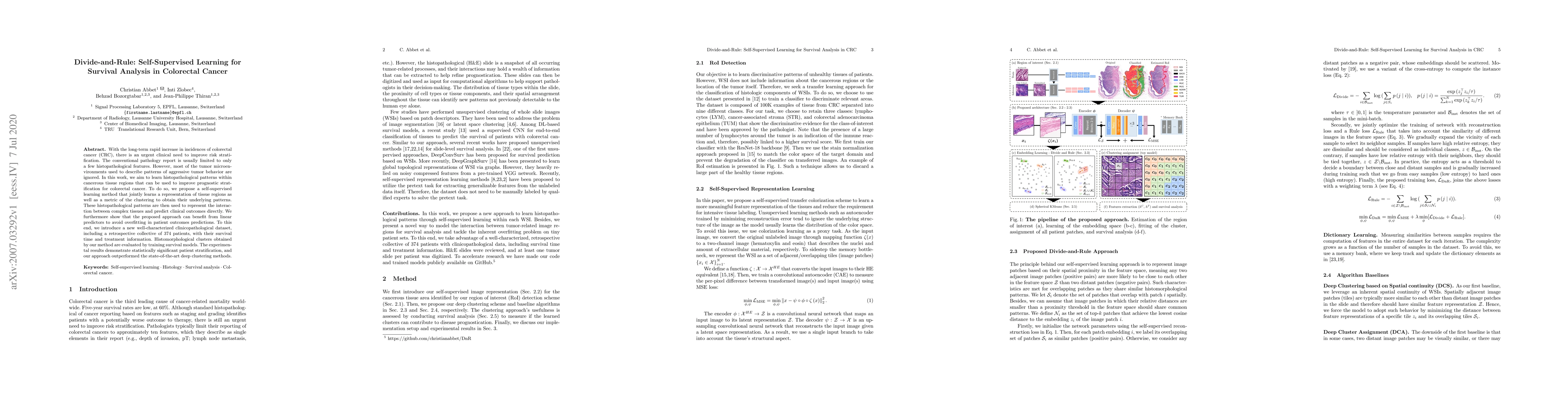
With the long-term rapid increase in incidences of colorectal cancer (CRC), there is an urgent clinical need to improve risk stratification. The conventional pathology report is usually limited to o...
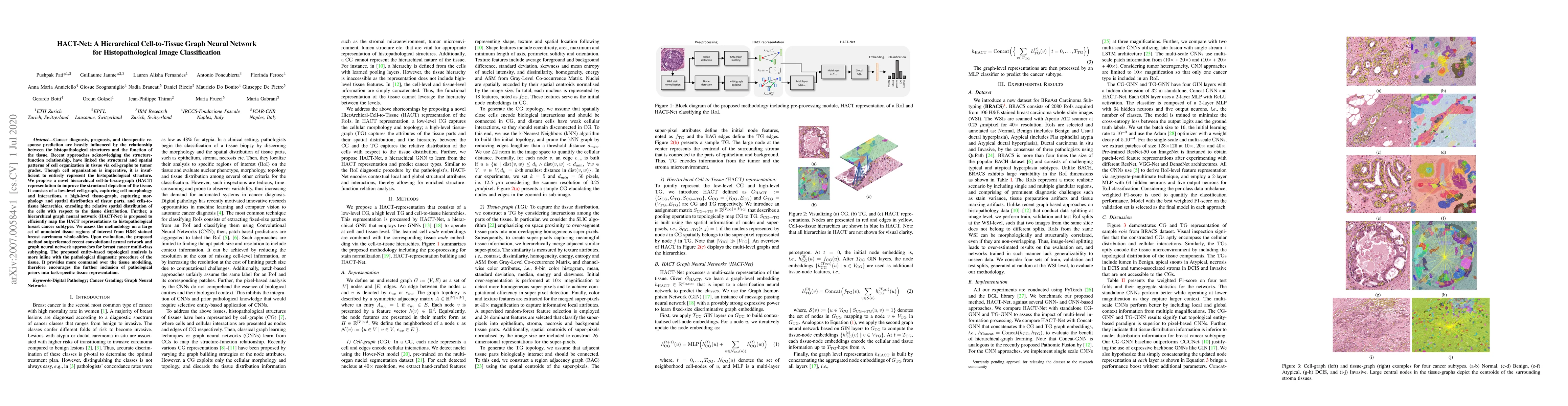
Cancer diagnosis, prognosis, and therapeutic response prediction are heavily influenced by the relationship between the histopathological structures and the function of the tissue. Recent approaches...
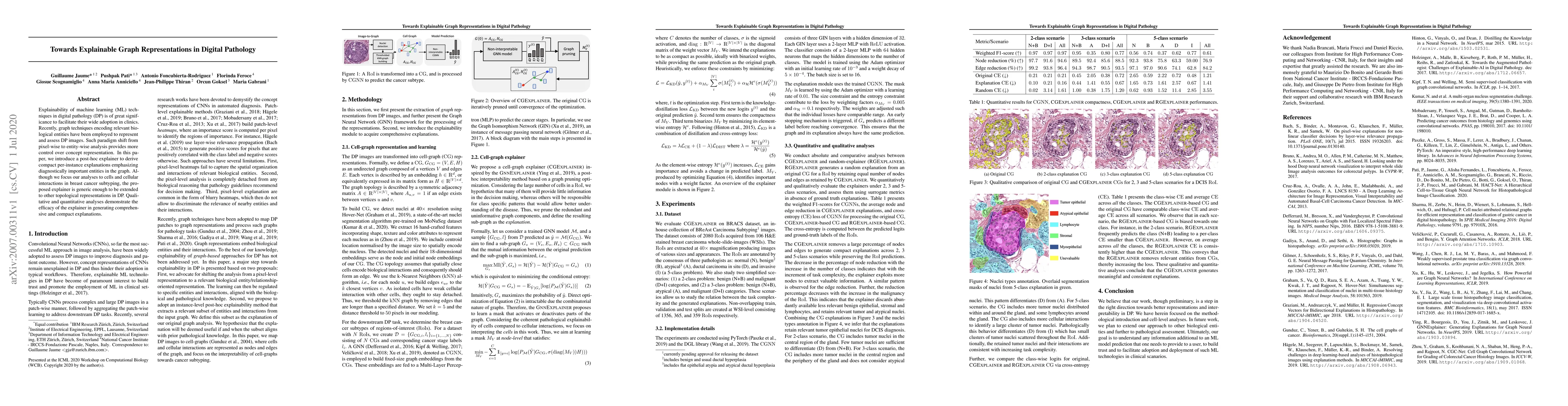
Explainability of machine learning (ML) techniques in digital pathology (DP) is of great significance to facilitate their wide adoption in clinics. Recently, graph techniques encoding relevant biolo...
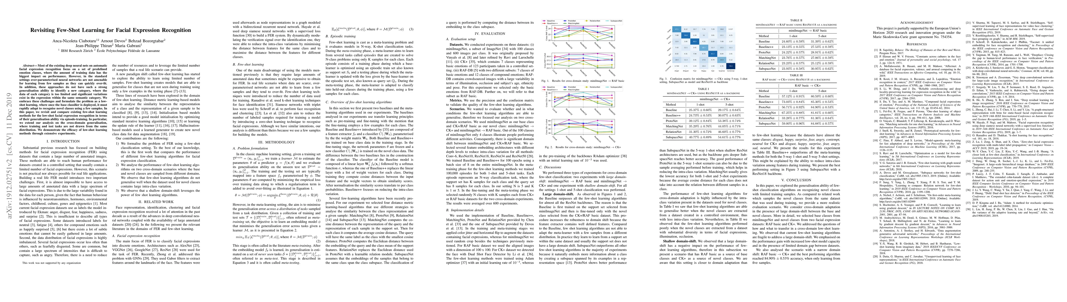
Most of the existing deep neural nets on automatic facial expression recognition focus on a set of predefined emotion classes, where the amount of training data has the biggest impact on performance...
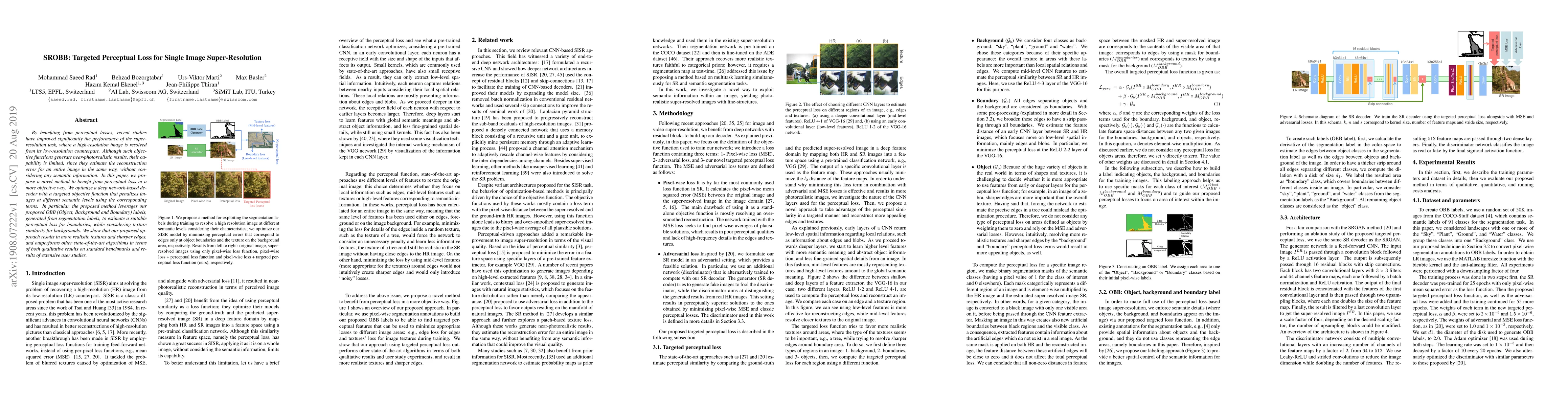
By benefiting from perceptual losses, recent studies have improved significantly the performance of the super-resolution task, where a high-resolution image is resolved from its low-resolution count...
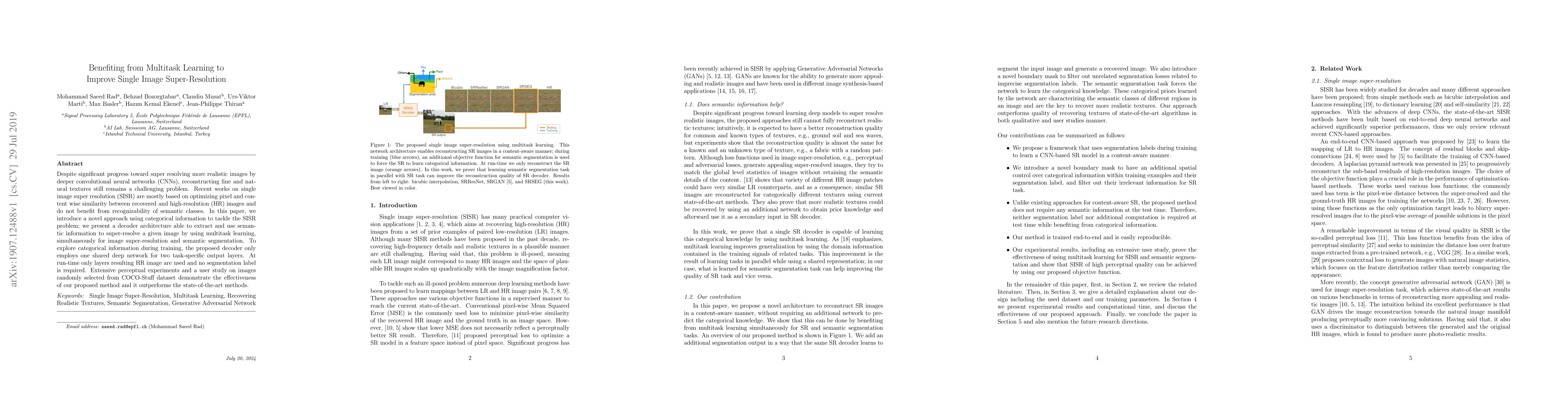
Despite significant progress toward super resolving more realistic images by deeper convolutional neural networks (CNNs), reconstructing fine and natural textures still remains a challenging problem...
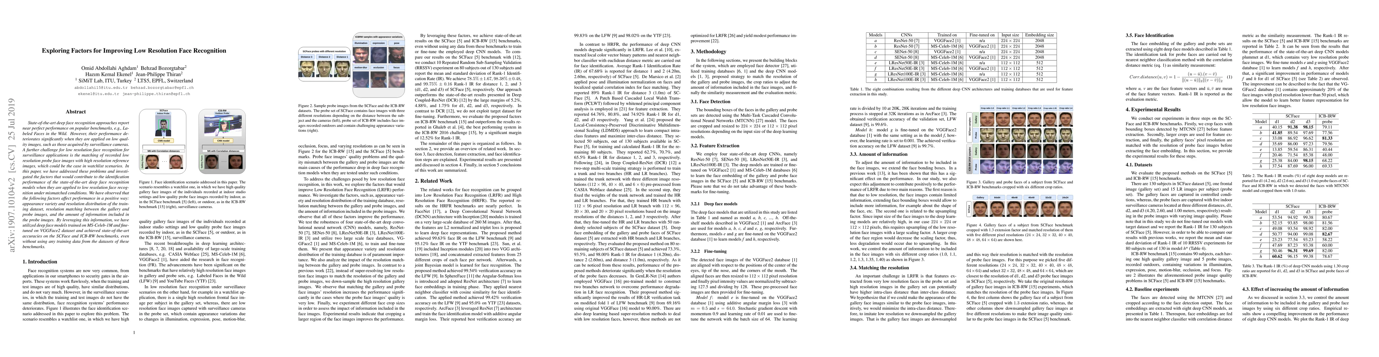
State-of-the-art deep face recognition approaches report near perfect performance on popular benchmarks, e.g., Labeled Faces in the Wild. However, their performance deteriorates significantly when t...
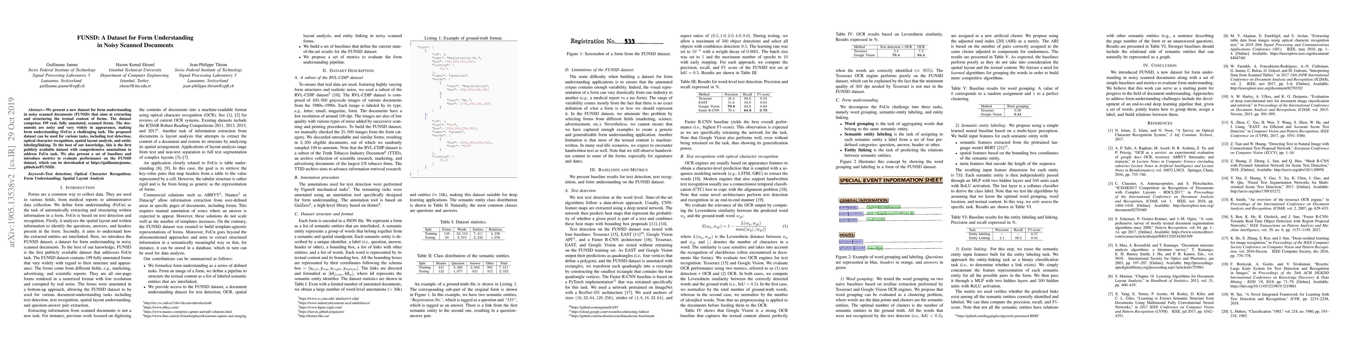
We present a new dataset for form understanding in noisy scanned documents (FUNSD) that aims at extracting and structuring the textual content of forms. The dataset comprises 199 real, fully annotat...
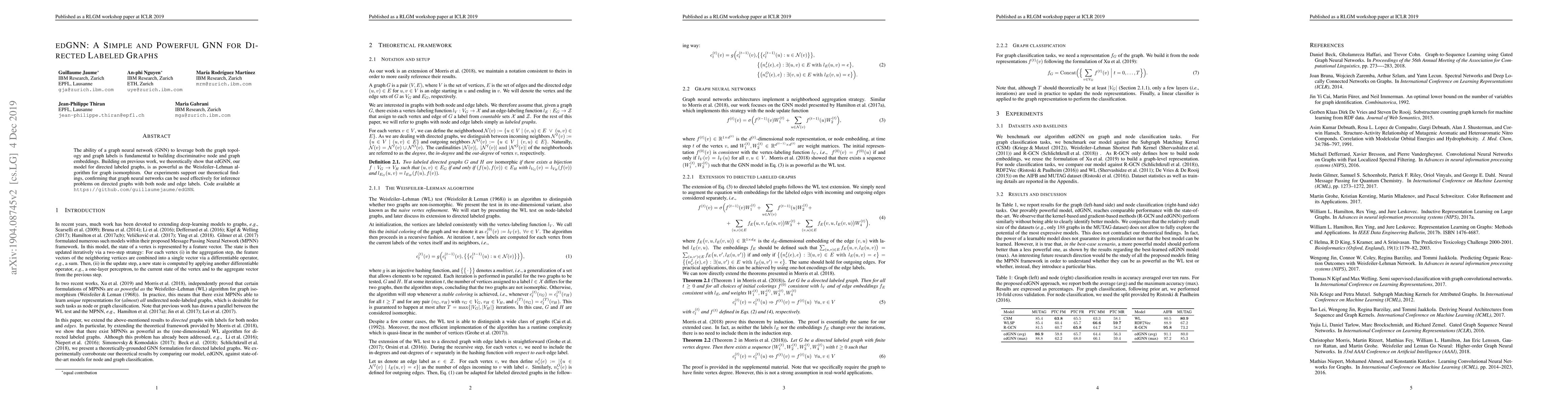
The ability of a graph neural network (GNN) to leverage both the graph topology and graph labels is fundamental to building discriminative node and graph embeddings. Building on previous work, we th...
High spatio-angular resolution diffusion MRI (dMRI) has been shown to provide accurate identification of complex neuronal fiber configurations, albeit, at the cost of long acquisition times. We prop...
The modern approaches for computer vision tasks significantly rely on machine learning, which requires a large number of quality images. While there is a plethora of image datasets with a single type ...
Diffusion Magnetic Resonance Imaging (dMRI) is a non-invasive method for depicting brain microstructure in vivo. Fiber orientation distributions (FODs) are mathematical representations extensively use...
Humans perceive the world through multisensory integration, blending the information of different modalities to adapt their behavior. Contrastive learning offers an appealing solution for multimodal s...
Quantifying the myelin sheath radius of myelinated axons in vivo is important for understanding, diagnosing, and monitoring various neurological disorders. Despite advancements in diffusion MRI (dMRI)...
Constrained Spherical Deconvolution (CSD) is crucial for estimating white matter fiber orientations using diffusion MRI data. A relevant parameter in CSD is the maximum order $l_{max}$ used in the sph...
In this paper, we address the challenge of few-shot classification in histopathology whole slide images (WSIs) by utilizing foundational vision-language models (VLMs) and slide-level prompt learning. ...
This paper introduces ReservoirTTA, a novel plug-in framework designed for prolonged test-time adaptation (TTA) in scenarios where the test domain continuously shifts over time, including cases where ...
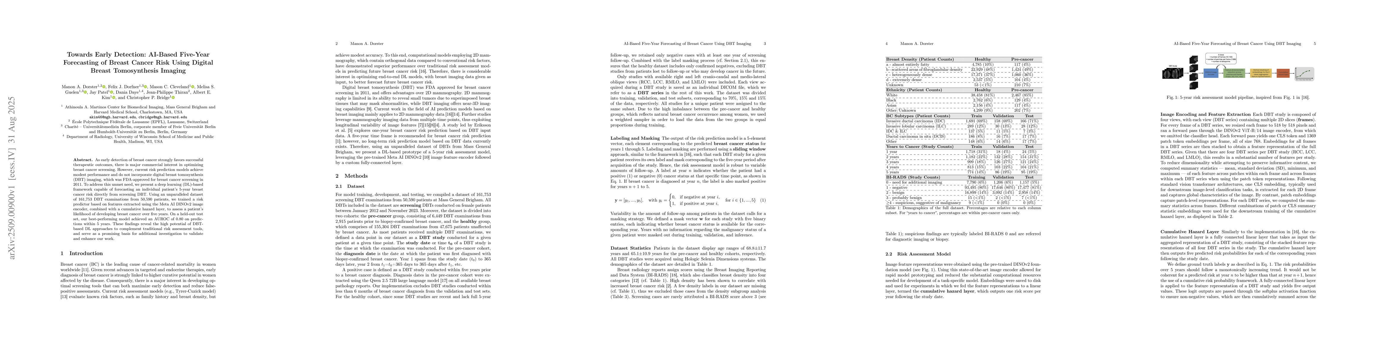
As early detection of breast cancer strongly favors successful therapeutic outcomes, there is major commercial interest in optimizing breast cancer screening. However, current risk prediction models a...
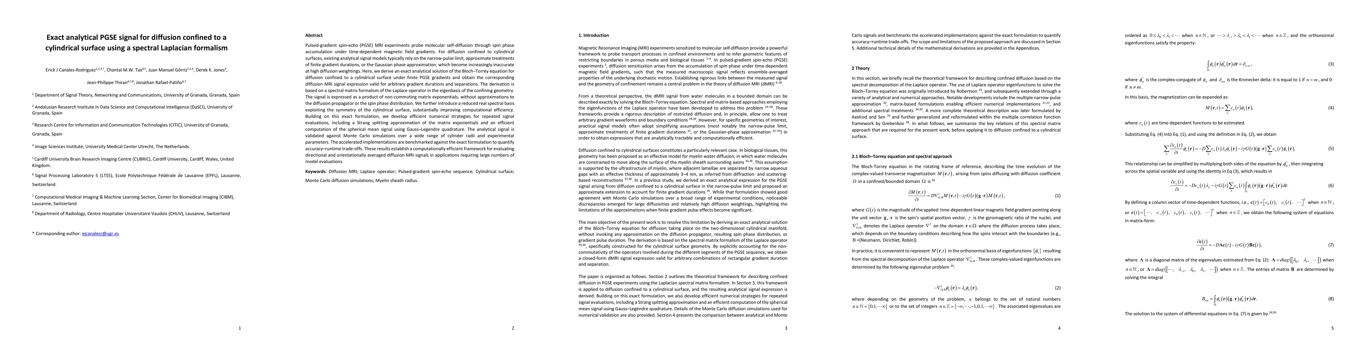
Pulsed-gradient spin-echo (PGSE) MRI experiments probe molecular self-diffusion through spin phase accumulation under time-dependent magnetic field gradients. For diffusion confined to cylindrical sur...
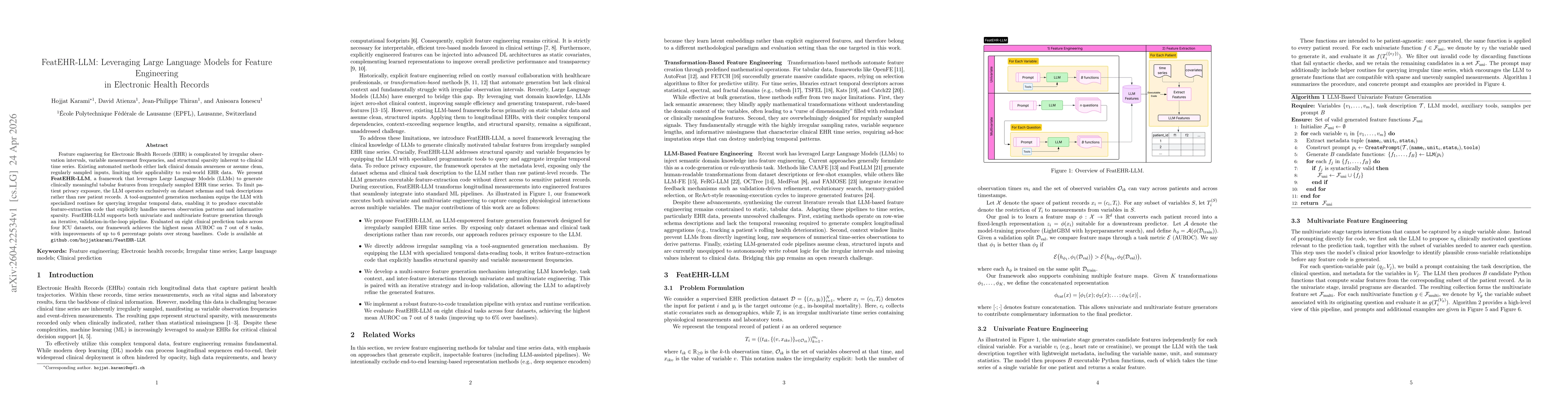
Feature engineering for Electronic Health Records (EHR) is complicated by irregular observation intervals, variable measurement frequencies, and structural sparsity inherent to clinical time series. E...