This study investigates a surface-based relaxation model to estimate axon radius using diffusion-relaxation MRI, calibrated with postmortem histology. The model successfully correlates intra-axonal T2 and T1 times with histological axon radius, enabling accurate predictions of axon radius in the corpus callosum using MRI data.
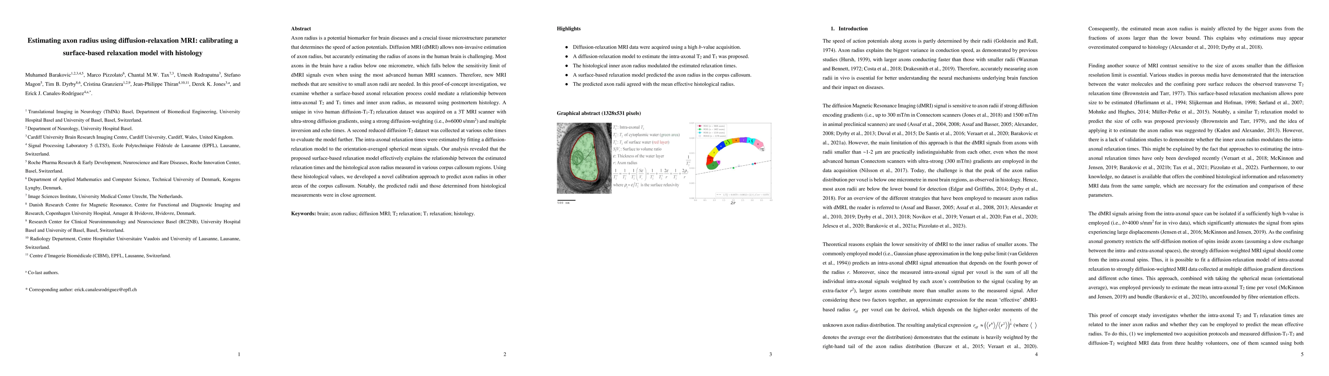
Axon radius is a potential biomarker for brain diseases and a crucial tissue microstructure parameter that determines the speed of action potentials. Diffusion MRI (dMRI) allows non-invasive estimation of axon radius, but accurately estimating the radius of axons in the human brain is challenging. Most axons in the brain have a radius below one micrometer, which falls below the sensitivity limit of dMRI signals even when using the most advanced human MRI scanners. Therefore, new MRI methods that are sensitive to small axon radii are needed. In this proof-of-concept investigation, we examine whether a surface-based axonal relaxation process could mediate a relationship between intra-axonal T2 and T1 times and inner axon radius, as measured using postmortem histology. A unique in vivo human diffusion-T1-T2 relaxation dataset was acquired on a 3T MRI scanner with ultra-strong diffusion gradients, using a strong diffusion-weighting (i.e., b = 6,000 s/mm$^2$) and multiple inversion and echo times. A second reduced diffusion-T2 dataset was collected at various echo times to evaluate the model further. The intra-axonal relaxation times were estimated by fitting a diffusion-relaxation model to the orientation-averaged spherical mean signals. Our analysis revealed that the proposed surface-based relaxation model effectively explains the relationship between the estimated relaxation times and the histological axon radius measured in various corpus callosum regions. Using these histological values, we developed a novel calibration approach to predict axon radius in other areas of the corpus callosum. Notably, the predicted radii and those determined from histological measurements were in close agreement.
Get AI-generated insights about this paper's methodology, results, and significance.
Current paper (gray), citations (green), references (blue)
Jean-Philippe Thiran, Derek K. Jones, Erick J. Canales-Rodríguez et al.
Daniel C. Alexander, Marco Palombo, Stefano B. Blumberg et al.
Ting Gong, Chiara Maffei, Evan Dann et al.
Title | Authors | Year | Actions |
---|---|---|---|
A diffusion MRI model for random walks confined on cylindrical surfaces: towards non-invasive quantification of myelin sheath radius
|
Erick J Canales-Rodr'iguez, C. Tax, E. Fischi-Gómez, Derek K. Jones, Jean-Philippe Thiran, Jonathan Rafael-Patiño
|
2024
|
View Paper |
A translational MRI approach to validate acute axonal damage detection as an early event in multiple sclerosis
|
Antonio Cerdán Cerdá, Nicola Toschi, C. Treaba, V. Barletta, Elena Herranz, Ambica Mehndiratta, J. Gómez-Sánchez, C. Mainero, S. De Santis
|
2024
|
View Paper |
Pore size estimation in axon-mimicking microfibers with diffusion-relaxation MRI
|
E. Canales-Rodríguez, M. Pizzolato, Feng-Lei Zhou, M. Barakovic, Jean-Philippe Thiran, Derek K. Jones, Geoffrey J. M. Parker, T. Dyrby
|
2023
|
View Paper |
Title | Authors | Year | Actions |
---|---|---|---|
Axial and radial axonal diffusivities and radii from single encoding strongly diffusion-weighted MRI
|
M. Pizzolato, E. Canales-Rodríguez, M. Andersson, T. Dyrby
|
2023
|
View Paper |
Characterization of b-value dependent T2 relaxation rates for probing neurite microstructure
|
L. Ning, C. Westin, Y. Rathi
|
2022
|
View Paper |
Towards a representative reference for MRI-based human axon radius assessment using light microscopy
|
L. Mordhorst, M. Morozova, Sebastian Papazoglou, Björn Fricke, J. M. Oeschger, Thibault Tabarin, Henriette Rusch, C. Jäger, Stefan Geyer, N. Weiskopf, M. Morawski, S. Mohammadi
|
2022
|
View Paper |
Axon diameter inferences in the human corpus callosum using oscillating gradient spin echo sequences.
|
Sheryl L. Herrera, Maxina Sheft, Morgan Mercredi, R. Buist, K. Matsuda, Melanie Martin
|
2021
|
View Paper |
Revisiting the T2 spectrum imaging inverse problem: Bayesian regularized non-negative least squares
|
E. Canales-Rodríguez, M. Pizzolato, Thomas Yu, G. Piredda, T. Hilbert, J. Raduà, Thomas Kober, J. Thiran
|
2021
|
View Paper |
Axonal T2 estimation using the spherical variance of the strongly diffusion-weighted MRI signal
|
M. Pizzolato, M. Andersson, Erick Jorge Canales-Rodríguez, J. Thiran, T. Dyrby
|
2021
|
View Paper |
Bundle-Specific Axon Diameter Index as a New Contrast to Differentiate White Matter Tracts
|
M. Barakovic, G. Girard, S. Schiavi, D. Romascano, M. Descoteaux, C. Granziera, Derek K. Jones, G. Innocenti, J. Thiran, Alessandro Daducci
|
2021
|
View Paper |
Age- and gender-related differences in brain tissue microstructure revealed by multi-component T2 relaxometry
|
E. Canales-Rodríguez, S. Alonso-Lana, N. Verdolini, S. Sarró, Isabel Feria, I. Montoro, B. Garcia-Ruiz, E. Jiménez, C. Varo, À. Albacete, I. Argila-Plaza, A. Lluch, C. Bonnín, E. Vilella, E. Vieta, E. Pomarol-Clotet, R. Salvador
|
2021
|
View Paper |
Measuring compartmental T2-orientational dependence in human brain white matter using a tiltable RF coil and diffusion-T2 correlation MRI
|
C. Tax, Elena Kleban, Maxime Chamberland, M. Barakovic, U. Rudrapatna, Derek K. Jones
|
2021
|
View Paper |
Validating Pore Size Estimates in a Complex Microfibre Environment on a Human MRI System
|
Chu-Chung Huang, C. Hsu, F. Zhou, S. Kusmia, Mark Drakesmith, G. Parker, Ching-Po Lin, Derek K. Jones
|
2021
|
View Paper |
Coaxial electrospun biomimetic copolymer fibres for application in diffusion magnetic resonance imaging
|
F. Zhou, D. McHugh, Zhanxiong Li, J. Gough, Gareth R. Williams, G. Parker
|
2021
|
View Paper |
The variability of MR axon radii estimates in the human white matter
|
J. Veraart, Erika P. Raven, L. Edwards, N. Weiskopf, Derek K. Jones
|
2021
|
View Paper |
Comparison of non-parametric T2 relaxometry methods for myelin water quantification
|
Erick Jorge Canales-Rodríguez, M. Pizzolato, G. Piredda, T. Hilbert, N. Kunz, C. Pot, Thomas Yu, R. Salvador, E. Pomarol-Clotet, T. Kober, J. Thiran, Alessandro Daducci
|
2021
|
View Paper |
Resolving bundle-specific intra-axonal T2 values within a voxel using diffusion-relaxation tract-based estimation
|
M. Barakovic, C. Tax, U. Rudrapatna, Maxime Chamberland, Jonathan Rafael-Patino, C. Granziera, J. Thiran, Alessandro Daducci, Erick Jorge Canales-Rodríguez, Derek K. Jones
|
2020
|
View Paper |
A simple estimate of axon size with diffusion MRI
|
K. Harkins, C. Beaulieu, Junzhong Xu, J. Gore, M. Does
|
2020
|
View Paper |
Axon diameter index estimation independent of fiber orientation distribution using high-gradient diffusion MRI
|
Q. Fan, A. Nummenmaa, T. Witzel, S. Huang
|
2020
|
View Paper |
Model-informed machine learning for multi-component T2 relaxometry
|
Thomas Yu, Erick Jorge Canales-Rodríguez, M. Pizzolato, G. Piredda, T. Hilbert, E. Fischi-Gómez, M. Weigel, M. Barakovic, M. Bach Cuadra, C. Granziera, T. Kober, J. Thiran
|
2020
|
View Paper |
Challenges for biophysical modeling of microstructure
|
I. Jelescu, M. Palombo, F. Bagnato, K. Schilling
|
2020
|
View Paper |
Sequences
|
Jordi Cortadella
|
2020
|
View Paper |
ActiveAxADD: Toward non‐parametric and orientationally invariant axon diameter distribution mapping using PGSE
|
D. Romascano, M. Barakovic, Jonathan Rafael-Patino, T. Dyrby, J. Thiran, Alessandro Daducci
|
2020
|
View Paper |
Axon morphology is modulated by the local environment and impacts the noninvasive investigation of its structure–function relationship
|
M. Andersson, H. Kjer, Jonathan Rafael-Patino, A. Pacureanu, B. Pakkenberg, J. Thiran, M. Ptito, M. Bech, A. Dahl, Vedrana Andersen Dahl, T. Dyrby
|
2020
|
View Paper |
Origin of orientation‐dependent R1 (=1/T1) relaxation in white matter
|
Felix Schyboll, U. Jaekel, Francesco Petruccione, H. Neeb
|
2020
|
View Paper |
Noninvasive quantification of axon radii using diffusion MRI
|
J. Veraart, Daniel Nunes, U. Rudrapatna, E. Fieremans, Derek K. Jones, D. Novikov, N. Shemesh
|
2020
|
View Paper |
Estimating axon conduction velocity in vivo from microstructural MRI
|
Mark Drakesmith, Mark Drakesmith, R. Harms, S. Rudrapatna, G. Parker, C. J. Evans, Derek K. Jones
|
2019
|
View Paper |
SciPy 1.0: fundamental algorithms for scientific computing in Python
|
Pauli Virtanen, R. Gommers, T. Oliphant, Matt Haberland, Tyler Reddy, D. Cournapeau, Evgeni Burovski, Pearu Peterson, Warren Weckesser, Jonathan Bright, S. J. van der Walt, M. Brett, Joshua Wilson, K. Millman, N. Mayorov, Andrew R. J. Nelson, E. Jones, Robert Kern, Eric Larson, C. Carey, İlhan Polat, Yu Feng, Eric W. Moore, J. Vanderplas, D. Laxalde, Josef Perktold, R. Cimrman, Ian Henriksen, E. Quintero, Charles R. Harris, A. Archibald, Antônio H. Ribeiro, Fabian Pedregosa, P. van Mulbregt, Aditya Alessandro Pietro Alex Andreas Andreas Anthony Ant Vijaykumar Bardelli Rothberg Hilboll Kloeckner Sco, A. Vijaykumar, Alessandro Pietro Bardelli, Alex Rothberg, A. Hilboll, Andre Kloeckner, A. Scopatz, Antony Lee, Ariel S. Rokem, C. N. Woods, Chad Fulton, Charles Masson, C. Häggström, Clark Fitzgerald, D. Nicholson, David R. Hagen, D. Pasechnik, E. Olivetti, Eric Martin, Eric Wieser, Fabrice Silva, F. Lenders, Florian Wilhelm, G. Young, Gavin A. Price, G. Ingold, Gregory E. Allen, Gregory R. Lee, H. Audren, I. Probst, J. Dietrich, J. Silterra, James T. Webber, J. Slavič, J. Nothman, J. Buchner, Johannes Kulick, Johannes L. Schönberger, J. V. De Miranda Cardoso, J. Reimer, J. Harrington, Juan Rodríguez, Juan Nunez-Iglesias, Justin Kuczynski, K. Tritz, M. Thoma, M. Newville, Matthias Kümmerer, Maximilian Bolingbroke, Michael Tartre, M. Pak, Nathaniel J. Smith, N. Nowaczyk, Nikolay Shebanov, O. Pavlyk, P. A. Brodtkorb, Perry Lee, R. McGibbon, Roman Feldbauer, Sam Lewis, S. Tygier, Scott Sievert, S. Vigna, Stefan Peterson, S. More, Tadeusz Pudlik, T. Oshima, T. Pingel, T. Robitaille, Thomas Spura, T. Jones, T. Cera, Tim Leslie, Tiziano Zito, Tom Krauss, U. Upadhyay, Y. Halchenko, Y. Vázquez-Baeza
|
2019
|
View Paper |
Imaging brain microstructure with diffusion MRI: practicality and applications
|
D. Alexander, T. Dyrby, M. Nilsson, Hui Zhang
|
2019
|
View Paper |
Diversity of Cortico-descending Projections: Histological and Diffusion MRI Characterization in the Monkey
|
G. Innocenti, R. Caminiti, E. Rouiller, G. Knott, T. Dyrby, M. Descoteaux, J. Thiran
|
2019
|
View Paper |
Observation of Angular Dependence of T1 in the Human White Matter at 3T.
|
Michael J. Knight, Robin A. Damion, R. Kauppinen
|
2019
|
View Paper |
Measuring intra‐axonal T2 in white matter with direction‐averaged diffusion MRI
|
Emilie T. McKinnon, J. Jensen
|
2018
|
View Paper |
Deficit of corpus callosum axons, reduced axon diameter and decreased area are markers of abnormal development of interhemispheric connections in autistic subjects
|
J. Wegiel, W. Kaczmarski, M. Flory, V. Martínez‐Cerdeño, T. Wisniewski, K. Nowicki, I. Kuchna, J. Wegiel
|
2018
|
View Paper |
Validation strategies for the interpretation of microstructure imaging using diffusion MRI
|
T. Dyrby, G. Innocenti, M. Bech, H. Lundell
|
2018
|
View Paper |
Transverse NMR relaxation in biological tissues
|
V. Kiselev, D. Novikov
|
2018
|
View Paper |
The Regulation of Axon Diameter: From Axonal Circumferential Contractility to Activity-Dependent Axon Swelling
|
A. R. Costa, Rita Pinto-Costa, S. C. Sousa, M. Sousa
|
2018
|
View Paper |
Searching for the neurite density with diffusion MRI: Challenges for biophysical modeling
|
Björn Lampinen, Filip Szczepankiewicz, Mikael Novén, D. van Westen, O. Hansson, E. Englund, J. Mårtensson, C. Westin, M. Nilsson
|
2018
|
View Paper |
The impact of fibre orientation on T1-relaxation and apparent tissue water content in white matter
|
Felix Schyboll, U. Jaekel, B. Weber, H. Neeb
|
2018
|
View Paper |
A biomimetic tumor tissue phantom for validating diffusion‐weighted MRI measurements
|
D. McHugh, F. Zhou, I. Wimpenny, G. Poologasundarampillai, J. Naish, P. L. Hubbard Cristinacce, G. Parker
|
2017
|
View Paper |
TE dependent Diffusion Imaging (TEdDI) distinguishes between compartmental T
2 relaxation times
|
J. Veraart, D. Novikov, E. Fieremans
|
2017
|
View Paper |
Magnetic Resonance Relaxation Anisotropy: Physical Principles and Uses in Microstructure Imaging
|
Michael J. Knight, Serena Dillon, Lina Jarutytė, R. Kauppinen
|
2017
|
View Paper |
Resolution limit of cylinder diameter estimation by diffusion MRI: The impact of gradient waveform and orientation dispersion
|
M. Nilsson, S. Lasič, I. Drobnjak, D. Topgaard, C. Westin
|
2017
|
View Paper |
Node of Ranvier length as a potential regulator of myelinated axon conduction speed
|
I. Arancibia‐Carcamo, Marc C. Ford, L. Cossell, K. Ishida, K. Tohyama, D. Attwell
|
2017
|
View Paper |
Quantifying brain microstructure with diffusion MRI: Theory and parameter estimation
|
D. Novikov, E. Fieremans, S. Jespersen, V. Kiselev
|
2016
|
View Paper |
An in vivo study of the orientation‐dependent and independent components of transverse relaxation rates in white matter
|
Rita Gil, Diana Khabipova, M. Zwiers, T. Hilbert, T. Kober, J. Marques
|
2016
|
View Paper |
Denoising of diffusion MRI using random matrix theory
|
J. Veraart, D. Novikov, D. Christiaens, B. Ades-aron, Jan Sijbers, E. Fieremans
|
2016
|
View Paper |
Mapping axonal density and average diameter using non-monotonic time-dependent gradient-echo MRI.
|
Daniel Nunes, Tomás L. Cruz, S. Jespersen, N. Shemesh
|
2016
|
View Paper |
Diffusion-mediated nuclear spin phase decoherence in cylindrically porous materials
|
Michael J. Knight, R. Kauppinen
|
2016
|
View Paper |
White matter microstructure from nonparametric axon diameter distribution mapping
|
D. Benjamini, M. Komlosh, L. Holtzclaw, U. Nevo, P. Basser
|
2016
|
View Paper |
Multi-compartment microscopic diffusion imaging
|
Enrico Kaden, Nathaniel D. Kelm, Robert P. Carson, M. Does, D. Alexander
|
2016
|
View Paper |
B0‐orientation dependent magnetic susceptibility‐induced white matter contrast in the human brainstem at 11.7T
|
M. Aggarwal, Y. Kageyama, Xu Li, Peter C.M. van Zijl
|
2016
|
View Paper |
Parametric Probability Distribution Functions for Axon Diameters of Corpus Callosum
|
F. Sepehrband, D. Alexander, K. Clark, N. Kurniawan, Zhengyi Yang, D. Reutens
|
2016
|
View Paper |
Including diffusion time dependence in the extra-axonal space improves in vivo estimates of axonal diameter and density in human white matter
|
S. Santis, Derek K. Jones, A. Roebroeck
|
2016
|
View Paper |
The microstructural correlates of T1 in white matter
|
K. Harkins, Junzhong Xu, A. Dula, Ke Li, W. Valentine, D. Gochberg, J. Gore, M. Does
|
2016
|
View Paper |
An integrated approach to correction for off-resonance effects and subject movement in diffusion MR imaging
|
J. Andersson, S. Sotiropoulos
|
2016
|
View Paper |
Towards higher sensitivity and stability of axon diameter estimation with diffusion‐weighted MRI
|
F. Sepehrband, D. Alexander, N. Kurniawan, D. Reutens, Zhengyi Yang
|
2016
|
View Paper |
Fiber ball imaging
|
J. Jensen, G. Glenn, J. Helpern
|
2016
|
View Paper |
In vivo mapping of human spinal cord microstructure at 300mT/m
|
T. Duval, J. McNab, K. Setsompop, T. Witzel, T. Schneider, S. Huang, B. Keil, Eric C. Klawiter, L. Wald, J. Cohen-Adad
|
2015
|
View Paper |
Mesoscopic structure of neuronal tracts from time-dependent diffusion
|
L. Burcaw, E. Fieremans, D. Novikov
|
2015
|
View Paper |
Quantitative mapping of the per‐axon diffusion coefficients in brain white matter
|
Enrico Kaden, F. Kruggel, D. Alexander
|
2015
|
View Paper |
Single-shot T1 mapping of the corpus callosum: a rapid characterization of fiber bundle anatomy
|
S. Hofer, Xiaoqing Wang, Volkert Roeloffs, J. Frahm
|
2015
|
View Paper |
PGSE, OGSE, and sensitivity to axon diameter in diffusion MRI: Insight from a simulation study
|
I. Drobnjak, Hui Zhang, A. Ianuş, Enrico Kaden, D. Alexander
|
2015
|
View Paper |
Nuclear magnetic resonance average pore-size estimations outside the fast-diffusion regime
|
M. Müller‐Petke, R. Dlugosch, J. Lehmann-Horn, M. Ronczka
|
2015
|
View Paper |
On the accuracy of T1 mapping: Searching for common ground
|
N. Stikov, M. Boudreau, I. Levesque, Christine L. Tardif, J. Barral, G. Pike
|
2015
|
View Paper |
The impact of gradient strength on in vivo diffusion MRI estimates of axon diameter
|
S. Huang, A. Nummenmaa, T. Witzel, T. Duval, J. Cohen-Adad, L. Wald, J. McNab
|
2015
|
View Paper |
Accelerated Microstructure Imaging via Convex Optimization (AMICO) from diffusion MRI data
|
Alessandro Daducci, Erick Jorge Canales-Rodríguez, Hui Zhang, T. Dyrby, D. Alexander, J. Thiran
|
2015
|
View Paper |
Biomimetic phantom for the validation of diffusion magnetic resonance imaging
|
P. Hubbard, F. Zhou, S. Eichhorn, G. Parker
|
2015
|
View Paper |
Mapping mean axon diameter and axonal volume fraction by MRI using temporal diffusion spectroscopy
|
Junzhong Xu, Hua Li, K. Harkins, Xiaoyu Jiang, Jingping Xie, Hakmook Kang, M. Does, J. Gore
|
2014
|
View Paper |
The diameter of cortical axons depends both on the area of origin and target.
|
G. Innocenti, A. Vercelli, R. Caminiti
|
2014
|
View Paper |
Jointly deriving NMR surface relaxivity and pore size distributions by NMR relaxation experiments on partially desaturated rocks
|
O. Mohnke
|
2014
|
View Paper |
Myelin and iron concentration in the human brain: A quantitative study of MRI contrast
|
Carsten Stüber, M. Morawski, A. Schäfer, C. Labadie, M. Wähnert, Christoph Leuze, Markus N. Streicher, N. Barapatre, K. Reimann, S. Geyer, D. Spemann, R. Turner
|
2014
|
View Paper |
Dipy, a library for the analysis of diffusion MRI data
|
E. Garyfallidis, M. Brett, B. Amirbekian, Ariel S. Rokem, S. Walt, M. Descoteaux, I. Nimmo-Smith
|
2014
|
View Paper |
Microanisotropy imaging: quantification of microscopic diffusion anisotropy and orientational order parameter by diffusion MRI with magic-angle spinning of the q-vector
|
S. Lasič, Filip Szczepankiewicz, Stefanie Eriksson, M. Nilsson, D. Topgaard
|
2014
|
View Paper |
Myelin water mapping by spatially regularized longitudinal relaxographic imaging at high magnetic fields
|
C. Labadie, Jing-Huei Lee, W. Rooney, S. Jarchow, M. Aubert-Frécon, C. Springer, H. Möller
|
2014
|
View Paper |
The CONNECT project: Combining macro- and micro-structure
|
Y. Assaf, D. Alexander, Derek K. Jones, A. Bizzi, Timothy Edward John Behrens, C. Clark, Y. Cohen, T. Dyrby, P. Huppi, T. Knösche, D. Bihan, G. Parker, C. Poupon
|
2013
|
View Paper |
Diffusion Weighted Image Denoising Using Overcomplete Local PCA
|
J. Manjón, P. Coupé, L. Concha, A. Buades, D. Louis Collins, Robles Montserrat
|
2013
|
View Paper |
One component? Two components? Three? The effect of including a nonexchanging “free” water component in multicomponent driven equilibrium single pulse observation of T1 and T2
|
S. Deoni, Lucy Matthews, S. Kolind
|
2013
|
View Paper |
Can T 2 -Spectroscopy Resolve Submicrometer Axon Diameters?
|
Enrico Kaden, D. Alexander
|
2013
|
View Paper |
Origin of B0 orientation dependent R2
* (=1/T2
*) in white matter
|
S. Oh, Young-Bo Kim, Z. Cho, Jongho Lee
|
2013
|
View Paper |
Contrast and stability of the axon diameter index from microstructure imaging with diffusion MRI
|
T. Dyrby, Lise V S⊘gaard, M. Hall, M. Ptito, D. Alexander
|
2012
|
View Paper |
Axon diameter mapping in the presence of orientation dispersion with diffusion MRI
|
Hui Zhang, P. Cristinacce, G. Parker, D. Alexander
|
2011
|
View Paper |
Correction of main and transmit magnetic field (B0 and B1) inhomogeneity effects in multicomponent‐driven equilibrium single‐pulse observation of T1 and T2
|
Sean C. L. Deoni
|
2011
|
View Paper |
Stereological estimation of the total number of myelinated callosal fibers in human subjects
|
J. Riise, B. Pakkenberg
|
2011
|
View Paper |
Quantitative MR imaging of brain iron: a postmortem validation study.
|
C. Langkammer, N. Krebs, W. Goessler, E. Scheurer, F. Ebner, K. Yen, F. Fazekas, S. Ropele
|
2010
|
View Paper |
Orientationally invariant indices of axon diameter and density from diffusion MRI
|
D. Alexander, P. Cristinacce, M. Hall, E. Moore, M. Ptito, G. Parker, T. Dyrby
|
2010
|
View Paper |
A robust methodology for in vivo T1 mapping
|
J. Barral, E. Gudmundson, N. Stikov, M. Etezadi‐Amoli, P. Stoica, D. Nishimura
|
2010
|
View Paper |
Multiexponential T2, magnetization transfer, and quantitative histology in white matter tracts of rat spinal cord
|
A. Dula, D. Gochberg, H. Valentine, W. Valentine, M. Does
|
2010
|
View Paper |
Evolution amplified processing with temporally dispersed slow neuronal connectivity in primates
|
R. Caminiti, H. Ghaziri, R. Galuske, P. Hof, G. Innocenti
|
2009
|
View Paper |
Evaluation of 14 nonlinear deformation algorithms applied to human brain MRI registration
|
A. Klein, J. Andersson, B. Ardekani, J. Ashburner, B. Avants, M. Chiang, G. Christensen, D. Collins, J. Gee, P. Hellier, J. Song, M. Jenkinson, C. Lepage, D. Rueckert, P. Thompson, T. Vercauteren, R. Woods, J. Mann, R. Parsey
|
2009
|
View Paper |
In vivo measurement of axon diameter distribution in the corpus callosum of rat brain.
|
D. Barazany, P. Basser, Y. Assaf
|
2009
|
View Paper |
A general framework for experiment design in diffusion MRI and its application in measuring direct tissue‐microstructure features
|
D. Alexander
|
2008
|
View Paper |
Axcaliber: A method for measuring axon diameter distribution from diffusion MRI
|
Y. Assaf, T. Blumenfeld-Katzir, Y. Yovel, P. Basser
|
2008
|
View Paper |
Symmetric diffeomorphic image registration with cross-correlation: Evaluating automated labeling of elderly and neurodegenerative brain
|
B. Avants, C. Epstein, M. Grossman, J. Gee
|
2008
|
View Paper |
Mapping hydration dynamics around a protein surface
|
Luyuan Zhang, Lijuan Wang, Ya-Ting Kao, Weihong Qiu, Yi Yang, Oghaghare K. Okobiah, D. Zhong
|
2007
|
View Paper |
Composite hindered and restricted model of diffusion (CHARMED) MR imaging of the human brain
|
Y. Assaf, P. Basser
|
2005
|
View Paper |
Advances in functional and structural MR image analysis and implementation as FSL
|
Stephen M. Smith, M. Jenkinson, M. Woolrich, C. Beckmann, Timothy Edward John Behrens, H. Johansen-Berg, P. Bannister, M. D. Luca, I. Drobnjak, D. Flitney, R. Niazy, James Saunders, J. Vickers, Yongyue Zhang, N. Stefano, J. Brady, P. Matthews
|
2004
|
View Paper |
New modeling and experimental framework to characterize hindered and restricted water diffusion in brain white matter
|
Y. Assaf, R. Freidlin, G. Rohde, P. Basser
|
2004
|
View Paper |
Water? What's so special about it?
|
J. Finney
|
2004
|
View Paper |
Water and proteins: a love-hate relationship.
|
Y. Levy, J. Onuchic
|
2004
|
View Paper |
How to correct susceptibility distortions in spin-echo echo-planar images: application to diffusion tensor imaging
|
J. Andersson, S. Skare, J. Ashburner
|
2003
|
View Paper |
Computer simulations in solid-state NMR. III. powder averaging
|
M. Edén
|
2003
|
View Paper |
Three‐pool model of white matter
|
J. Lancaster, T. Andrews, L. J. Hardies, S. Dodd, P. Fox
|
2003
|
View Paper |
The basis of anisotropic water diffusion in the nervous system – a technical review
|
Christian Beaulieu
|
2002
|
View Paper |
Anti-myelin-associated glycoprotein antibodies alter neurofilament spacing.
|
M. Lunn, T. Crawford, R. Hughes, J. Griffin, K. Sheikh
|
2002
|
View Paper |
Aquaporins in Brain: Distribution, Physiology, and Pathophysiology
|
J. Badaut, F. Lasbennes, P. Magistretti, L. Regli
|
2002
|
View Paper |
Neurofilament-dependent Radial Growth of Motor Axons and Axonal Organization of Neurofilaments Does Not Require the Neurofilament Heavy Subunit (NF-H) or Its Phosphorylation
|
M. Rao, Megan K. Houseweart, T. L. Williamson, T. Crawford, J. Folmer, D. Cleveland
|
1998
|
View Paper |
Mri: Use of the inversion recovery pulse sequence
|
G. Bydder, J. Hajnal, I. Young
|
1998
|
View Paper |
Determination of surface relaxivity from NMR diffusion measurements.
|
W. Slijkerman, J. Hofman
|
1998
|
View Paper |
MRI: use of the inversion recovery pulse sequence.
|
G. Bydder, J. Hajnal, I. Young
|
1998
|
View Paper |
Age-related changes in fibre composition of the human corpus callosum: sex differences.
|
F. Aboitiz, Eugenio Rodriguez, R. Olivares, E. Zaidel
|
1996
|
View Paper |
The rician distribution of noisy mri data
|
H. Gudbjartsson, S. Patz
|
1995
|
View Paper |
In vivo visualization of myelin water in brain by magnetic resonance
|
A. MacKay, K. Whittall, Julian Adler, David K. B. Li, D. Paty, D. Graeb
|
1994
|
View Paper |
Morphology of Callosal Axons Interconnecting Areas 17 and 18 of the Cat
|
J. Houzel, C. Milleret, G. Innocenti
|
1994
|
View Paper |
Evaluation of restricted diffusion in cylinders. Phosphocreatine in rabbit leg muscle.
|
P. Vangelderen, D. Despres, P. Vanzijl, C. Moonen
|
1994
|
View Paper |
Fiber composition of the human corpus callosum
|
F. Aboitiz, A. Scheibel, R. Fisher, E. Zaidel
|
1992
|
View Paper |
Elemental composition and water content of myelinated axons and glial cells in rat central nervous system
|
R. LoPachin, C. M. Castiglia, A. Saubermann
|
1991
|
View Paper |
Cytological and quantitative characteristics of four cerebral commissures in the rhesus monkey
|
A. LaMantia, P. Rakić
|
1990
|
View Paper |
Neurofilament gene expression: a major determinant of axonal caliber.
|
P. Hoffman, D. Cleveland, J. Griffin, Phillip W. Landes, N. Cowan, D. Price
|
1987
|
View Paper |
Measurement of spin-lattice relaxation times in nuclear magnetic resonance imaging
|
I. Pykett, B. R. Rosen, F. Buonanno, T. Brady, R. Brooks
|
1983
|
View Paper |
Diffusion of water in the endosperm tissue of wheat grains as studied by pulsed field gradient nuclear magnetic resonance.
|
P. Callaghan, K. Jolley, J. Lelièvre
|
1979
|
View Paper |
Importance of classical diffusion in NMR studies of water in biological cells
|
K. Brownstein, C. E. Tarr
|
1979
|
View Paper |
Spin-lattice relaxation in a system governed by diffusion
|
K. Brownstein, C. E. Tarr
|
1977
|
View Paper |
Changes of action potential shape and velocity for changing core conductor geometry.
|
S. Goldstein, W. Rall
|
1974
|
View Paper |
Relative conduction velocities of small myelinated and non-myelinated fibres in the central nervous system.
|
S. Waxman, S. Waxman, M. Bennett
|
1972
|
View Paper |
Axon caliber related to neurofilaments and microtubules in sciatic nerve fibers of rats and mice
|
R. Friede, T. Samorajski
|
1970
|
View Paper |
Reaction Rates by Nuclear Magnetic Resonance
|
H. Mcconnell
|
1958
|
View Paper |
Nuclear Magnetic Resonance Studies in Multiple Phase Systems: Lifetime of a Water Molecule in an Adsorbing Phase on Silica Gel
|
J. R. Zimmerman, W. Brittin
|
1957
|
View Paper |
A theory of the effects of fibre size in medullated nerve
|
W. Rushton
|
1951
|
View Paper |
CONDUCTION VELOCITY AND DIAMETER OF NERVE FIBERS
|
J. Hursh
|
1939
|
View Paper |
Measuring compartmental T2-orientational dependence in 22
|
2021
|
||
Microstructural Imaging of the Human Brain with A ‘Super-Scanner’: 10 Key Advantages of Ultra-Strong Gradients for Diffusion MRI The PIs and Colleagues of the National Facility for In Vivo MR Imaging of Human Tissue Microstructure
|
DC Alexander, F. Dell'Acqua, DJ McHugh, KL Miller, Parker Gjm, US Rudrapatna, Tax Cmw
|
2018
|
View Paper |
In vivo correlation between axon diameter and conduction velocity in the human brain
|
Assaf Horowitz, D. Barazany, I. Tavor, Moran Bernstein, G. Yovel, Y. Assaf
|
2014
|
View Paper |
White Matter Structure: A Microscopist’s View
|
J. Edgar, I. Griffiths
|
2009
|
View Paper |
The ‘wet mind’: water and functional neuroimaging
|
D. Le Bihan
|
2007
|
View Paper |
Absolute pore size distribution from NMR
|
2007
|
||
Computer simulations in solid-state NMR. I. Spin dynamics theory
|
M. Edén
|
2003
|
View Paper |
in vivo.
|
M. Drumm
|
1999
|
View Paper |
Water in biological systems: The NMR picture,” in Hydration processes in biology: theoretical and experimental approaches, ed
|
1999
|
||
Diffusion measurement in sandstone core: NMR determination of surface-to-volume ratio and surface relaxivity.
|
M. Hürlimann, L. Latour, C. Sotak, C. Sotak
|
1994
|
View Paper |
Translational diffusion in model membranes studied by NMR.
|
1977
|
||
Roche Pharma Research & Early Development, Neuroscience and Rare Diseases
|
|||
Basel (RC2NB), University Hospital and University of Lausanne, Lausanne, Switzerland
|
|||
studiesofwaterinbiologicalcells
|
|||
Kongens Lyngby, Denmark
|
Showing first page preview. Click "Open Full PDF" to view the complete paper.
Your comment contains inappropriate language and has been rejected.
Are you sure you want to remove this tag? This action cannot be undone.
Someone just favorited a paper!
Comments (0)