Academic Profile
Statistics
Similar Authors
Papers on arXiv
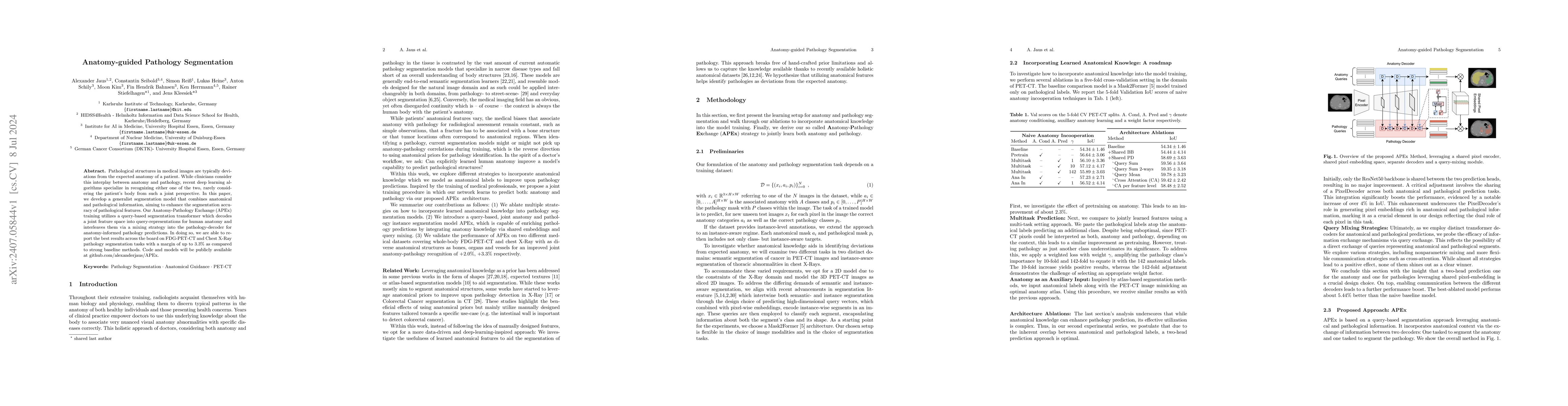
Pathological structures in medical images are typically deviations from the expected anatomy of a patient. While clinicians consider this interplay between anatomy and pathology, recent deep learning ...
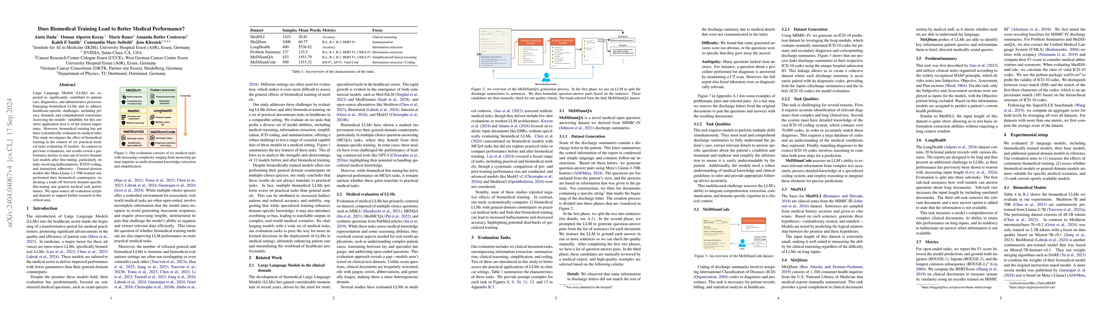
Large Language Models (LLMs) are expected to significantly contribute to patient care, diagnostics, and administrative processes. Emerging biomedical LLMs aim to address healthcare-specific challenges...
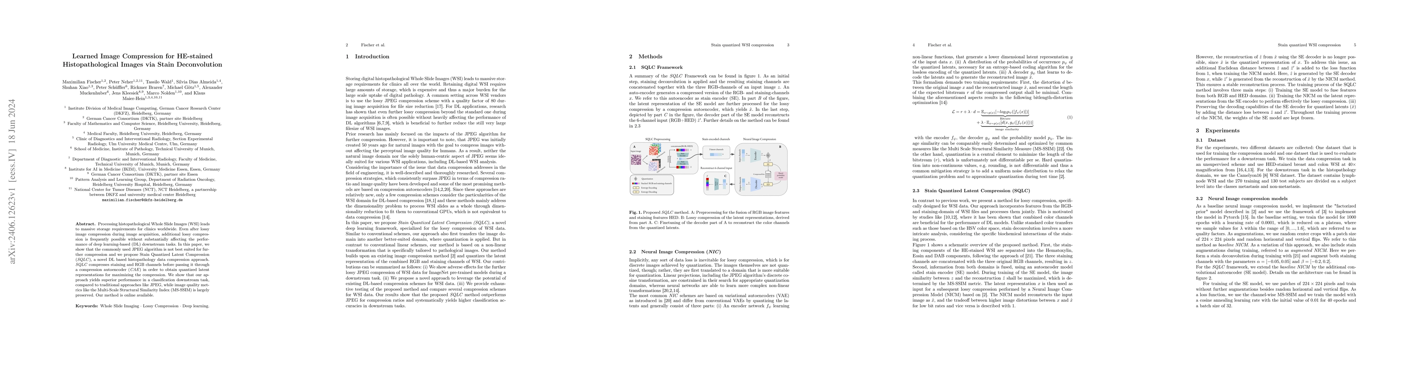
Processing histopathological Whole Slide Images (WSI) leads to massive storage requirements for clinics worldwide. Even after lossy image compression during image acquisition, additional lossy compres...
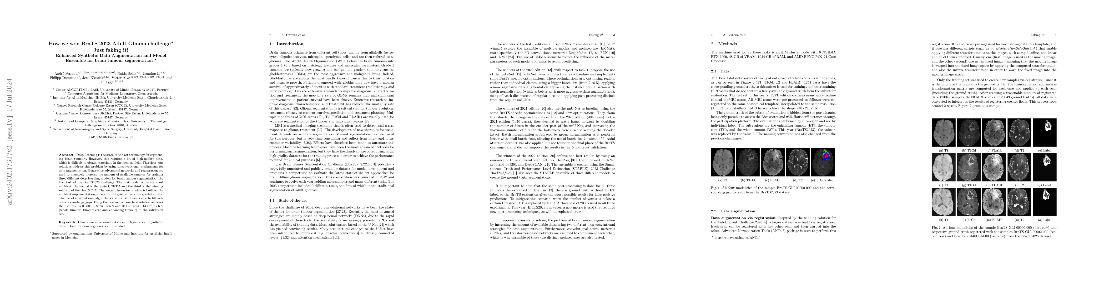
Deep Learning is the state-of-the-art technology for segmenting brain tumours. However, this requires a lot of high-quality data, which is difficult to obtain, especially in the medical field. Therefo...
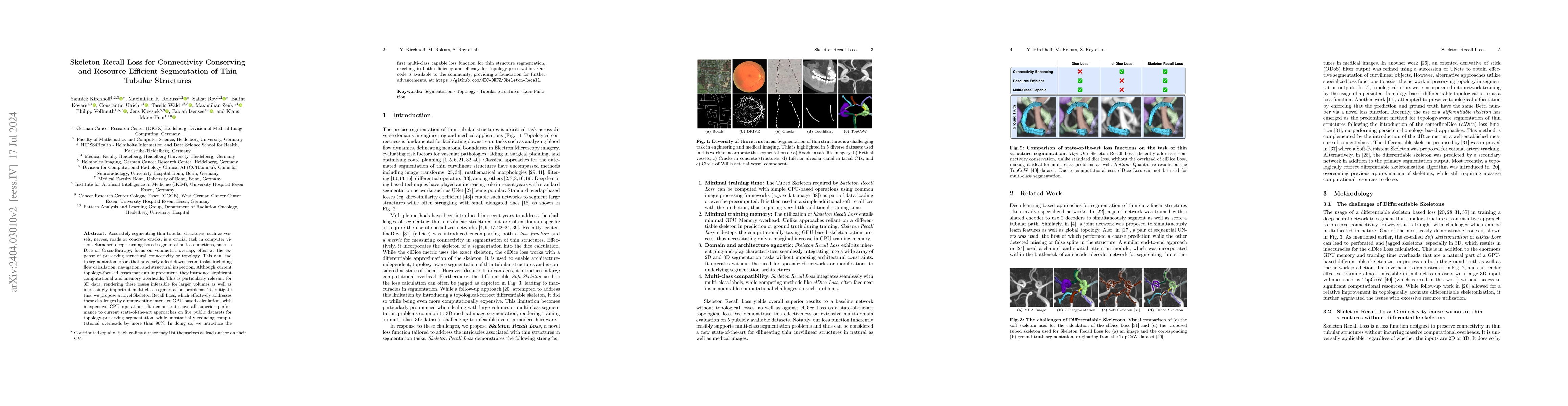
Accurately segmenting thin tubular structures, such as vessels, nerves, roads or concrete cracks, is a crucial task in computer vision. Standard deep learning-based segmentation loss functions, such a...
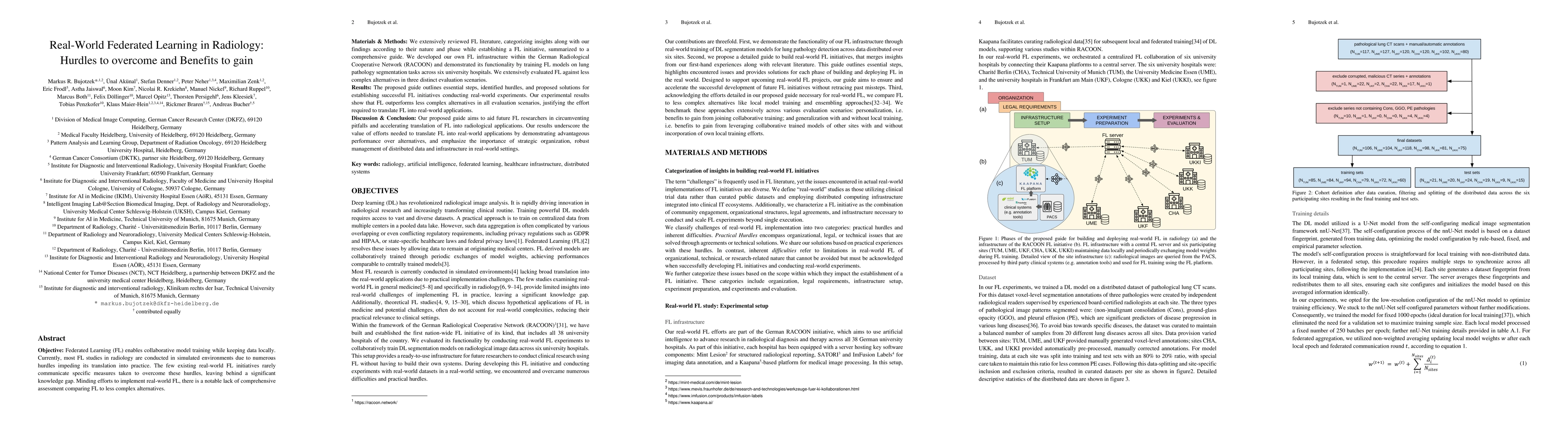
Objective: Federated Learning (FL) enables collaborative model training while keeping data locally. Currently, most FL studies in radiology are conducted in simulated environments due to numerous hu...
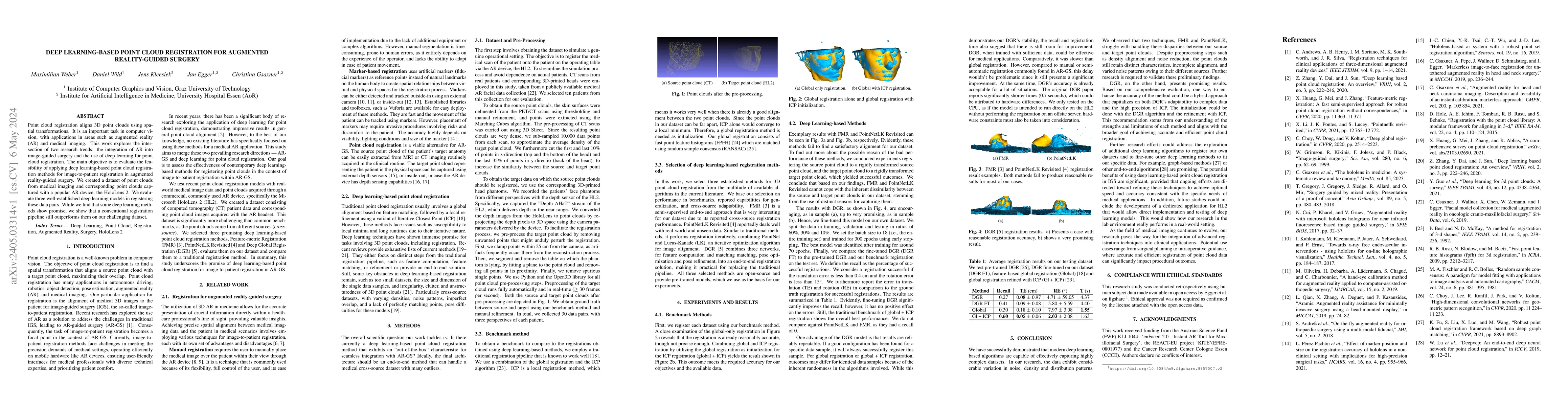
Point cloud registration aligns 3D point clouds using spatial transformations. It is an important task in computer vision, with applications in areas such as augmented reality (AR) and medical imagi...
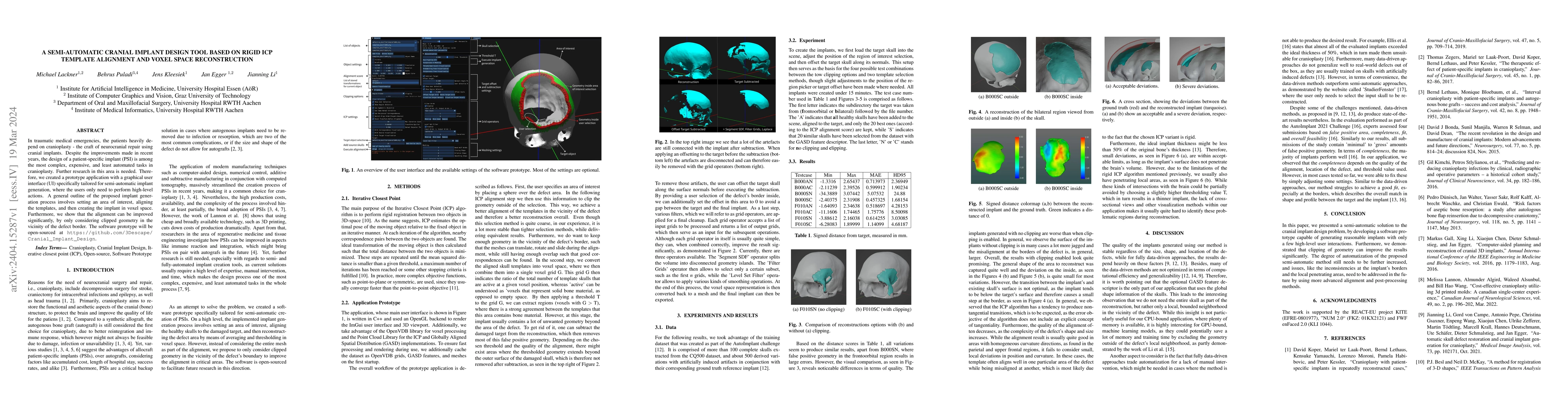
In traumatic medical emergencies, the patients heavily depend on cranioplasty - the craft of neurocranial repair using cranial implants. Despite the improvements made in recent years, the design of ...
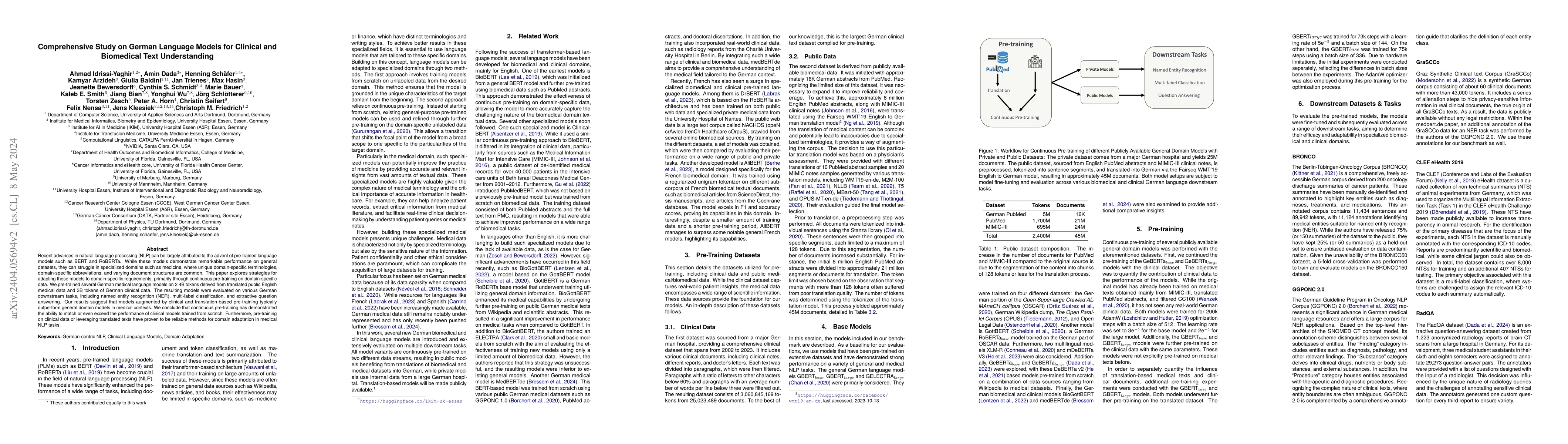
Recent advances in natural language processing (NLP) can be largely attributed to the advent of pre-trained language models such as BERT and RoBERTa. While these models demonstrate remarkable perfor...
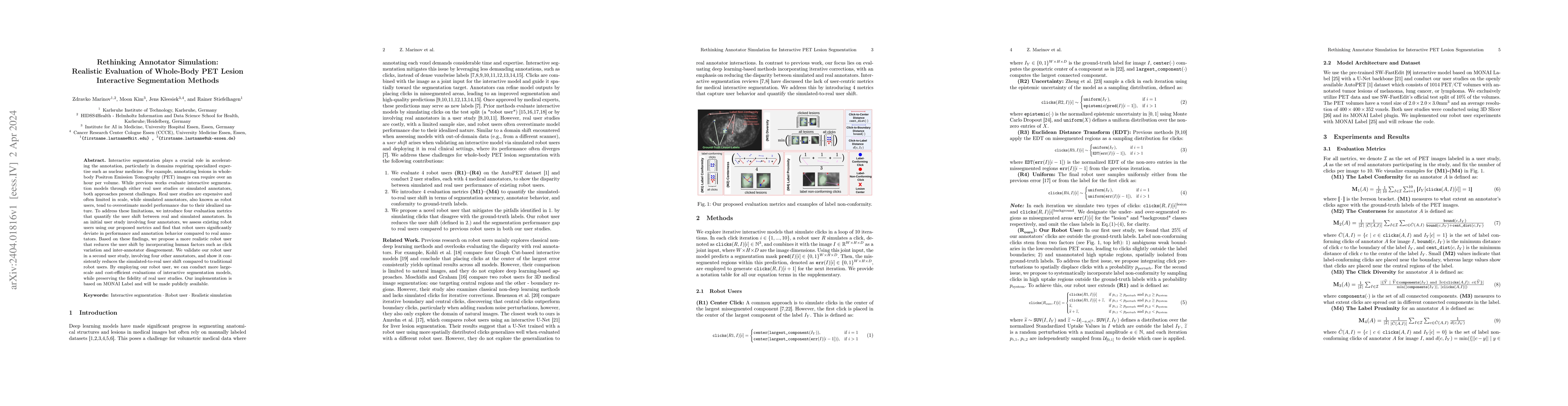
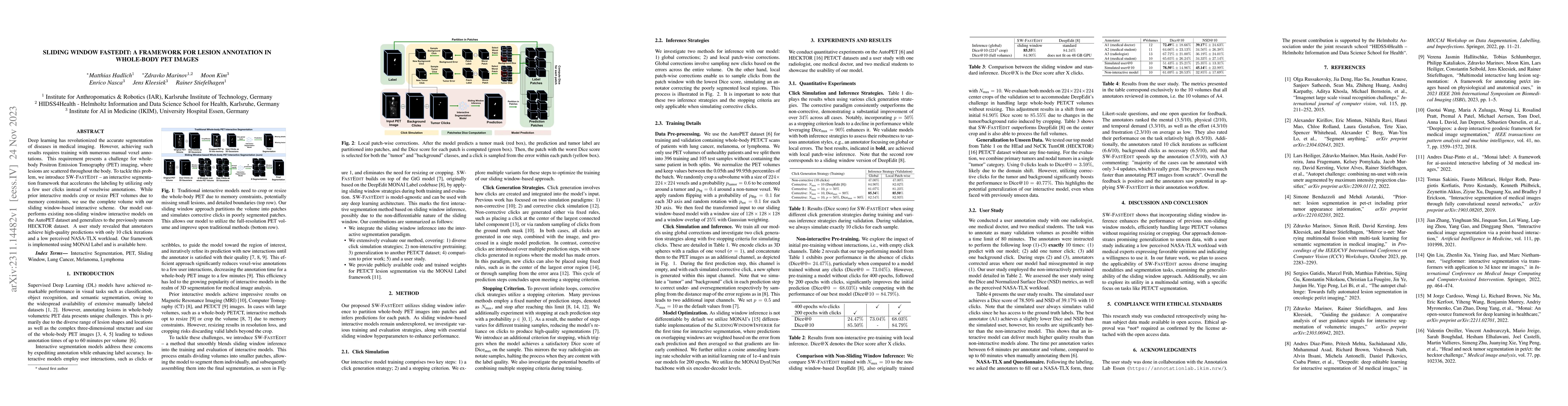
Interactive segmentation plays a crucial role in accelerating the annotation, particularly in domains requiring specialized expertise such as nuclear medicine. For example, annotating lesions in who...
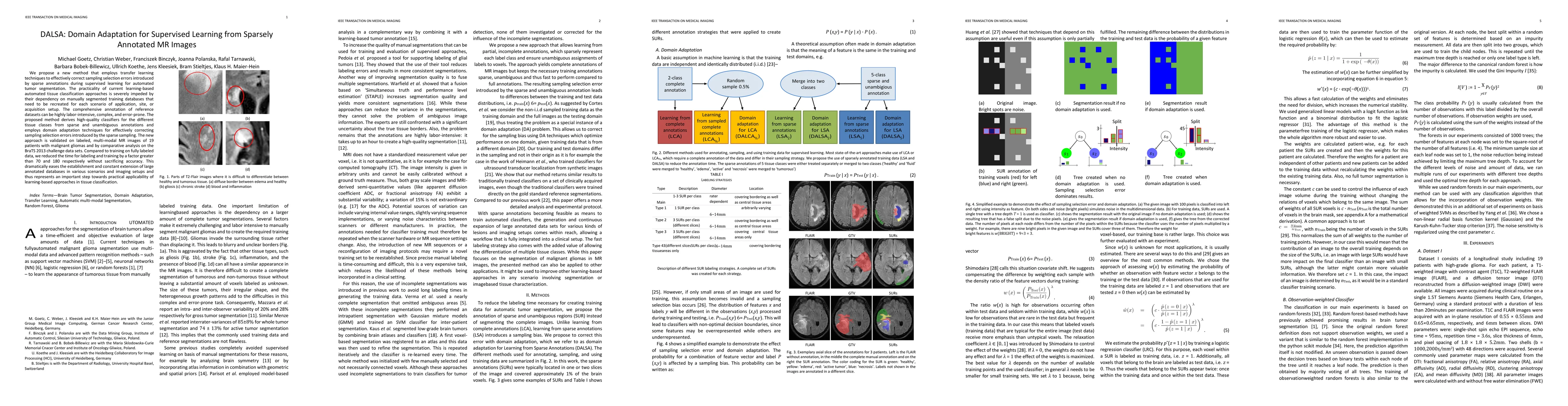
We propose a new method that employs transfer learning techniques to effectively correct sampling selection errors introduced by sparse annotations during supervised learning for automated tumor seg...
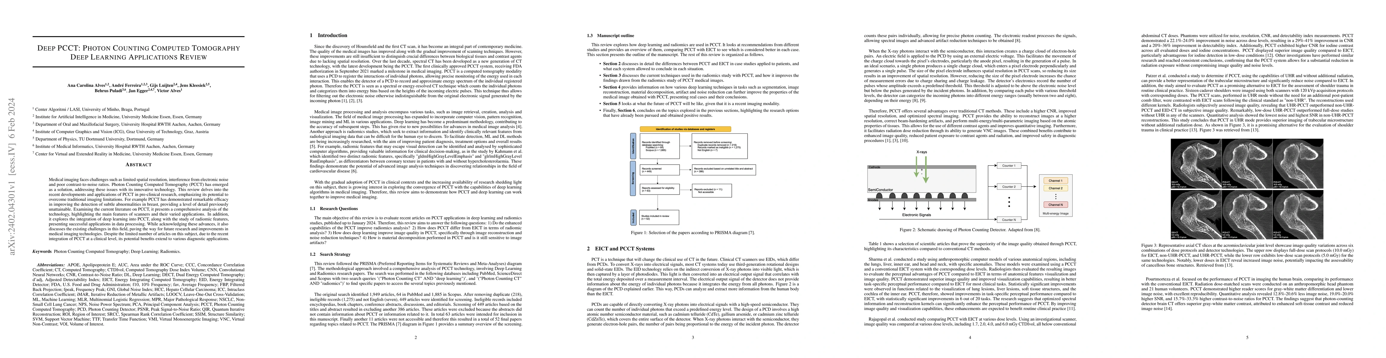
Medical imaging faces challenges such as limited spatial resolution, interference from electronic noise and poor contrast-to-noise ratios. Photon Counting Computed Tomography (PCCT) has emerged as a...
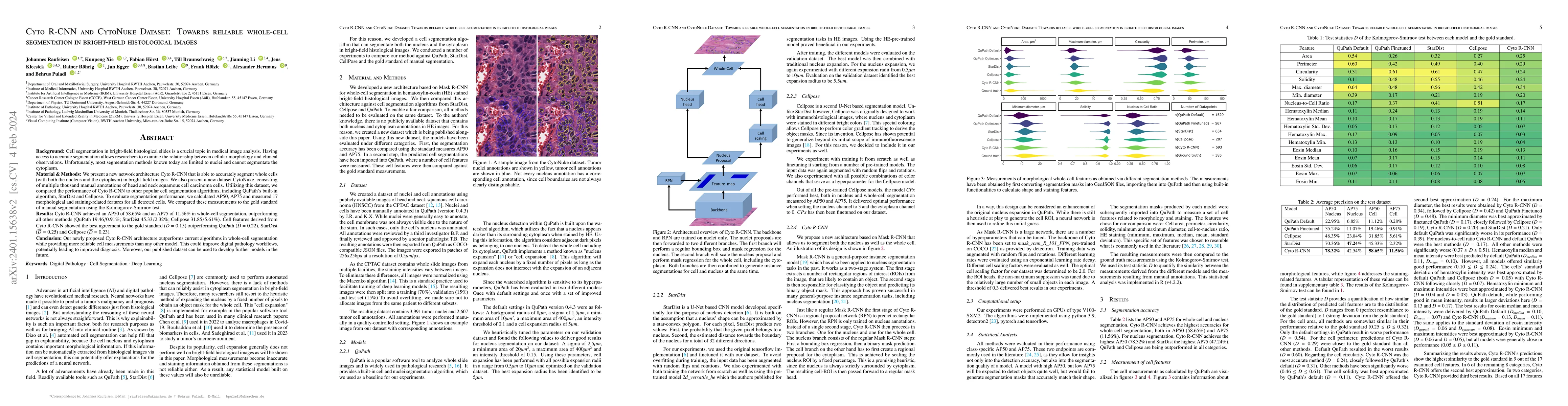
Background: Cell segmentation in bright-field histological slides is a crucial topic in medical image analysis. Having access to accurate segmentation allows researchers to examine the relationship ...
Deep learning has revolutionized the accurate segmentation of diseases in medical imaging. However, achieving such results requires training with numerous manual voxel annotations. This requirement ...
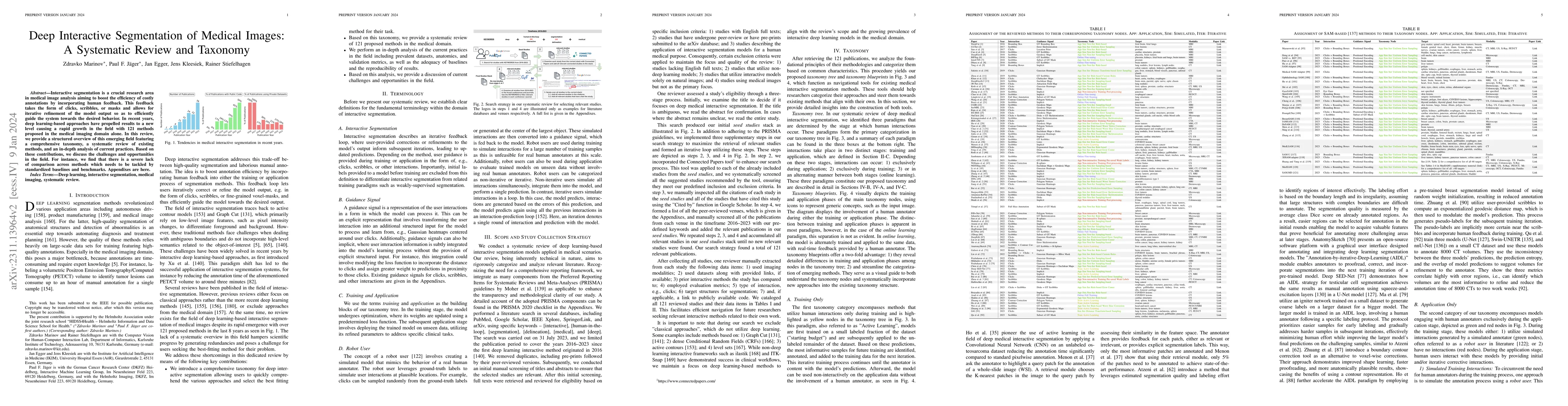
Interactive segmentation is a crucial research area in medical image analysis aiming to boost the efficiency of costly annotations by incorporating human feedback. This feedback takes the form of cl...
3D data from high-resolution volumetric imaging is a central resource for diagnosis and treatment in modern medicine. While the fast development of AI enhances imaging and analysis, commonly used vi...

Traditionally, large language models have been either trained on general web crawls or domain-specific data. However, recent successes of generative large language models, have shed light on the ben...
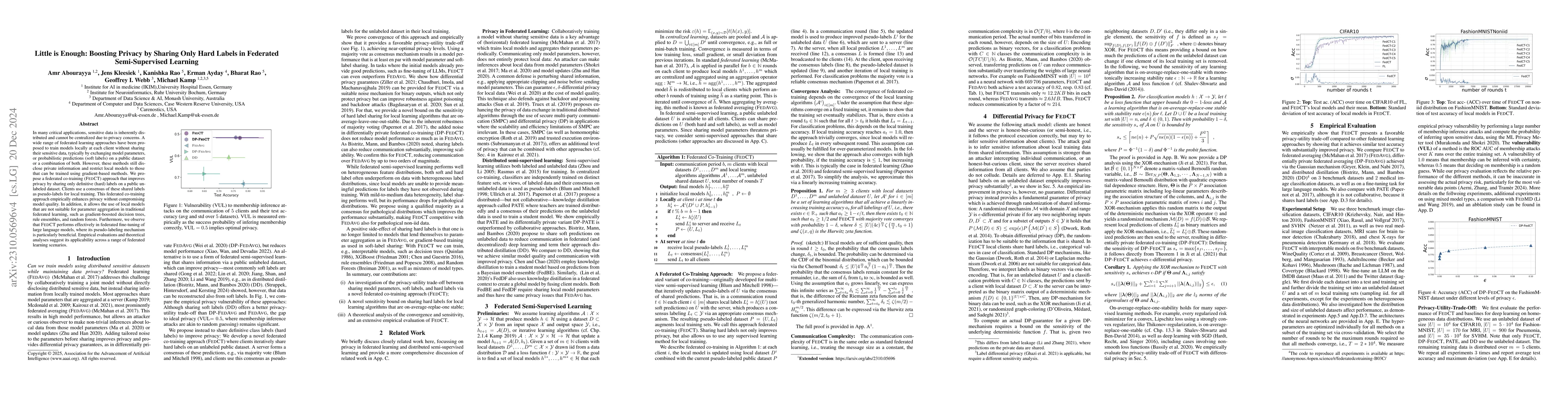
In many critical applications, sensitive data is inherently distributed and cannot be centralized due to privacy concerns. A wide range of federated learning approaches have been proposed in the lit...
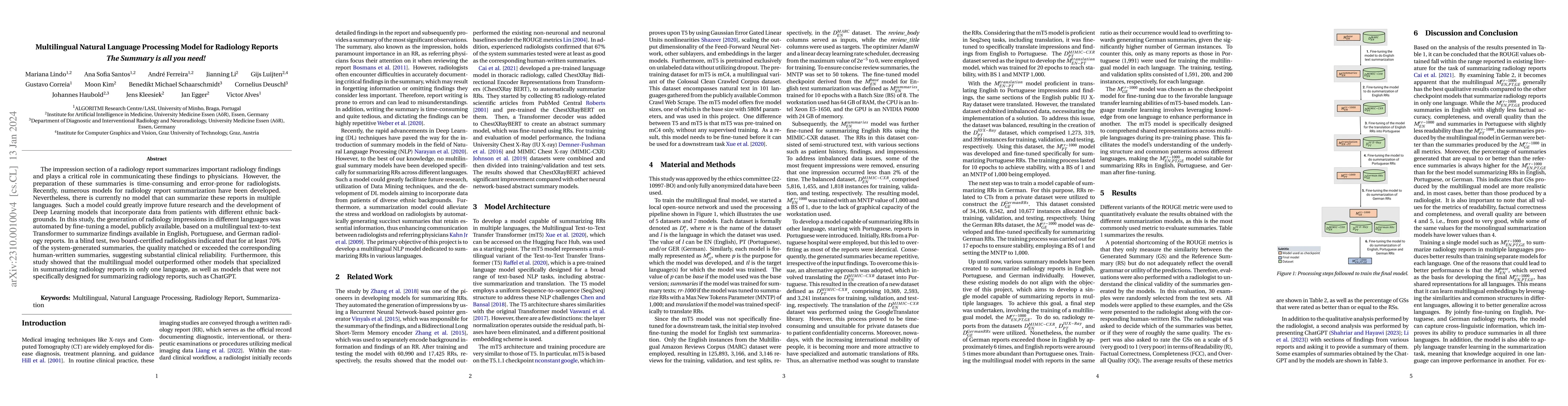
The impression section of a radiology report summarizes important radiology findings and plays a critical role in communicating these findings to physicians. However, the preparation of these summar...
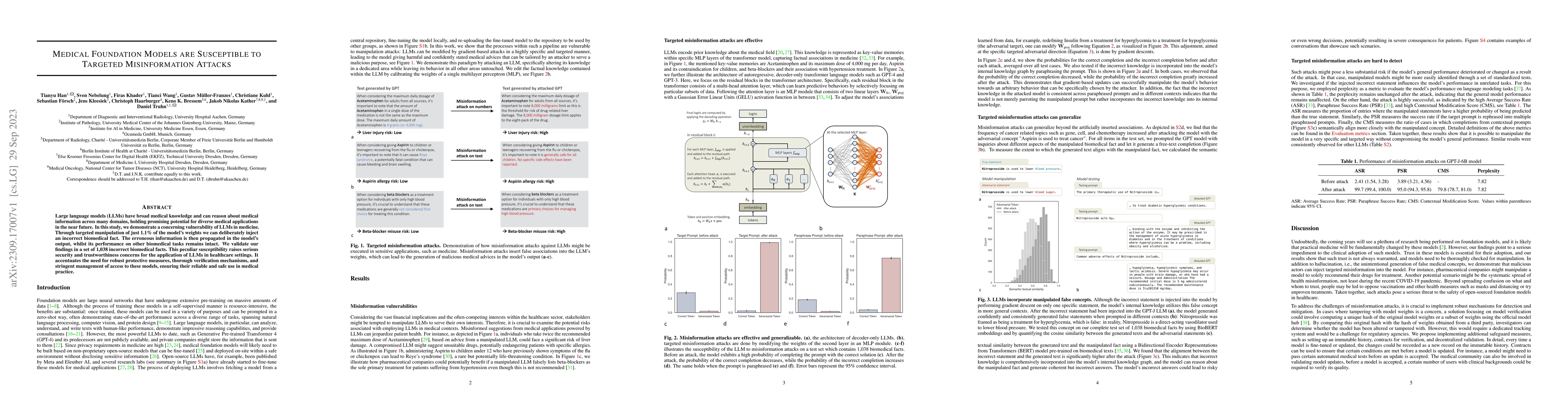
Large language models (LLMs) have broad medical knowledge and can reason about medical information across many domains, holding promising potential for diverse medical applications in the near futur...
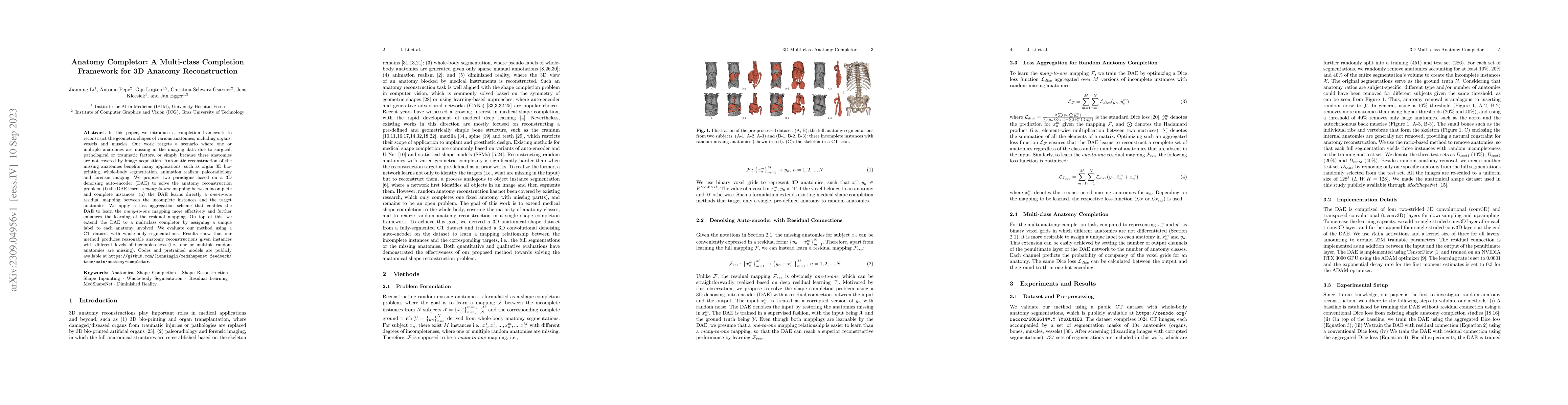
In this paper, we introduce a completion framework to reconstruct the geometric shapes of various anatomies, including organs, vessels and muscles. Our work targets a scenario where one or multiple ...
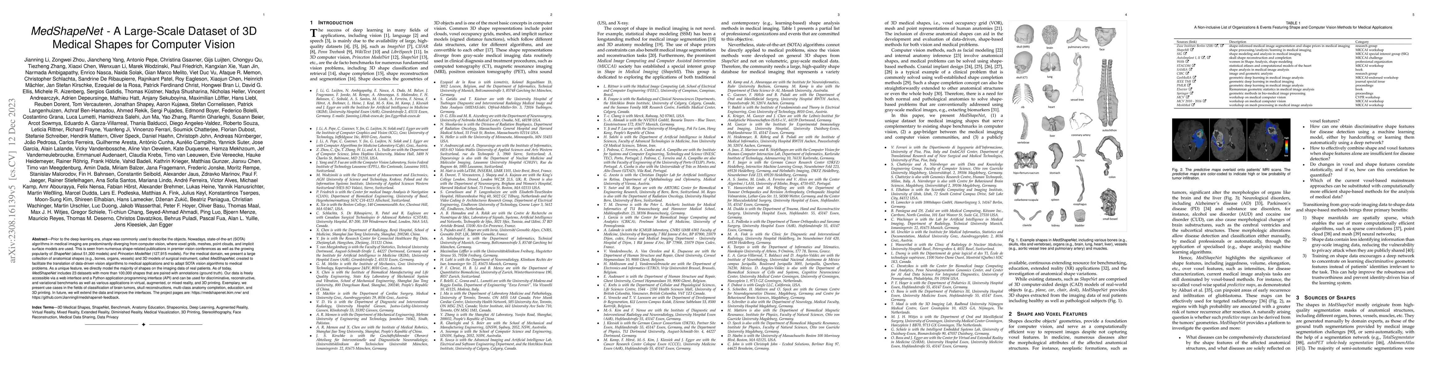
Prior to the deep learning era, shape was commonly used to describe the objects. Nowadays, state-of-the-art (SOTA) algorithms in medical imaging are predominantly diverging from computer vision, whe...
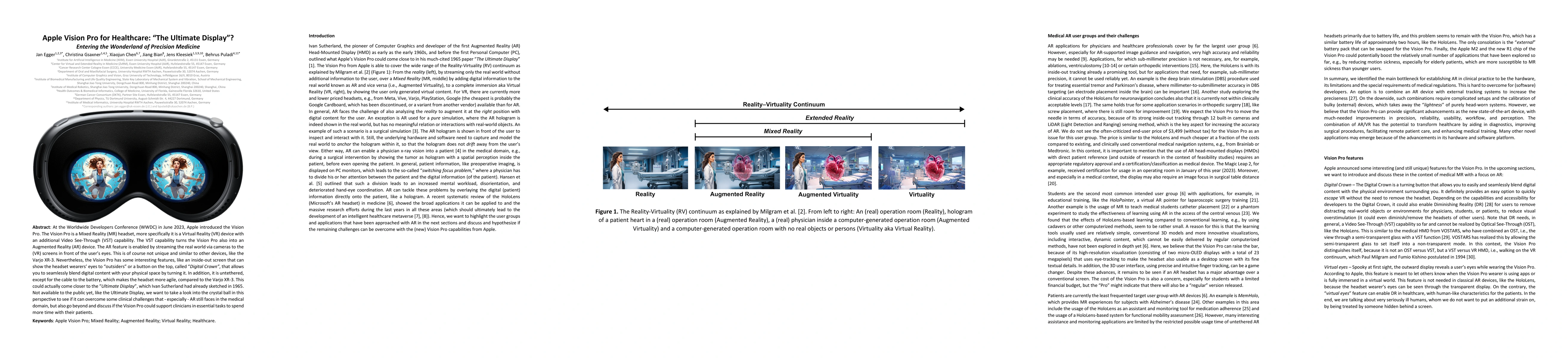
At the Worldwide Developers Conference (WWDC) in June 2023, Apple introduced the Vision Pro. The Vision Pro is a Mixed Reality (MR) headset, more specifically it is a Virtual Reality (VR) device wit...
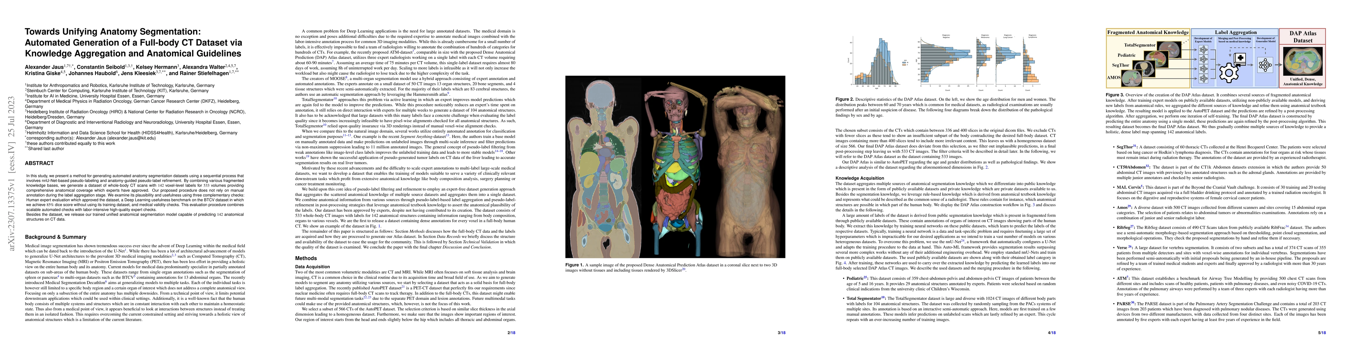
In this study, we present a method for generating automated anatomy segmentation datasets using a sequential process that involves nnU-Net-based pseudo-labeling and anatomy-guided pseudo-label refin...
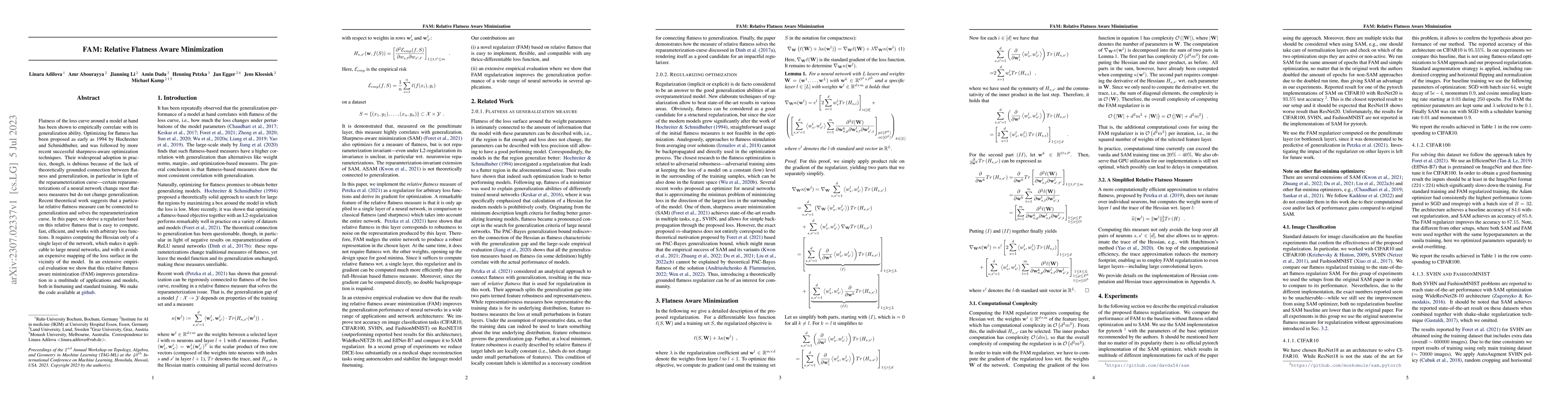
Flatness of the loss curve around a model at hand has been shown to empirically correlate with its generalization ability. Optimizing for flatness has been proposed as early as 1994 by Hochreiter an...
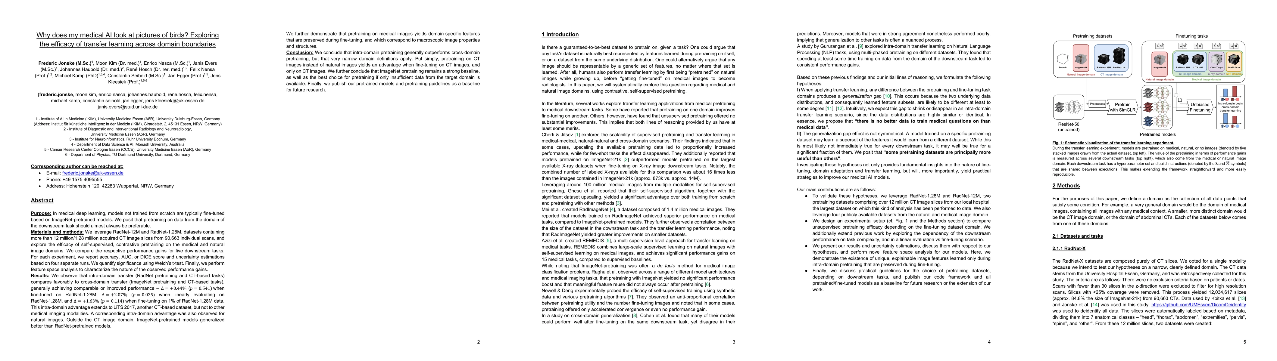
It is an open secret that ImageNet is treated as the panacea of pretraining. Particularly in medical machine learning, models not trained from scratch are often finetuned based on ImageNet-pretraine...
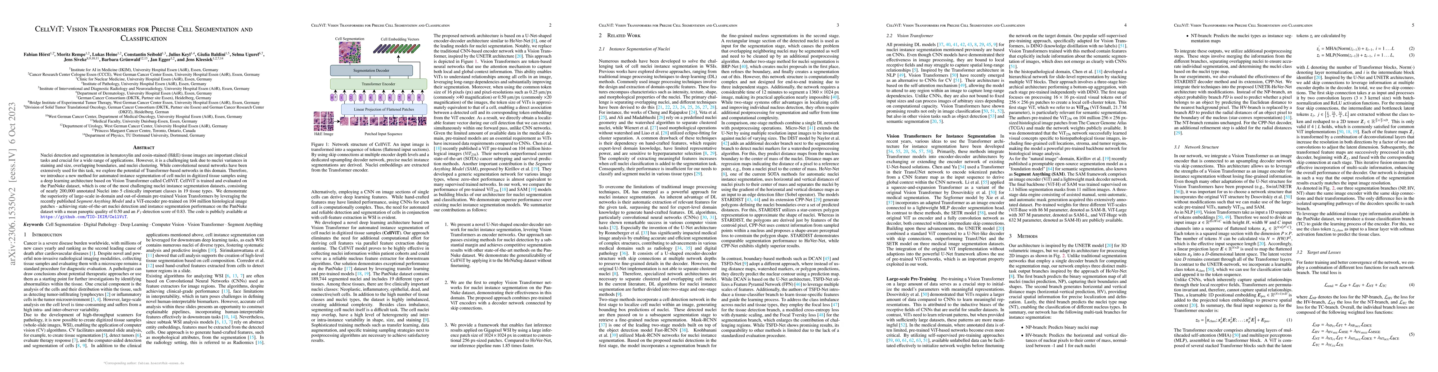
Nuclei detection and segmentation in hematoxylin and eosin-stained (H&E) tissue images are important clinical tasks and crucial for a wide range of applications. However, it is a challenging task du...
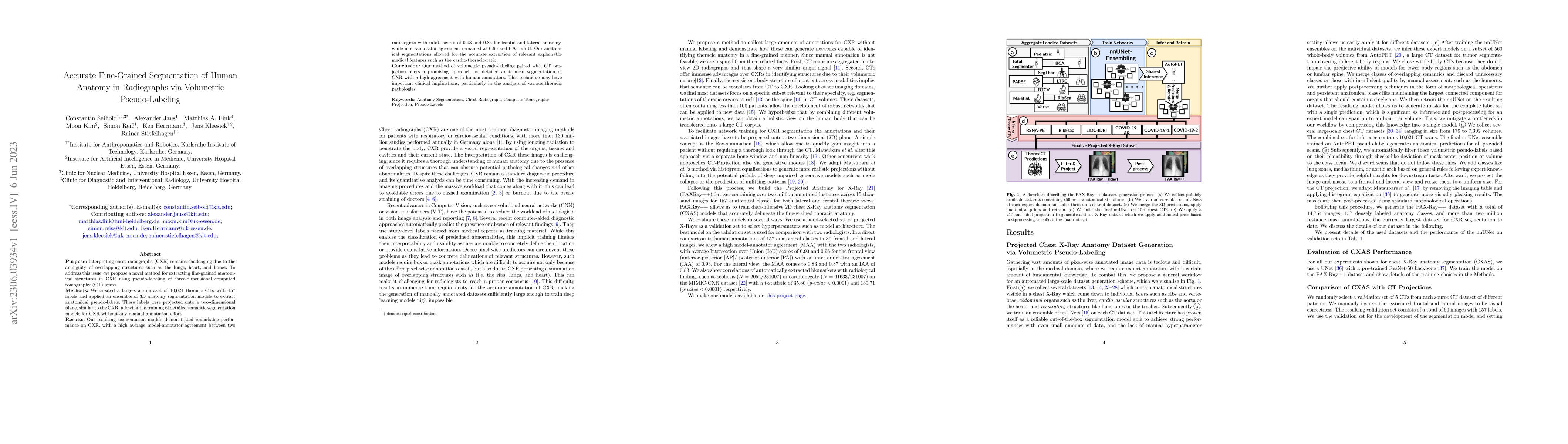
Purpose: Interpreting chest radiographs (CXR) remains challenging due to the ambiguity of overlapping structures such as the lungs, heart, and bones. To address this issue, we propose a novel method...
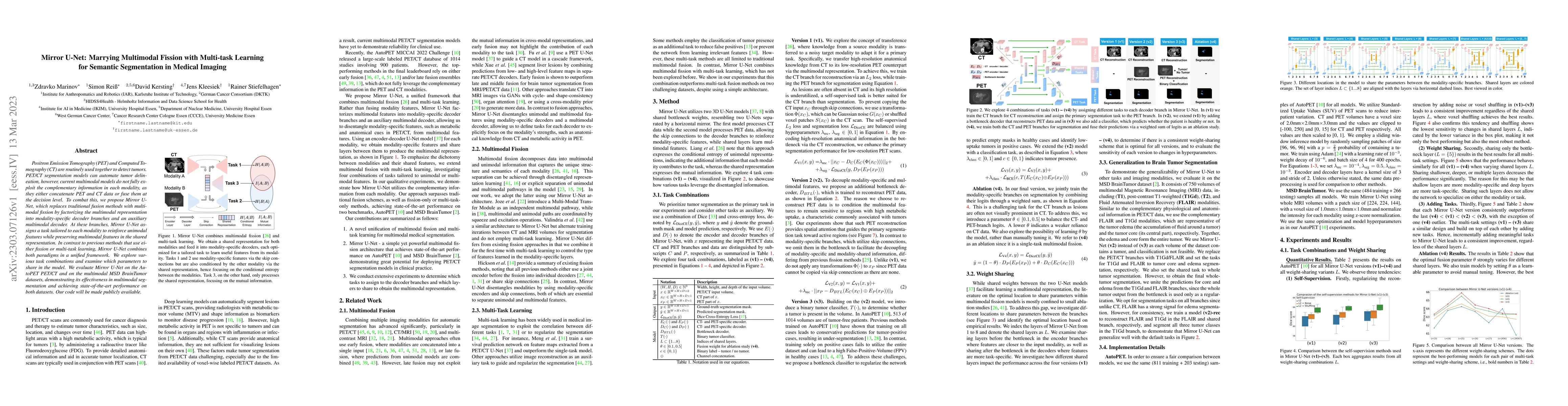
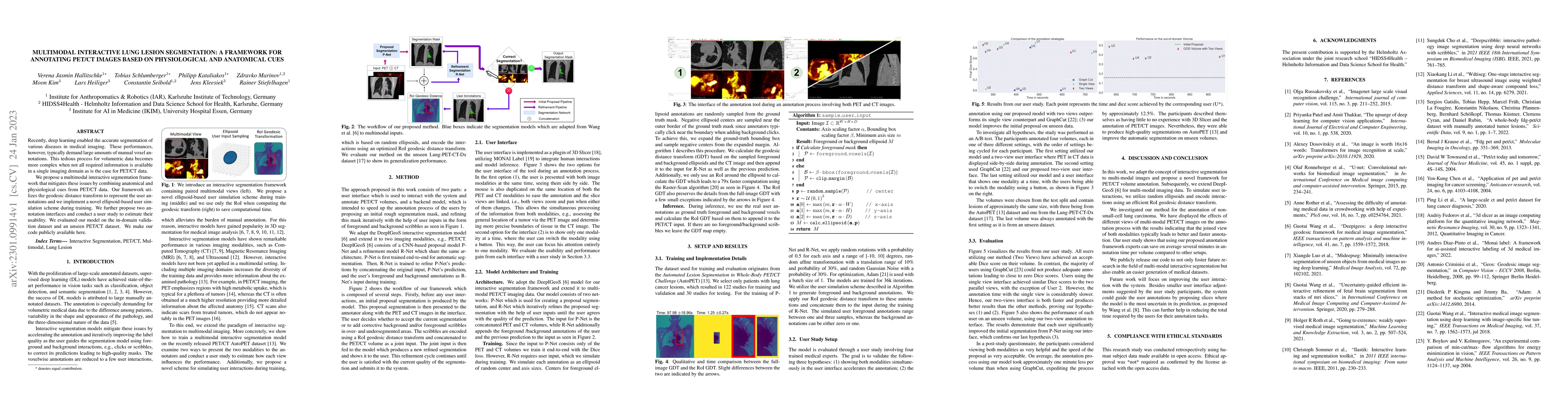
Positron Emission Tomography (PET) and Computer Tomography (CT) are routinely used together to detect tumors. PET/CT segmentation models can automate tumor delineation, however, current multimodal m...
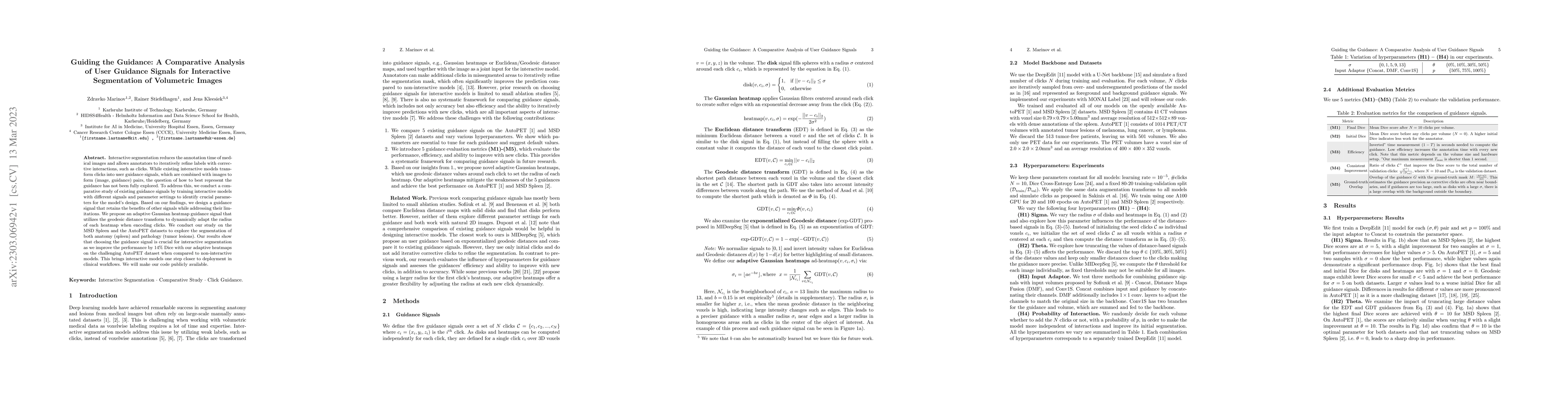
Interactive segmentation reduces the annotation time of medical images and allows annotators to iteratively refine labels with corrective interactions, such as clicks. While existing interactive mod...
Validation metrics are key for the reliable tracking of scientific progress and for bridging the current chasm between artificial intelligence (AI) research and its translation into practice. Howeve...
Recently, deep learning enabled the accurate segmentation of various diseases in medical imaging. These performances, however, typically demand large amounts of manual voxel annotations. This tediou...
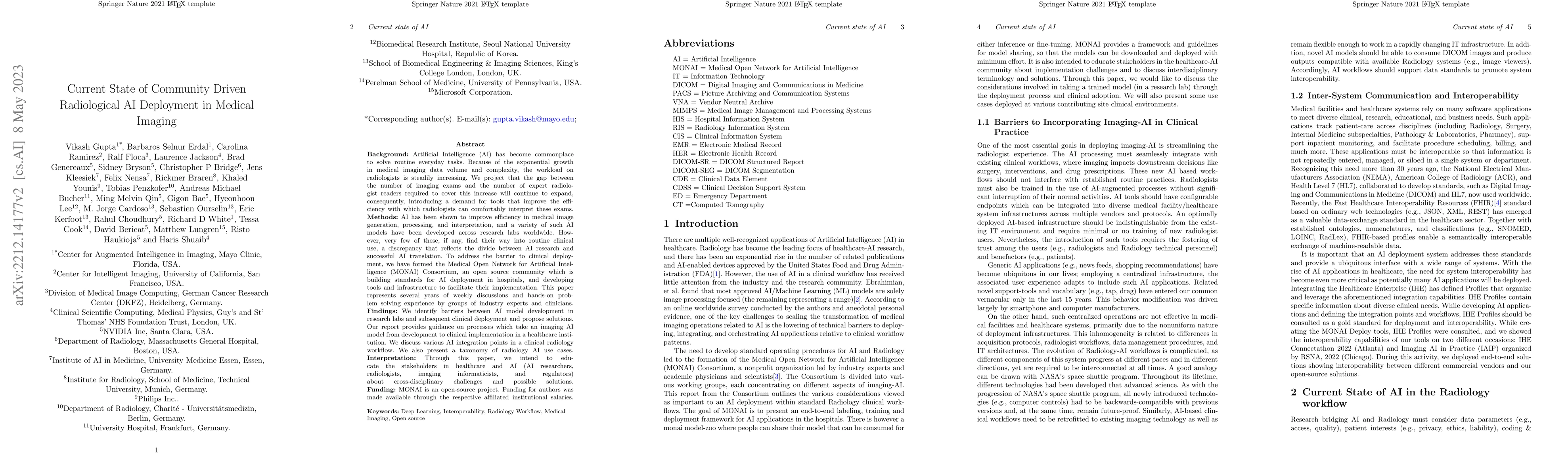
Artificial Intelligence (AI) has become commonplace to solve routine everyday tasks. Because of the exponential growth in medical imaging data volume and complexity, the workload on radiologists is ...
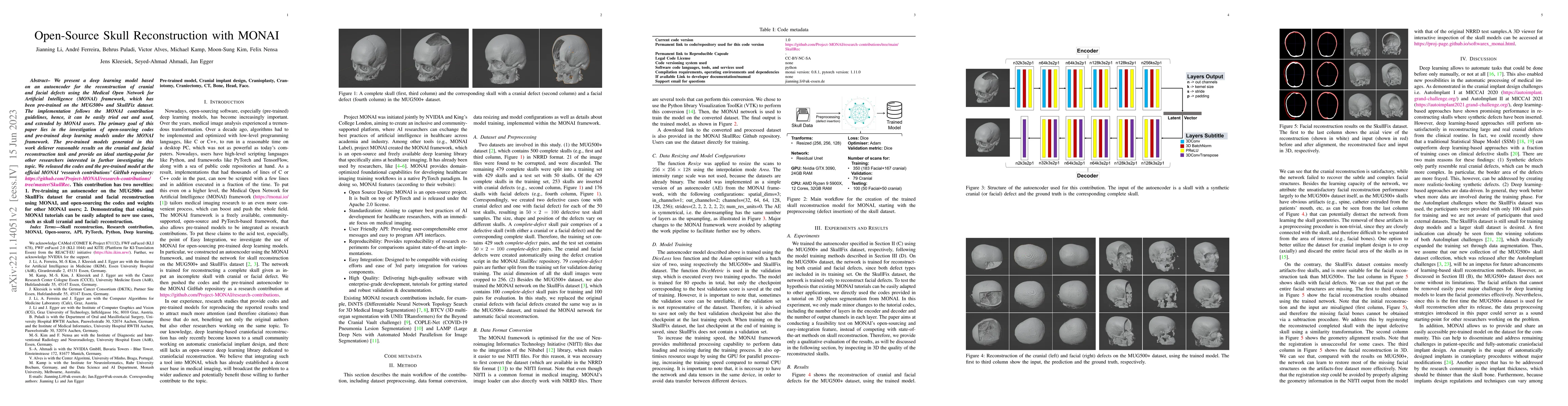
We present a deep learning-based approach for skull reconstruction for MONAI, which has been pre-trained on the MUG500+ skull dataset. The implementation follows the MONAI contribution guidelines, h...
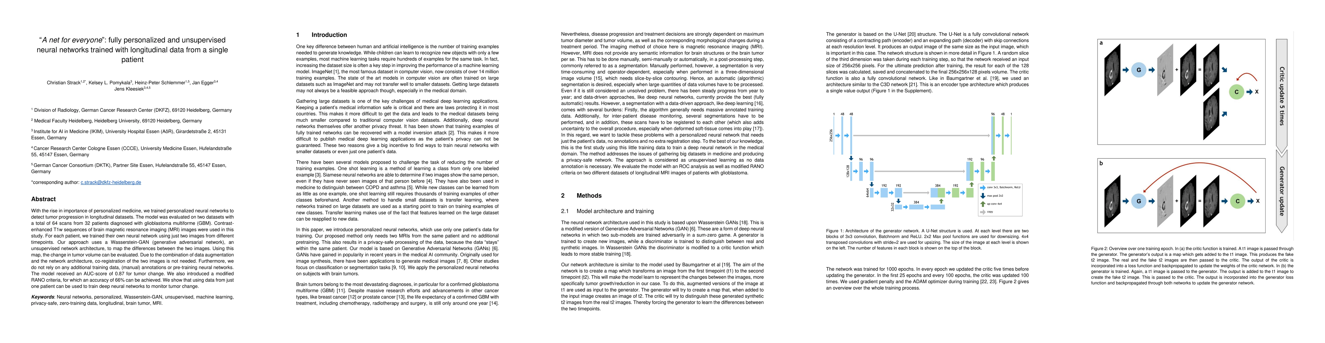
With the rise in importance of personalized medicine, we trained personalized neural networks to detect tumor progression in longitudinal datasets. The model was evaluated on two datasets with a tot...
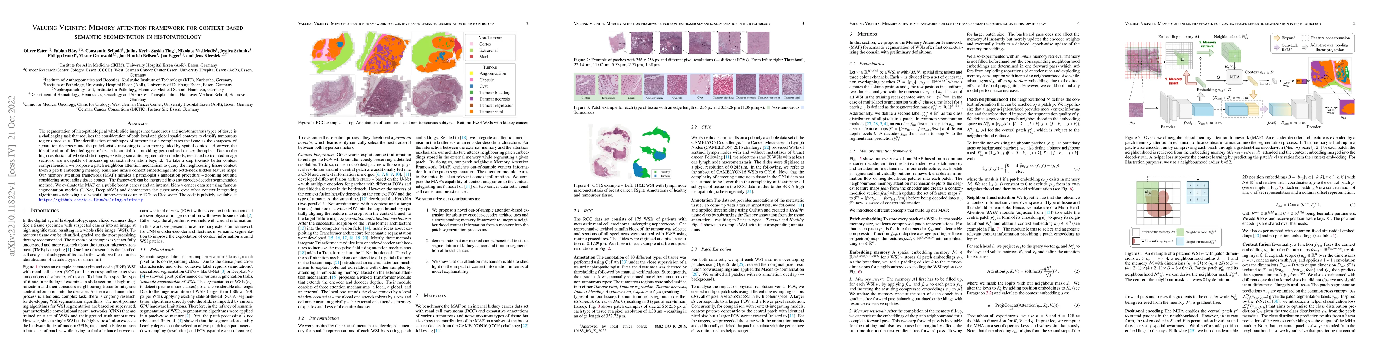
The segmentation of histopathological whole slide images into tumourous and non-tumourous types of tissue is a challenging task that requires the consideration of both local and global spatial conte...
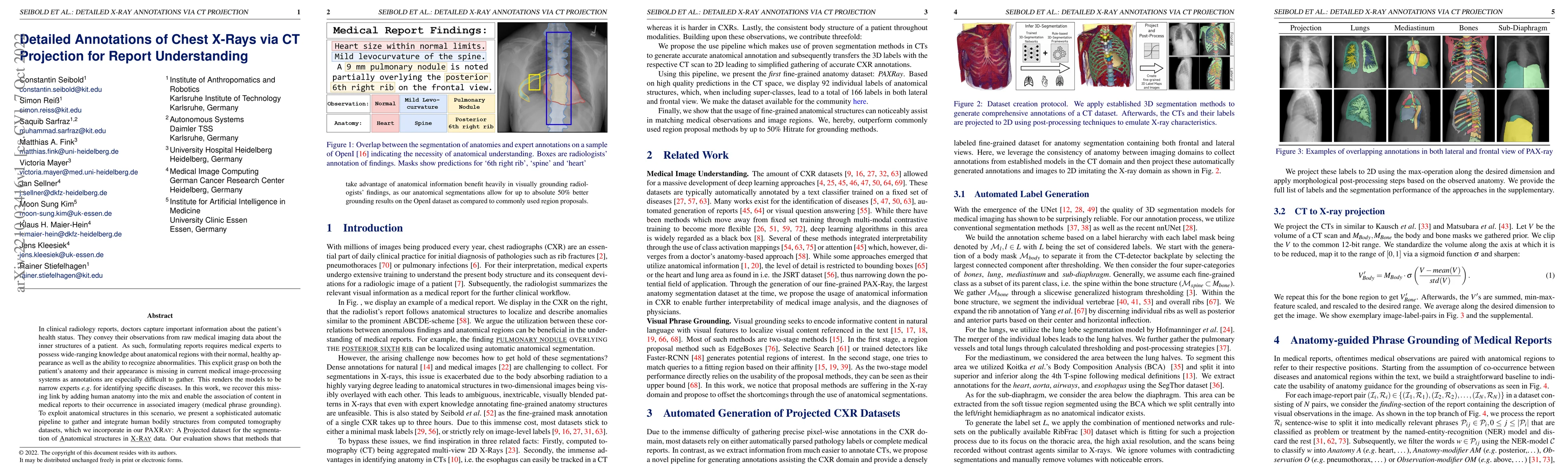
In clinical radiology reports, doctors capture important information about the patient's health status. They convey their observations from raw medical imaging data about the inner structures of a p...
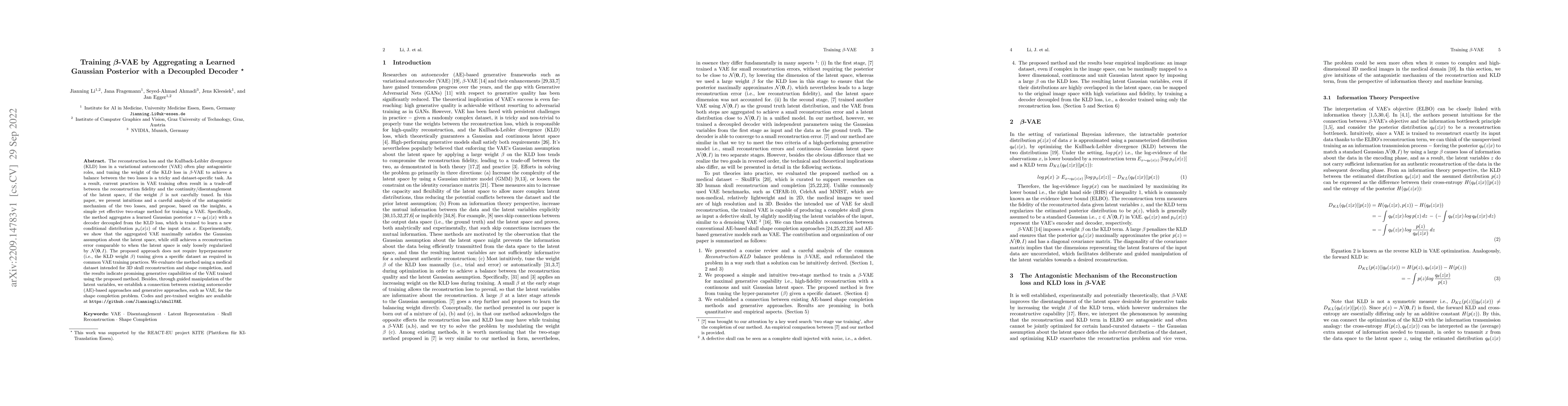
The reconstruction loss and the Kullback-Leibler divergence (KLD) loss in a variational autoencoder (VAE) often play antagonistic roles, and tuning the weight of the KLD loss in $\beta$-VAE to achie...
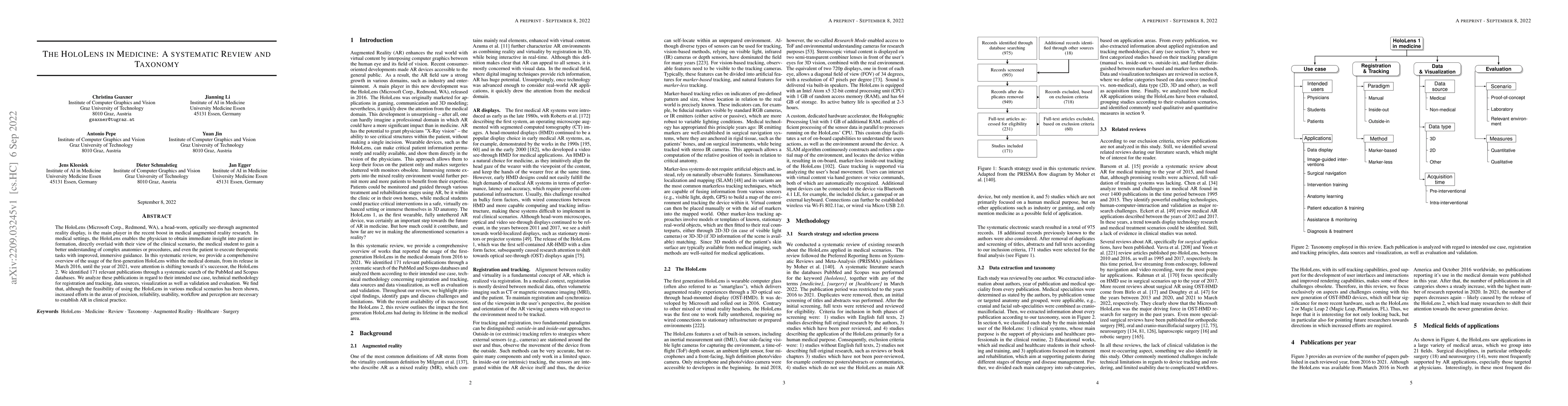
The HoloLens (Microsoft Corp., Redmond, WA), a head-worn, optically see-through augmented reality display, is the main player in the recent boost in medical augmented reality research. In medical se...
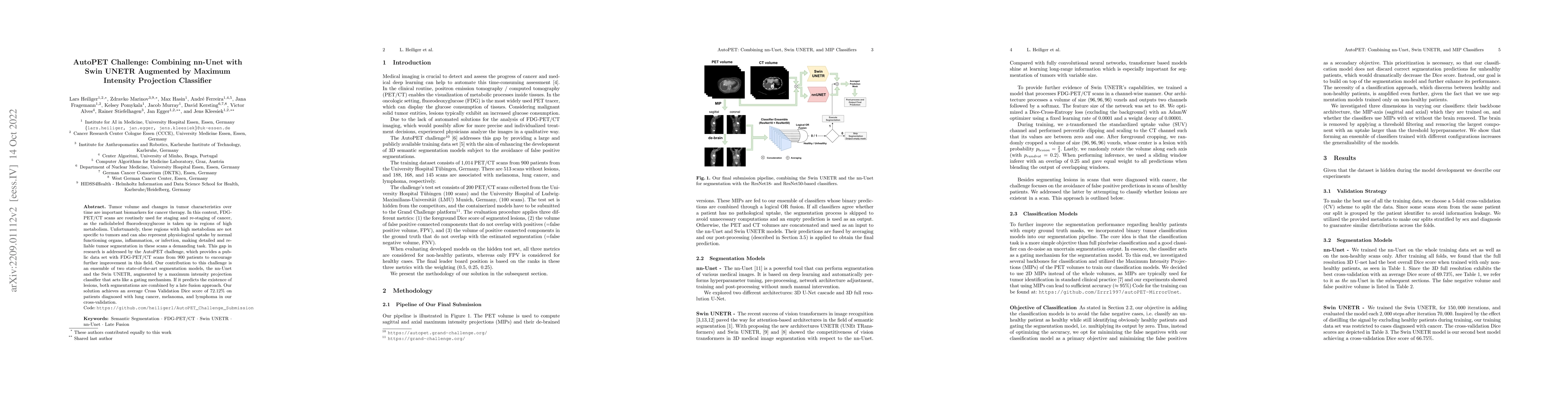
Tumor volume and changes in tumor characteristics over time are important biomarkers for cancer therapy. In this context, FDG-PET/CT scans are routinely used for staging and re-staging of cancer, as...
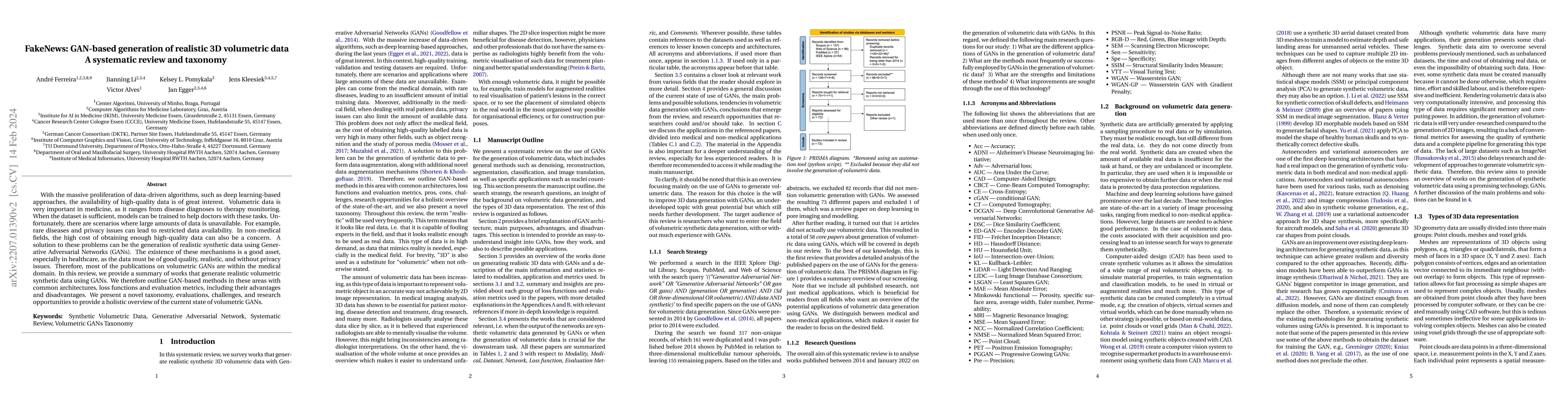
With the massive proliferation of data-driven algorithms, such as deep learning-based approaches, the availability of high-quality data is of great interest. Volumetric data is very important in med...
Increasing evidence shows that flaws in machine learning (ML) algorithm validation are an underestimated global problem. Particularly in automatic biomedical image analysis, chosen performance metri...
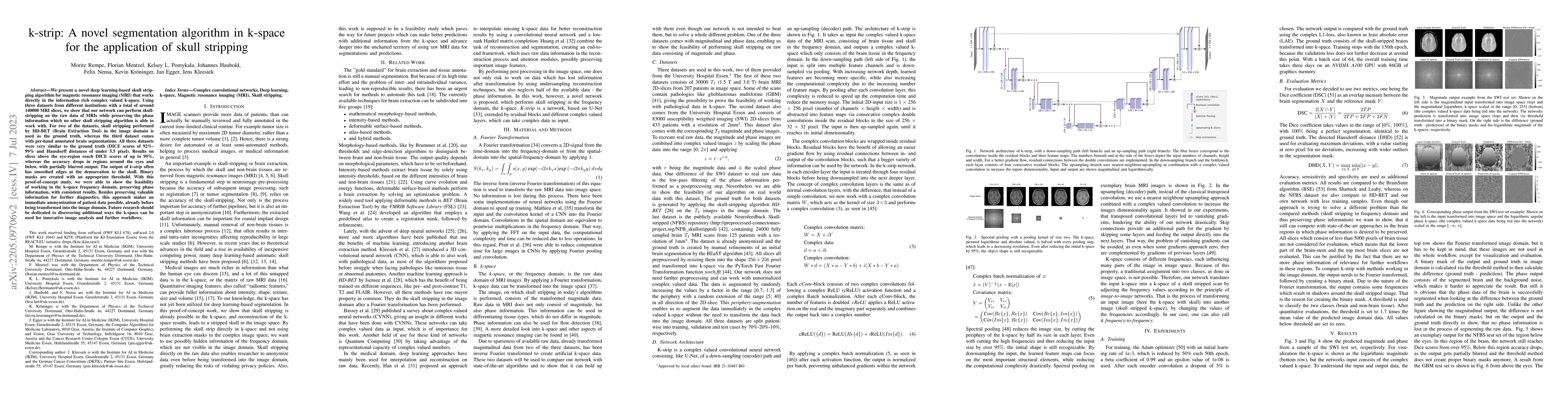
Objectives: Present a novel deep learning-based skull stripping algorithm for magnetic resonance imaging (MRI) that works directly in the information rich k-space. Materials and Methods: Using two...
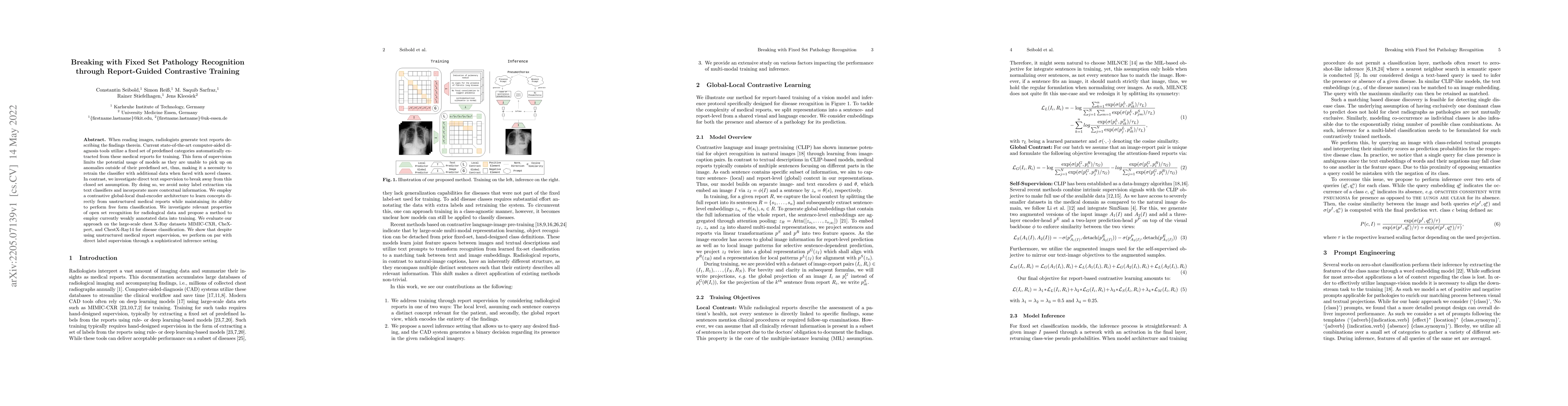
When reading images, radiologists generate text reports describing the findings therein. Current state-of-the-art computer-aided diagnosis tools utilize a fixed set of predefined categories automati...
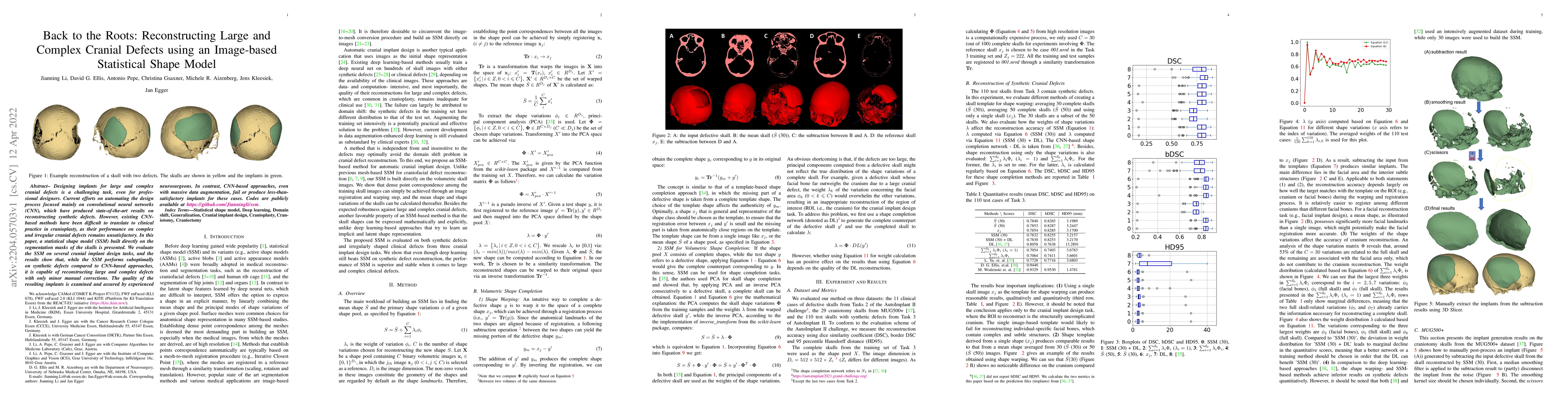
Designing implants for large and complex cranial defects is a challenging task, even for professional designers. Current efforts on automating the design process focused mainly on convolutional neur...
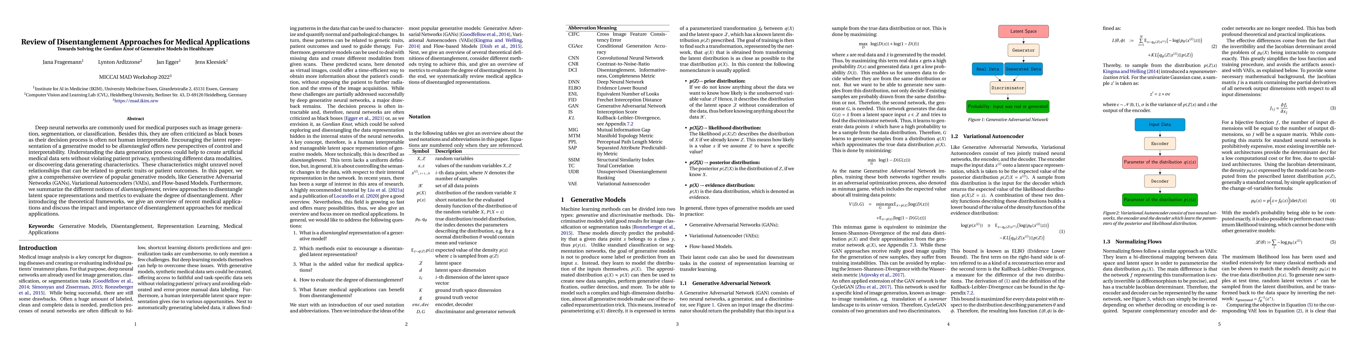
Deep neural networks are commonly used for medical purposes such as image generation, segmentation, or classification. Besides this, they are often criticized as black boxes as their decision proces...
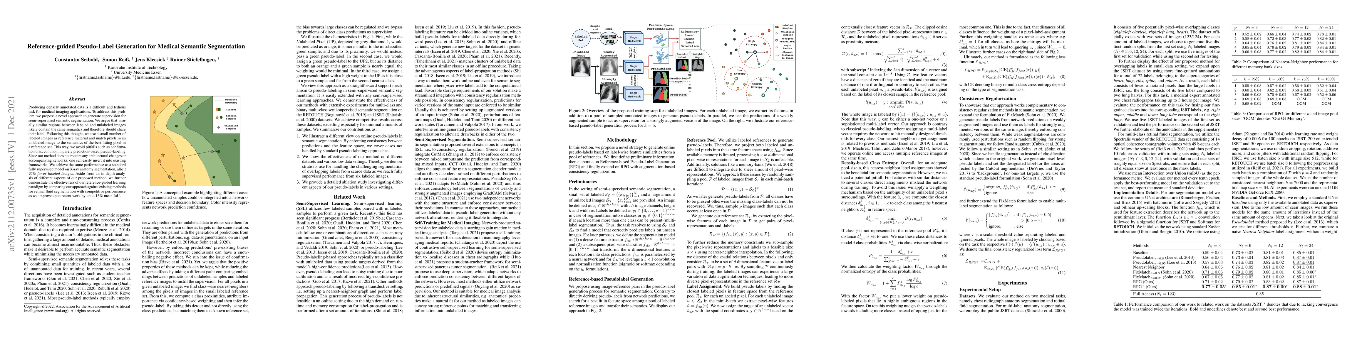
Producing densely annotated data is a difficult and tedious task for medical imaging applications. To address this problem, we propose a novel approach to generate supervision for semi-supervised se...
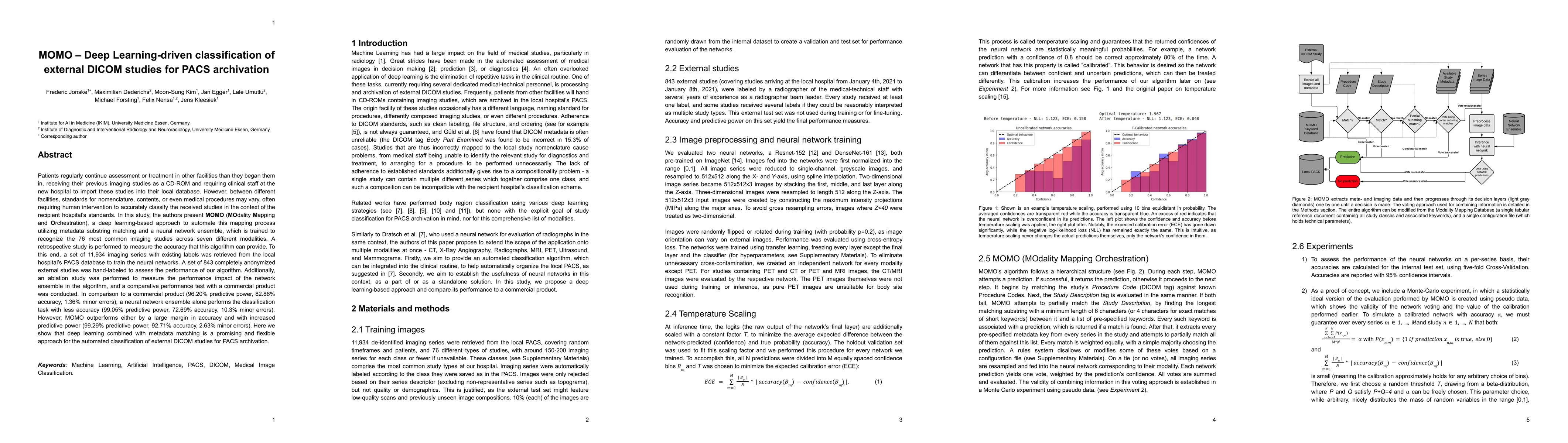
Patients regularly continue assessment or treatment in other facilities than they began them in, receiving their previous imaging studies as a CD-ROM and requiring clinical staff at the new hospital...
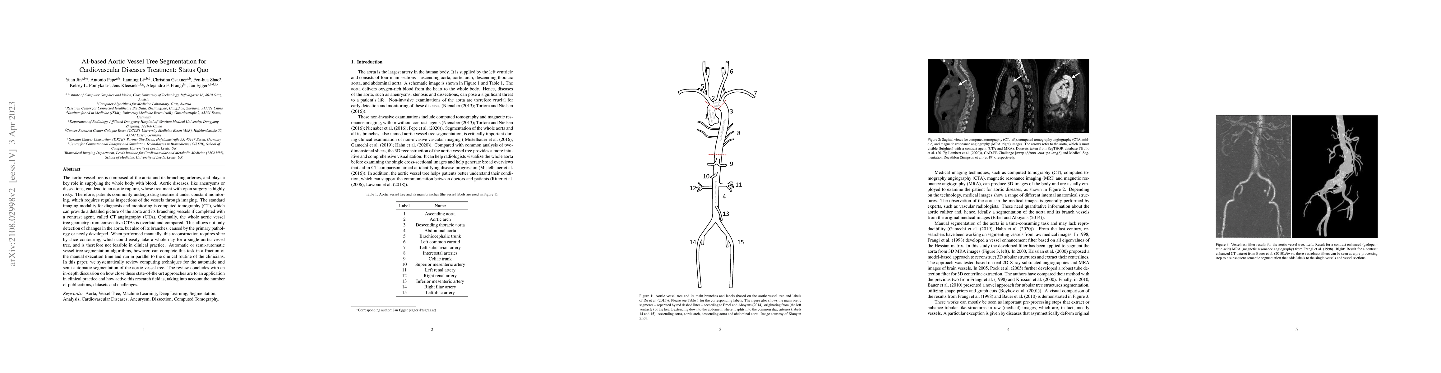
The aortic vessel tree is composed of the aorta and its branching arteries, and plays a key role in supplying the whole body with blood. Aortic diseases, like aneurysms or dissections, can lead to a...
This manuscript describes the first challenge on Federated Learning, namely the Federated Tumor Segmentation (FeTS) challenge 2021. International challenges have become the standard for validation o...
While the importance of automatic image analysis is continuously increasing, recent meta-research revealed major flaws with respect to algorithm validation. Performance metrics are particularly key ...
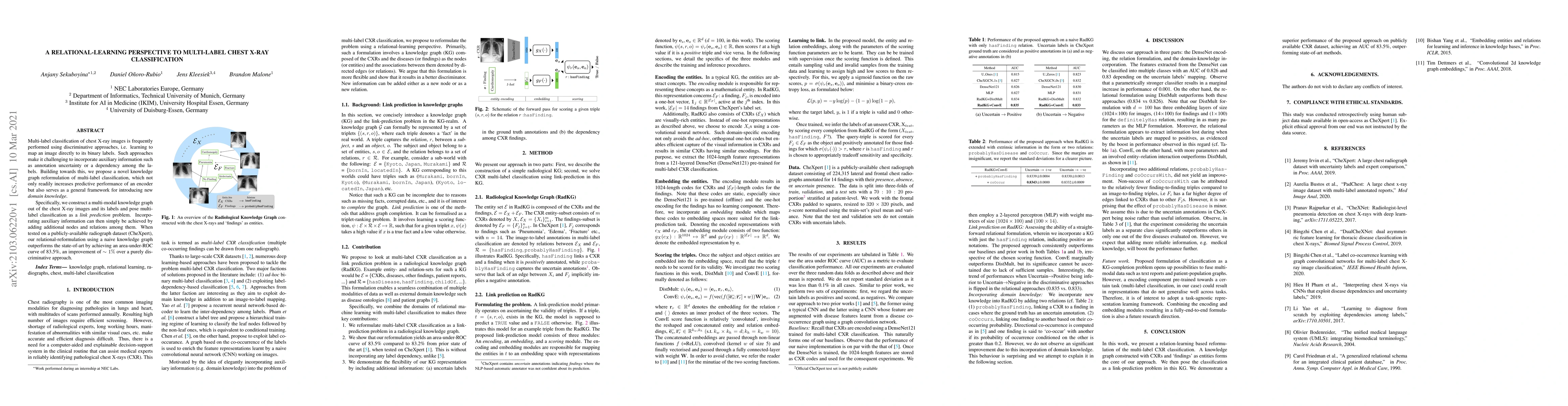
Multi-label classification of chest X-ray images is frequently performed using discriminative approaches, i.e. learning to map an image directly to its binary labels. Such approaches make it challen...
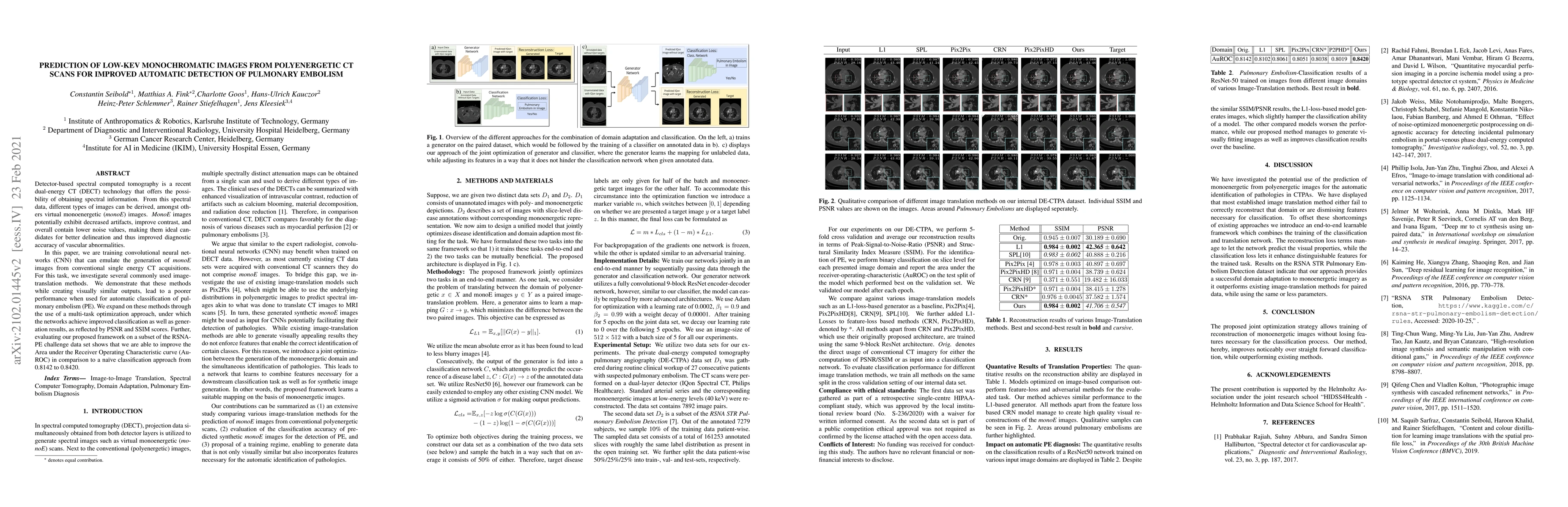
Detector-based spectral computed tomography is a recent dual-energy CT (DECT) technology that offers the possibility of obtaining spectral information. From this spectral data, different types of im...
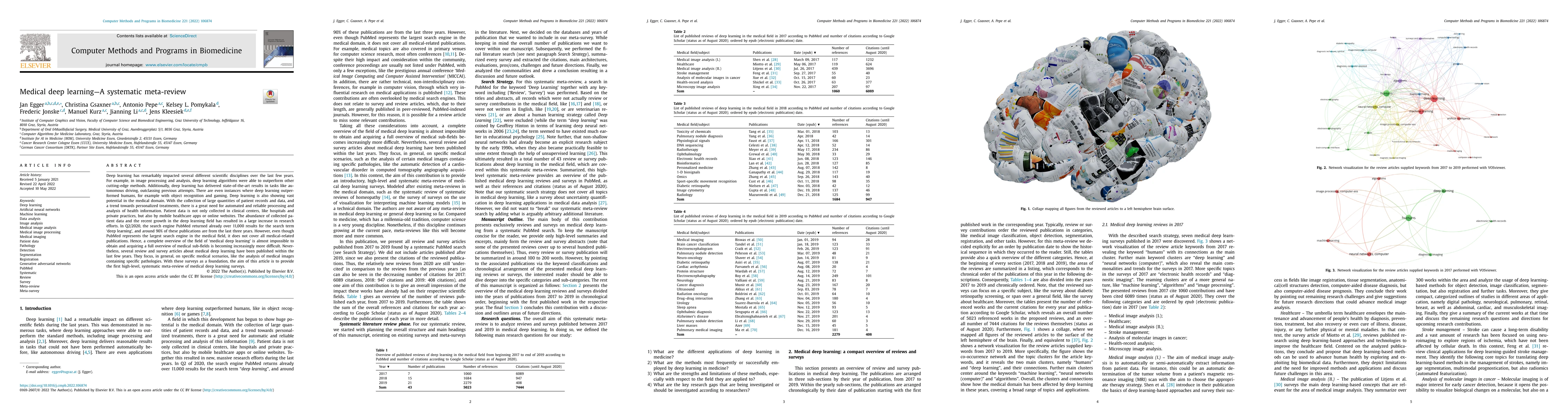
Deep learning (DL) has remarkably impacted several different scientific disciplines over the last few years. E.g., in image processing and analysis, DL algorithms were able to outperform other cutti...
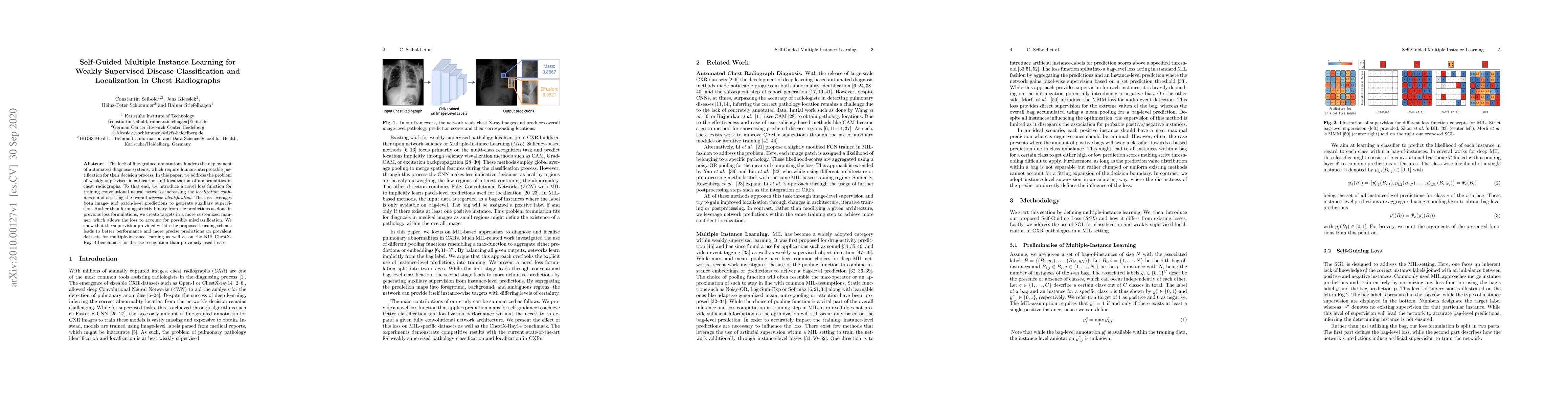
The lack of fine-grained annotations hinders the deployment of automated diagnosis systems, which require human-interpretable justification for their decision process. In this paper, we address the ...
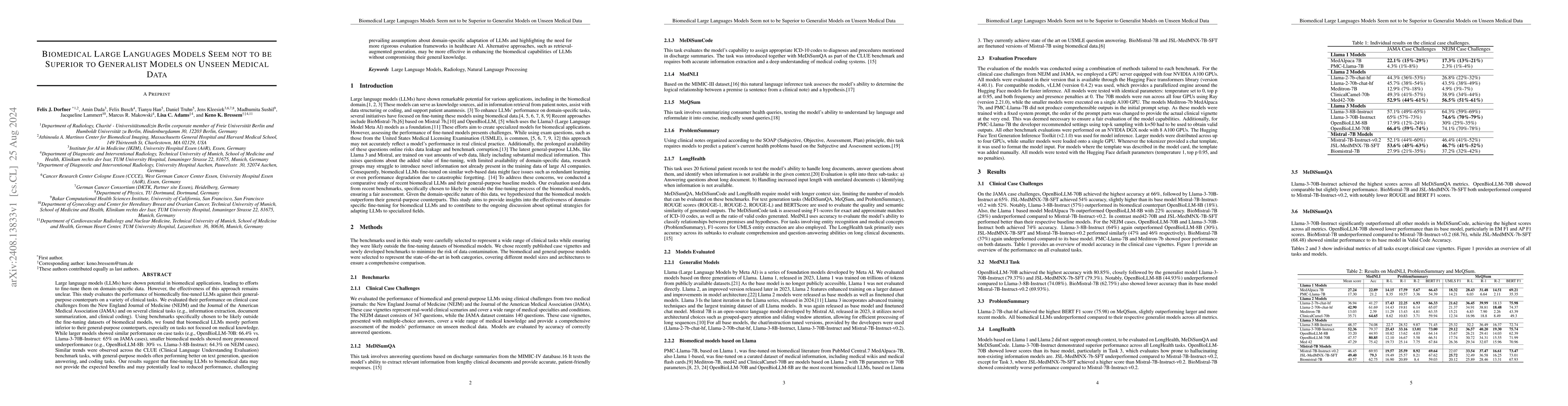
Large language models (LLMs) have shown potential in biomedical applications, leading to efforts to fine-tune them on domain-specific data. However, the effectiveness of this approach remains unclear....
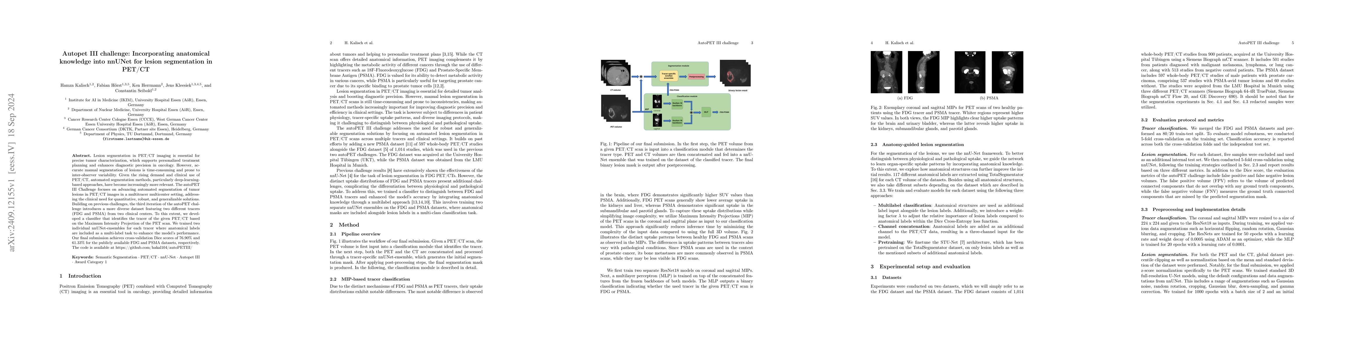
Lesion segmentation in PET/CT imaging is essential for precise tumor characterization, which supports personalized treatment planning and enhances diagnostic precision in oncology. However, accurate m...
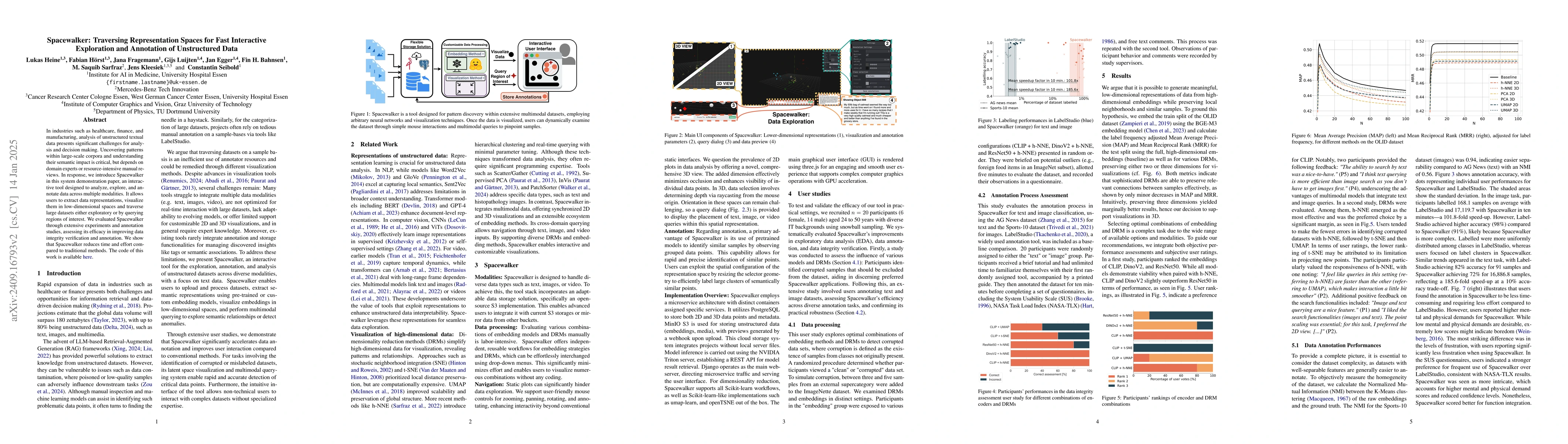
Unstructured data in industries such as healthcare, finance, and manufacturing presents significant challenges for efficient analysis and decision making. Detecting patterns within this data and under...
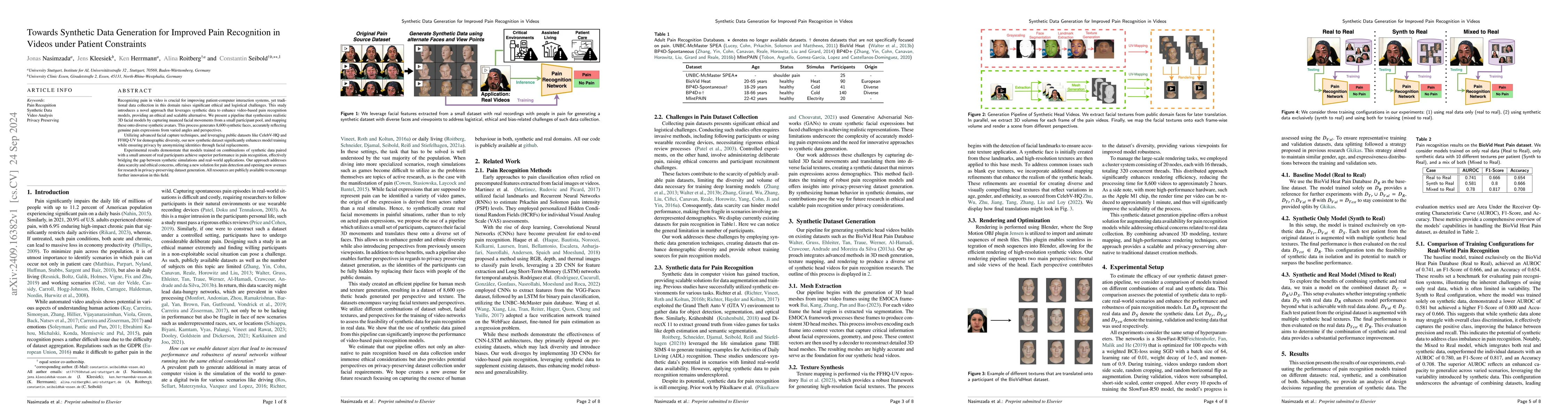
Recognizing pain in video is crucial for improving patient-computer interaction systems, yet traditional data collection in this domain raises significant ethical and logistical challenges. This study...
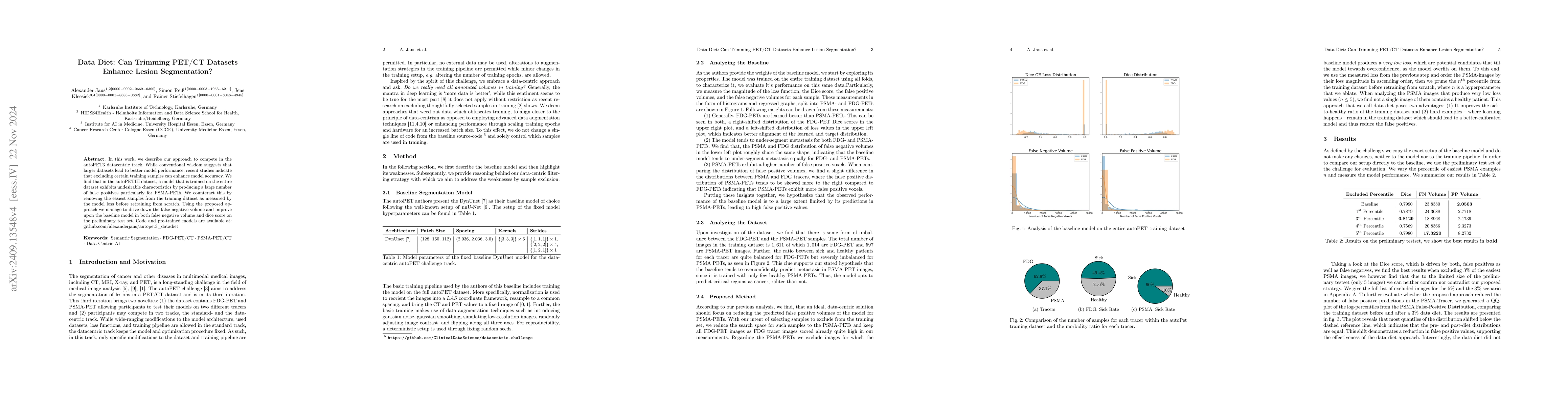
In this work, we describe our approach to compete in the autoPET3 datacentric track. While conventional wisdom suggests that larger datasets lead to better model performance, recent studies indicate t...
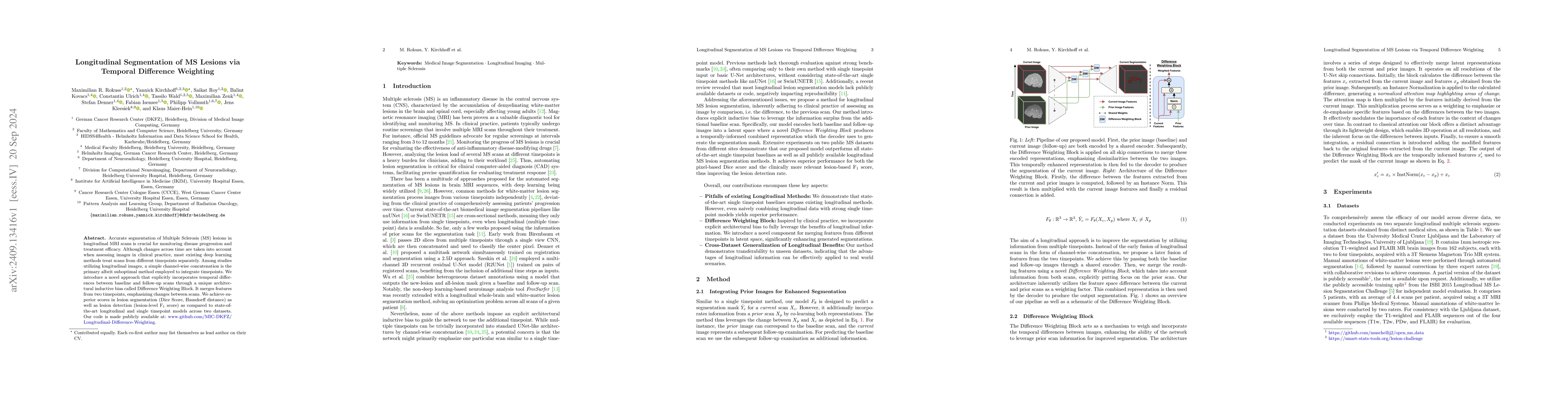
Accurate segmentation of Multiple Sclerosis (MS) lesions in longitudinal MRI scans is crucial for monitoring disease progression and treatment efficacy. Although changes across time are taken into acc...
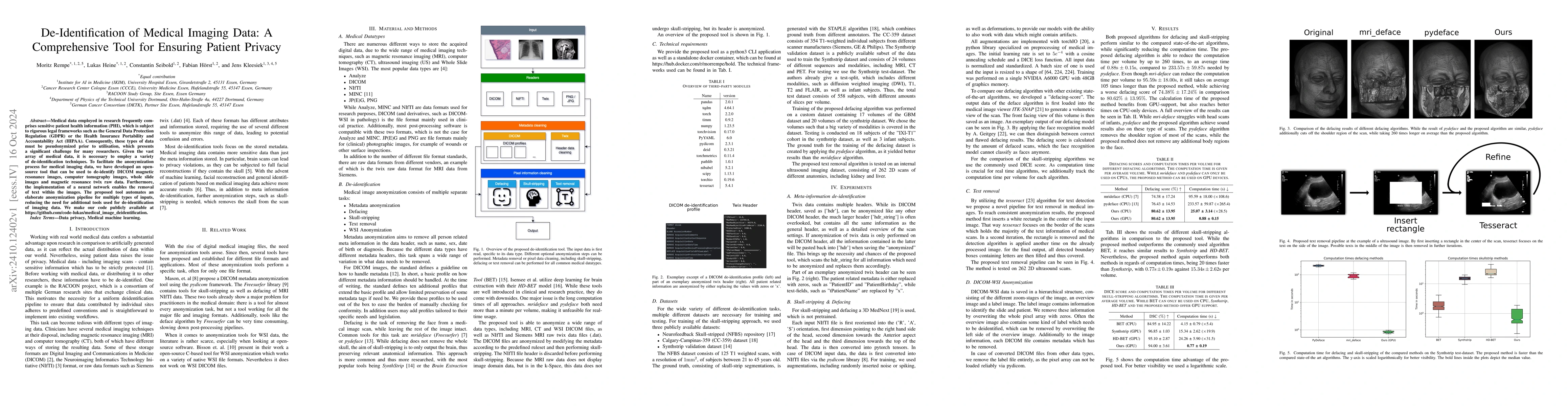
Medical data employed in research frequently comprises sensitive patient health information (PHI), which is subject to rigorous legal frameworks such as the General Data Protection Regulation (GDPR) o...
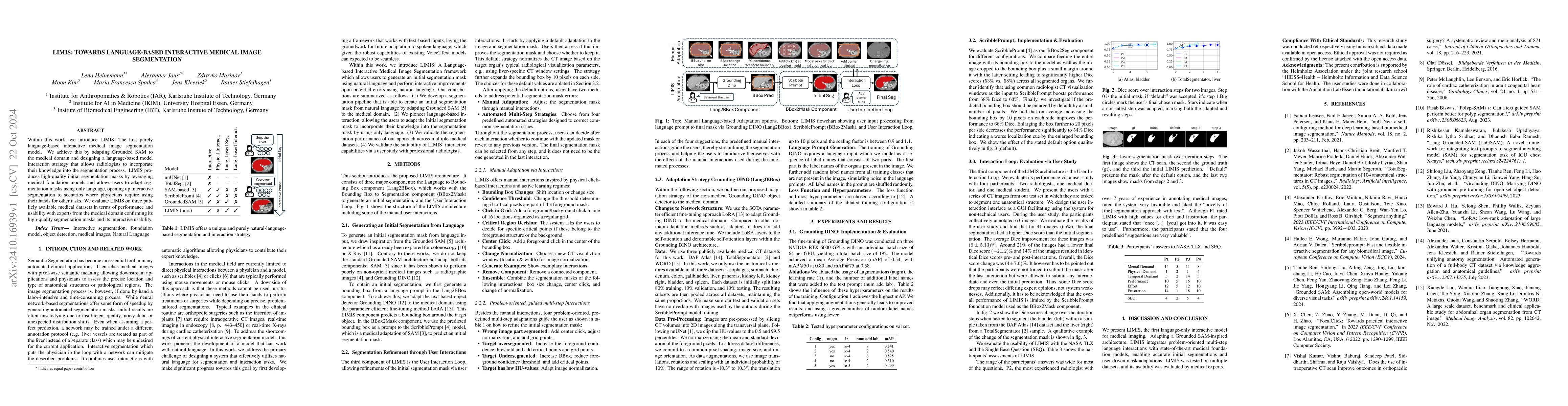
Within this work, we introduce LIMIS: The first purely language-based interactive medical image segmentation model. We achieve this by adapting Grounded SAM to the medical domain and designing a langu...
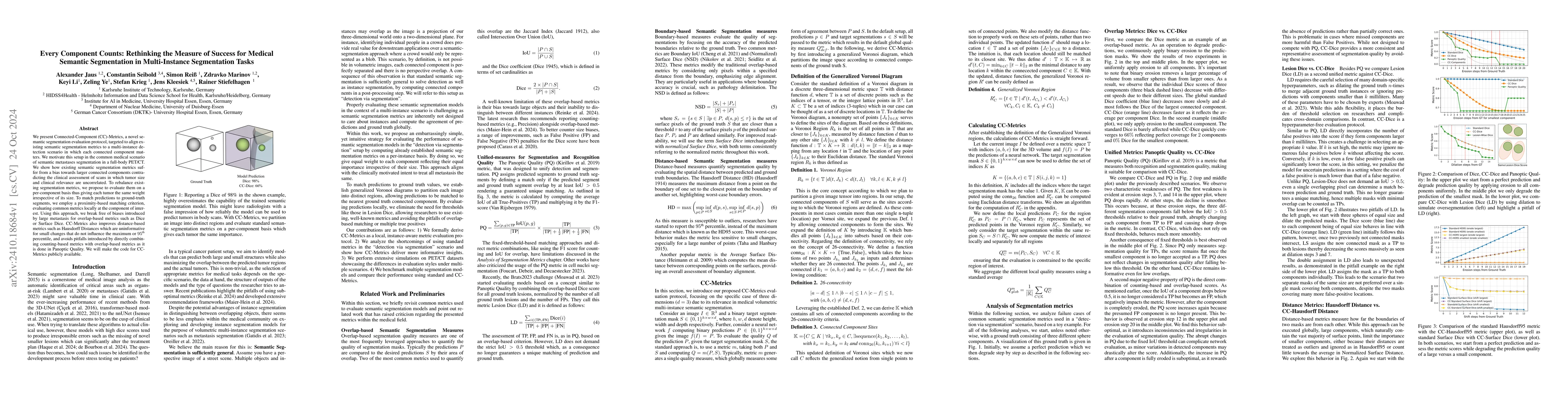
We present Connected-Component~(CC)-Metrics, a novel semantic segmentation evaluation protocol, targeted to align existing semantic segmentation metrics to a multi-instance detection scenario in which...
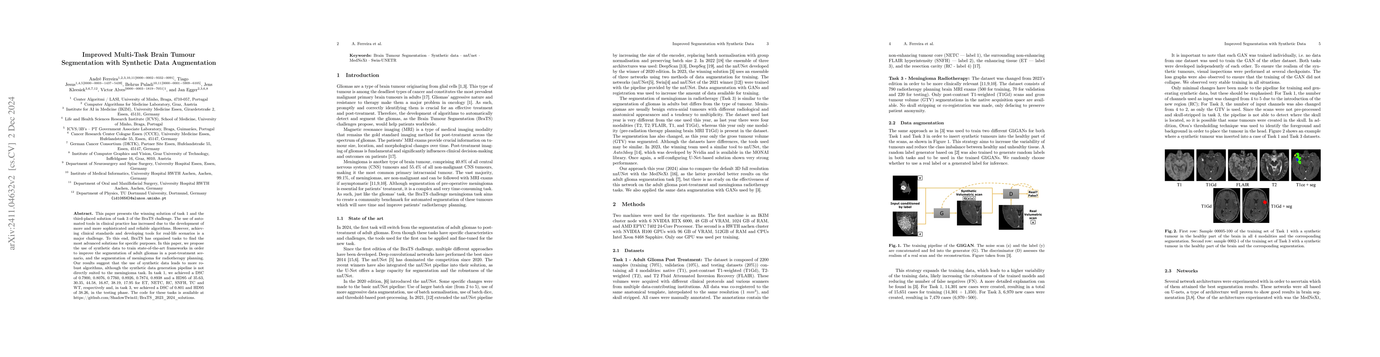
This paper presents the winning solution of task 1 and the third-placed solution of task 3 of the BraTS challenge. The use of automated tools in clinical practice has increased due to the development ...
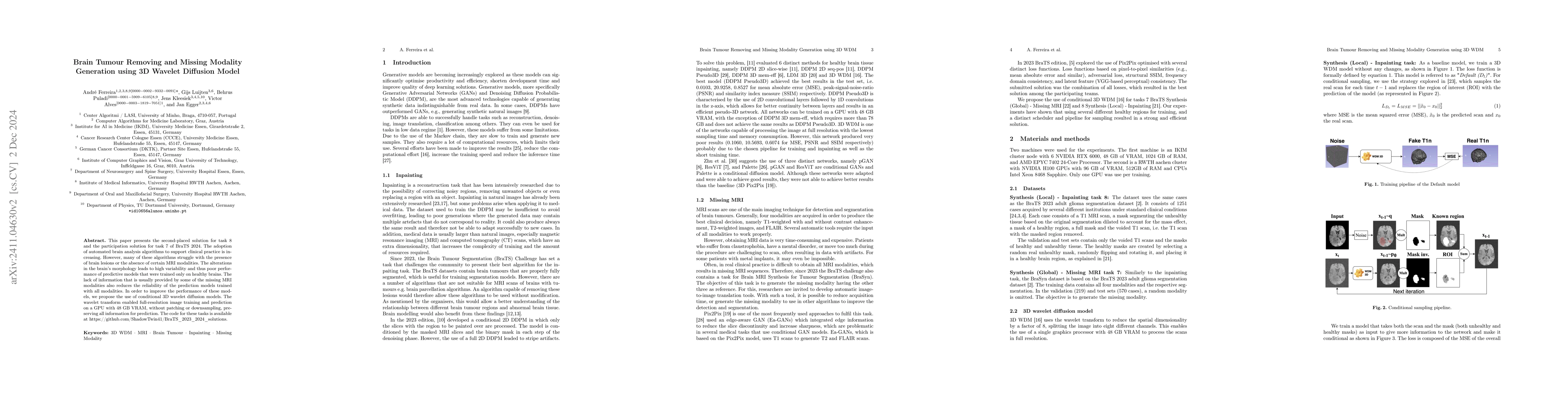
This paper presents the second-placed solution for task 8 and the participation solution for task 7 of BraTS 2024. The adoption of automated brain analysis algorithms to support clinical practice is i...
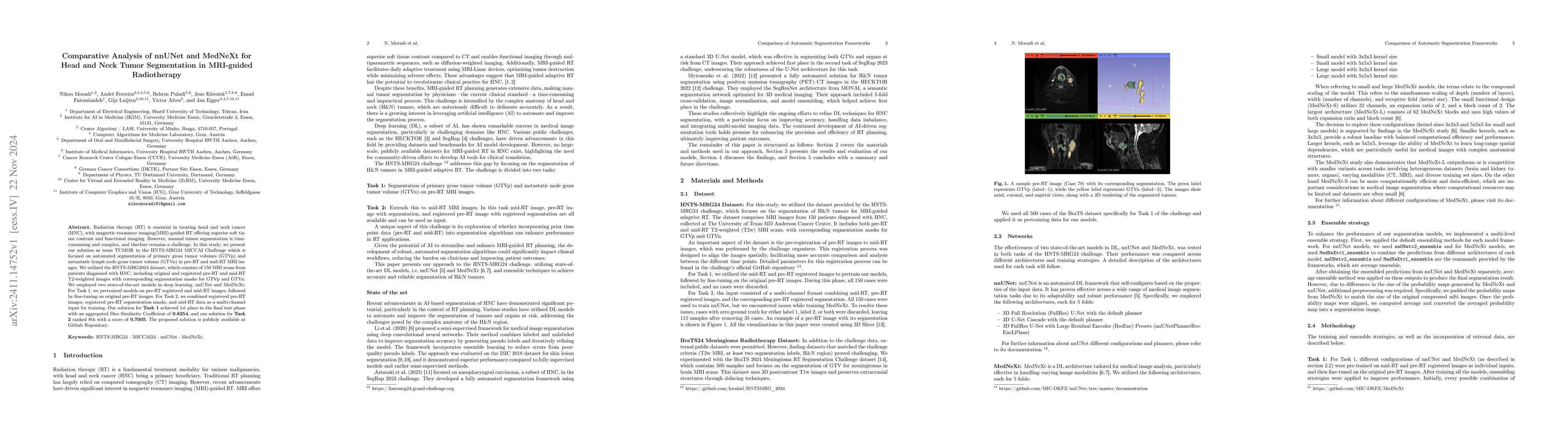
Radiation therapy (RT) is essential in treating head and neck cancer (HNC), with magnetic resonance imaging(MRI)-guided RT offering superior soft tissue contrast and functional imaging. However, manua...
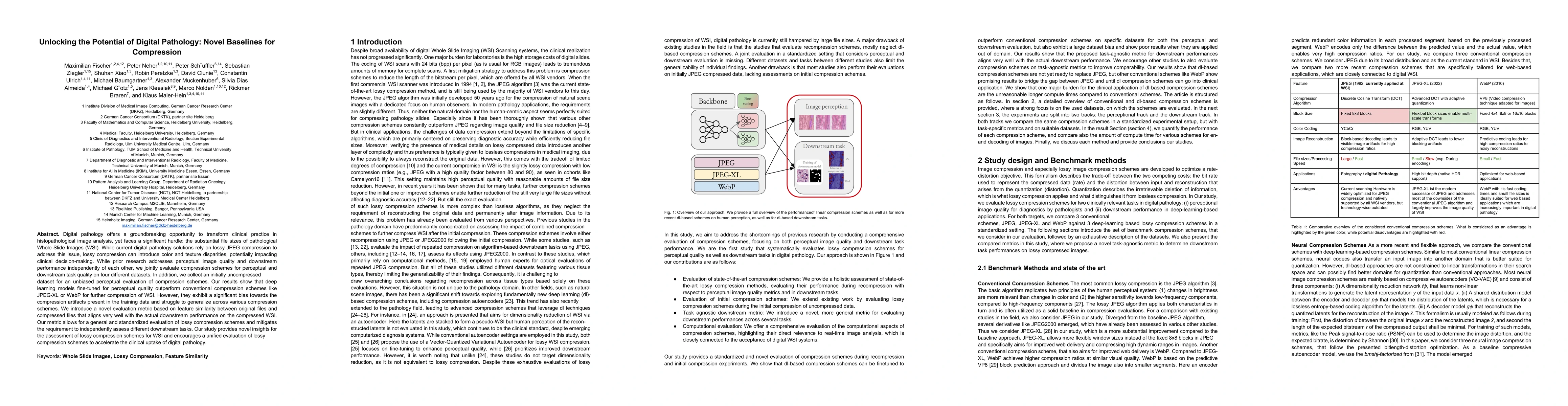
Digital pathology offers a groundbreaking opportunity to transform clinical practice in histopathological image analysis, yet faces a significant hurdle: the substantial file sizes of pathological Who...
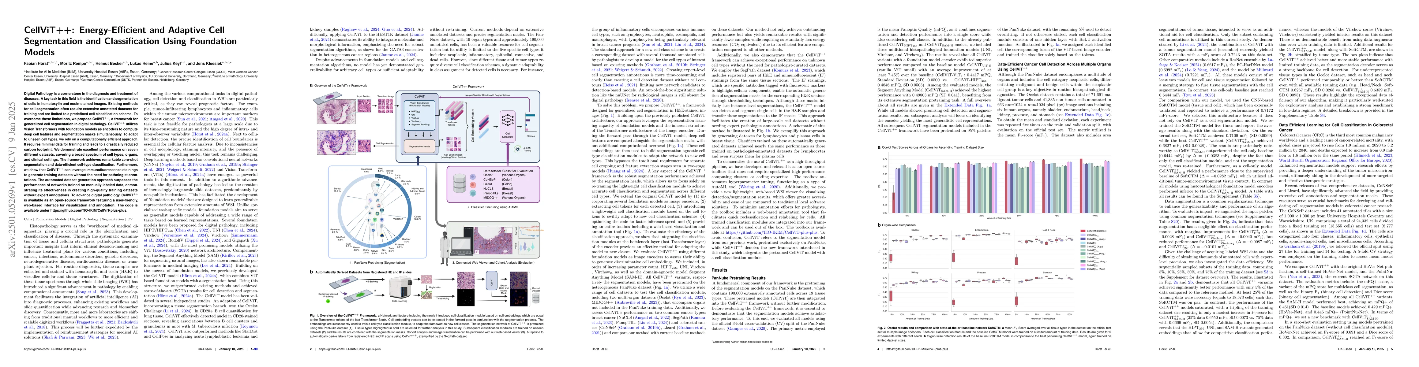
Digital Pathology is a cornerstone in the diagnosis and treatment of diseases. A key task in this field is the identification and segmentation of cells in hematoxylin and eosin-stained images. Existin...
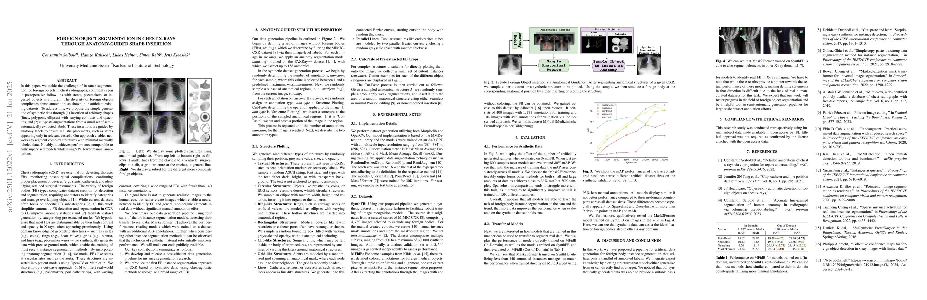
In this paper, we tackle the challenge of instance segmentation for foreign objects in chest radiographs, commonly seen in postoperative follow-ups with stents, pacemakers, or ingested objects in chil...
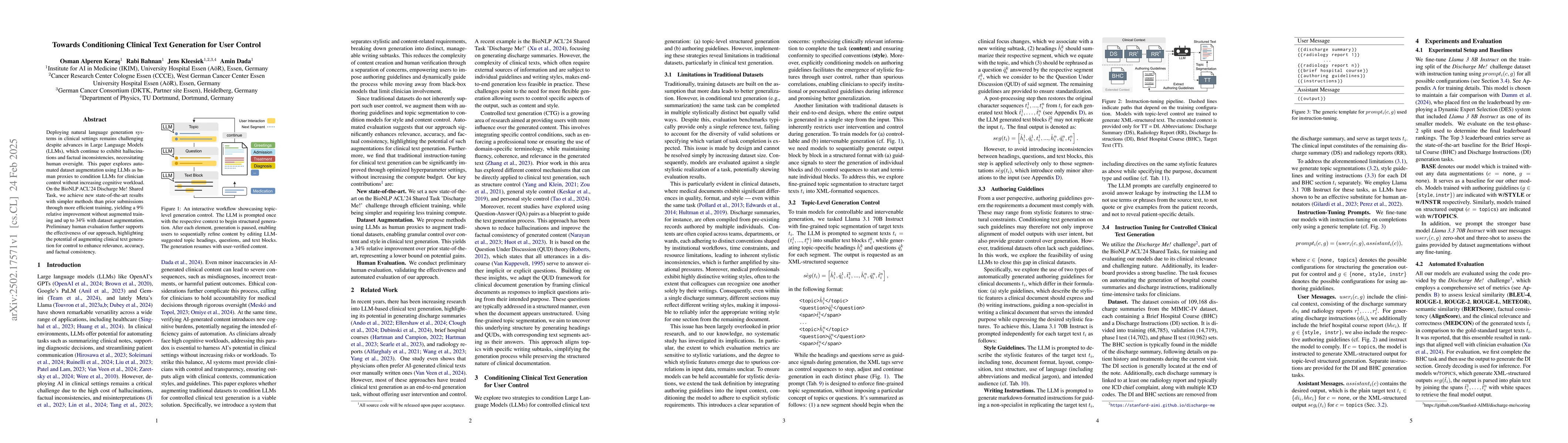
Deploying natural language generation systems in clinical settings remains challenging despite advances in Large Language Models (LLMs), which continue to exhibit hallucinations and factual inconsiste...

While increasing patients' access to medical documents improves medical care, this benefit is limited by varying health literacy levels and complex medical terminology. Large language models (LLMs) of...
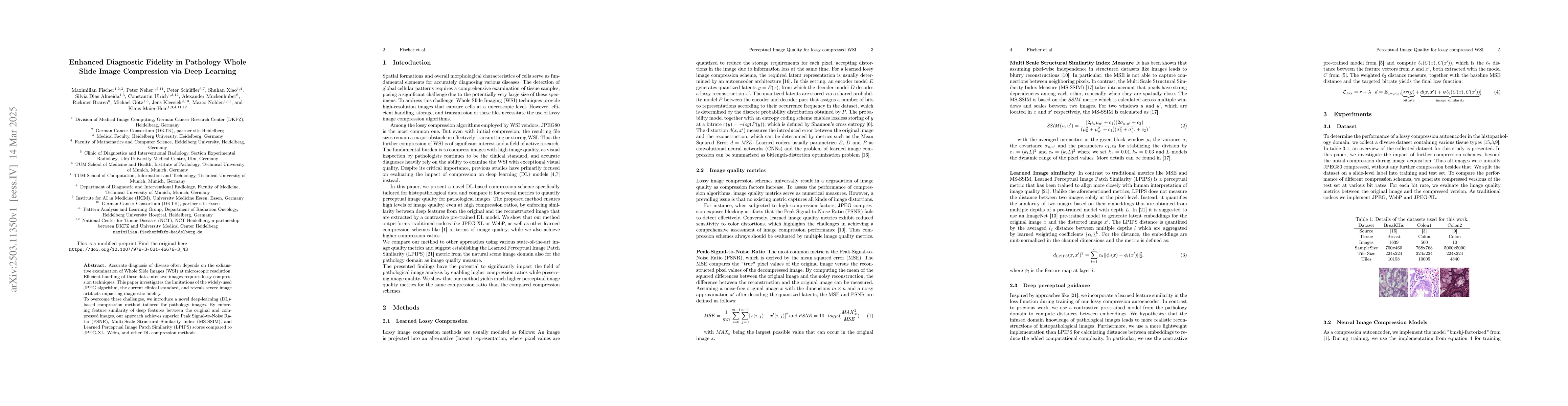
Accurate diagnosis of disease often depends on the exhaustive examination of Whole Slide Images (WSI) at microscopic resolution. Efficient handling of these data-intensive images requires lossy compre...
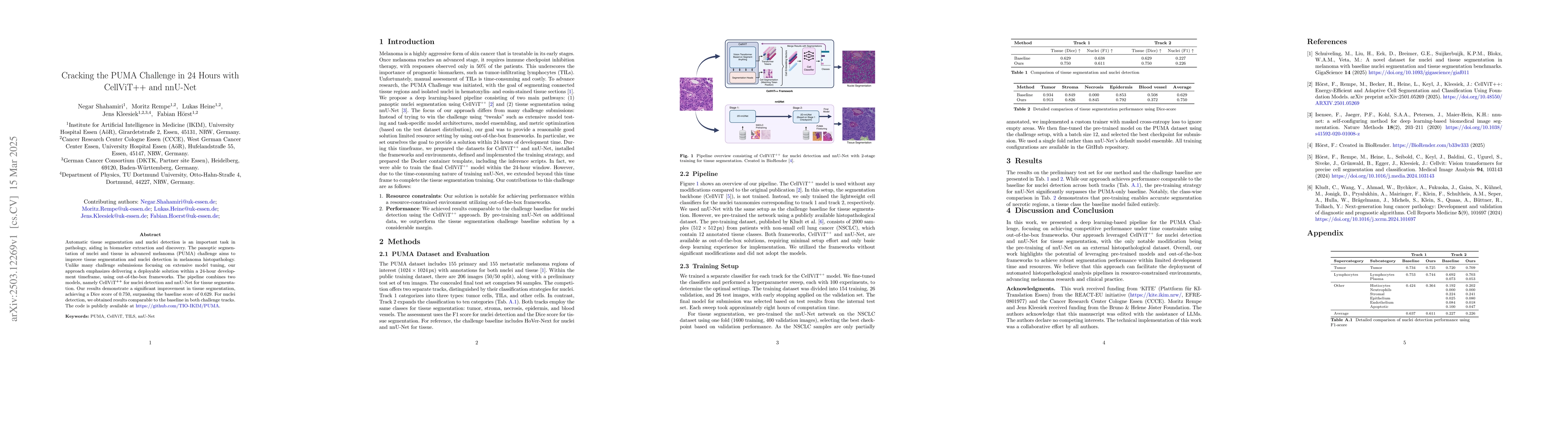
Automatic tissue segmentation and nuclei detection is an important task in pathology, aiding in biomarker extraction and discovery. The panoptic segmentation of nuclei and tissue in advanced melanoma ...
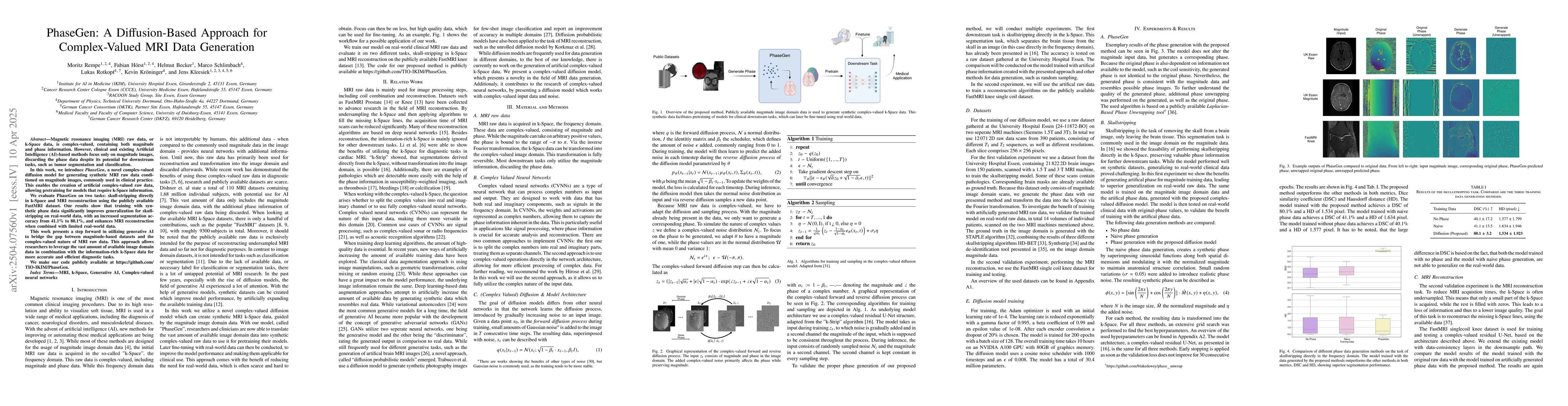
Magnetic resonance imaging (MRI) raw data, or k-Space data, is complex-valued, containing both magnitude and phase information. However, clinical and existing Artificial Intelligence (AI)-based method...
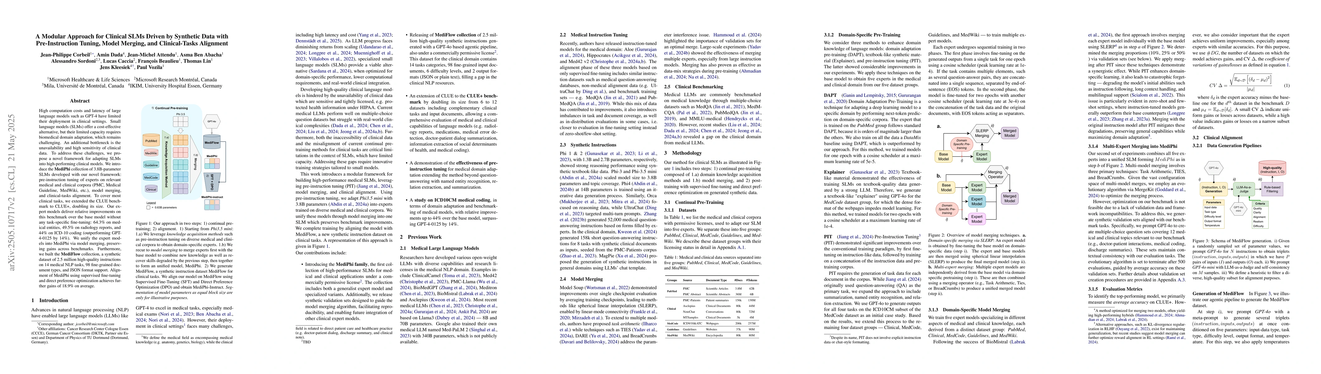
High computation costs and latency of large language models such as GPT-4 have limited their deployment in clinical settings. Small language models (SLMs) offer a cost-effective alternative, but their...
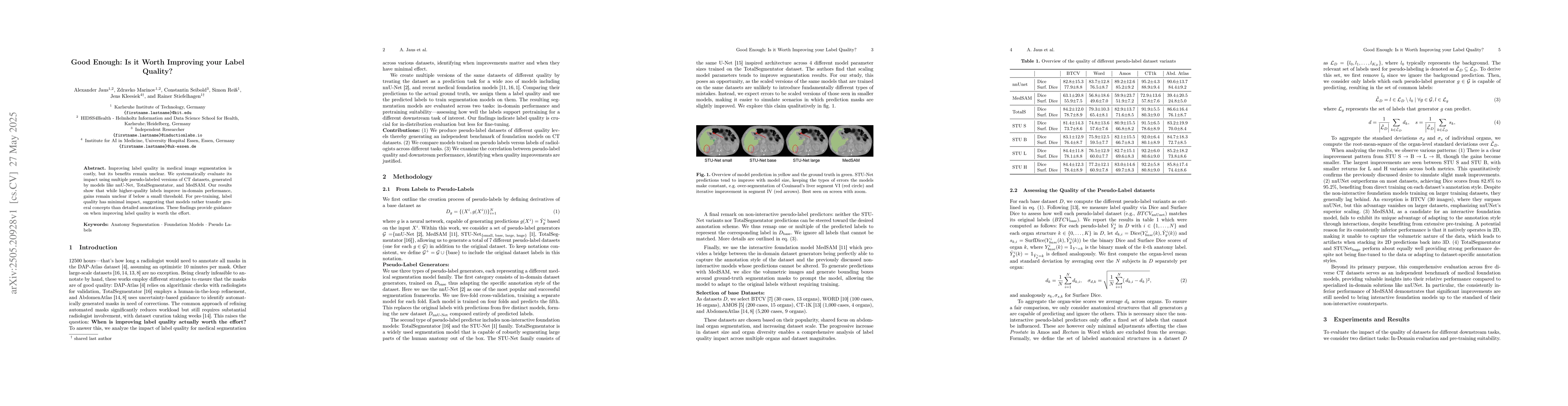
Improving label quality in medical image segmentation is costly, but its benefits remain unclear. We systematically evaluate its impact using multiple pseudo-labeled versions of CT datasets, generated...
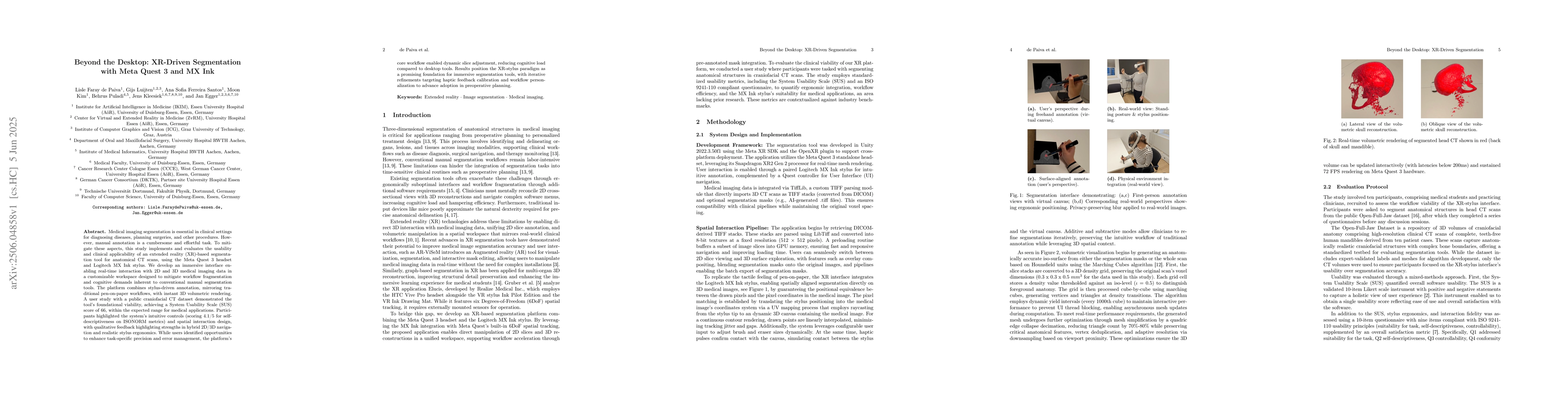
Medical imaging segmentation is essential in clinical settings for diagnosing diseases, planning surgeries, and other procedures. However, manual annotation is a cumbersome and effortful task. To miti...
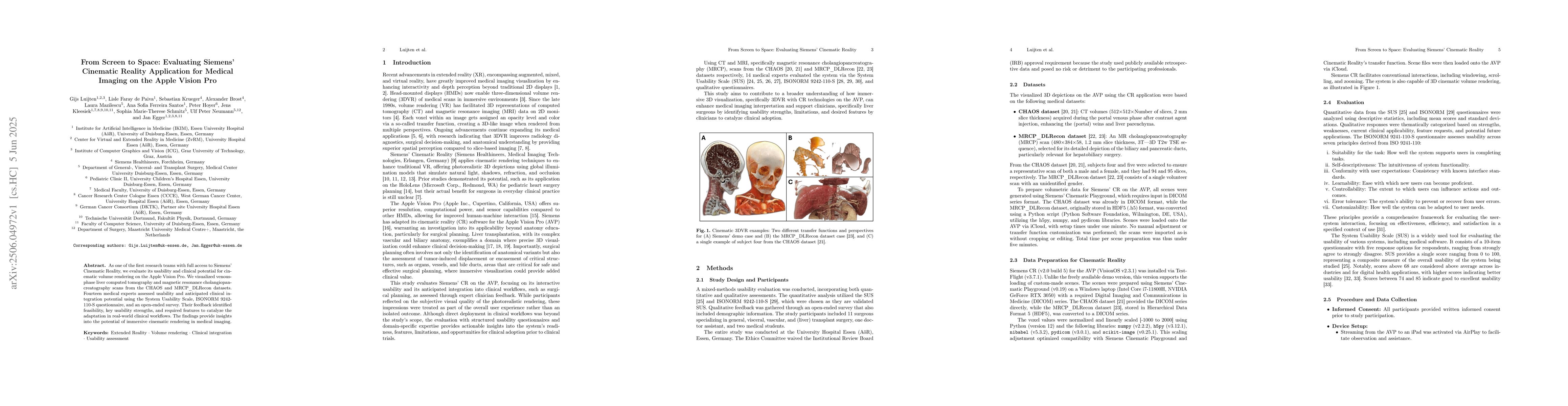
As one of the first research teams with full access to Siemens' Cinematic Reality, we evaluate its usability and clinical potential for cinematic volume rendering on the Apple Vision Pro. We visualize...
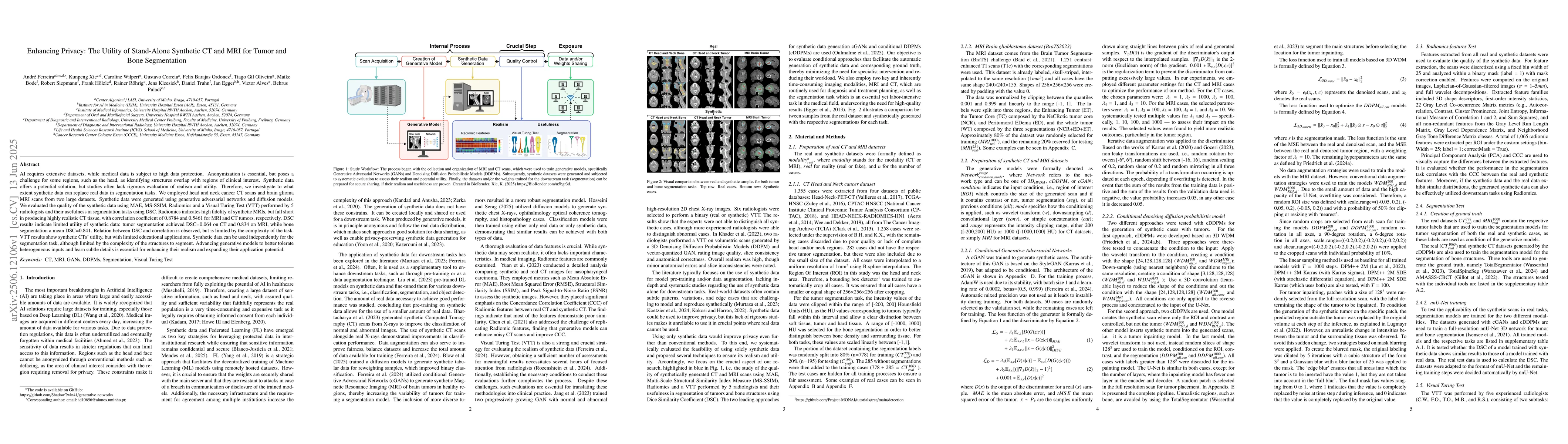
AI requires extensive datasets, while medical data is subject to high data protection. Anonymization is essential, but poses a challenge for some regions, such as the head, as identifying structures o...
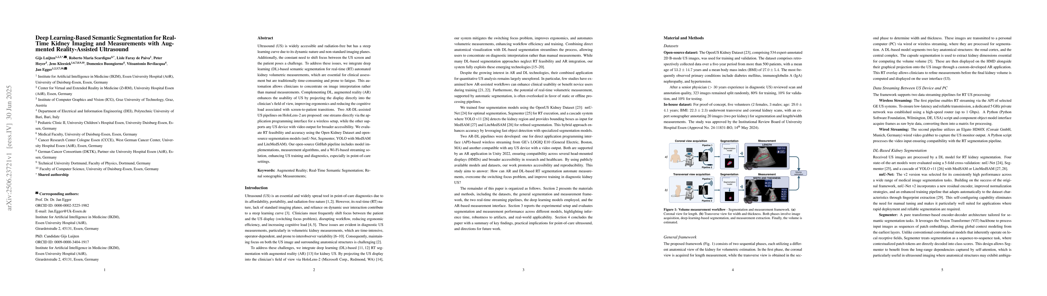
Ultrasound (US) is widely accessible and radiation-free but has a steep learning curve due to its dynamic nature and non-standard imaging planes. Additionally, the constant need to shift focus between...
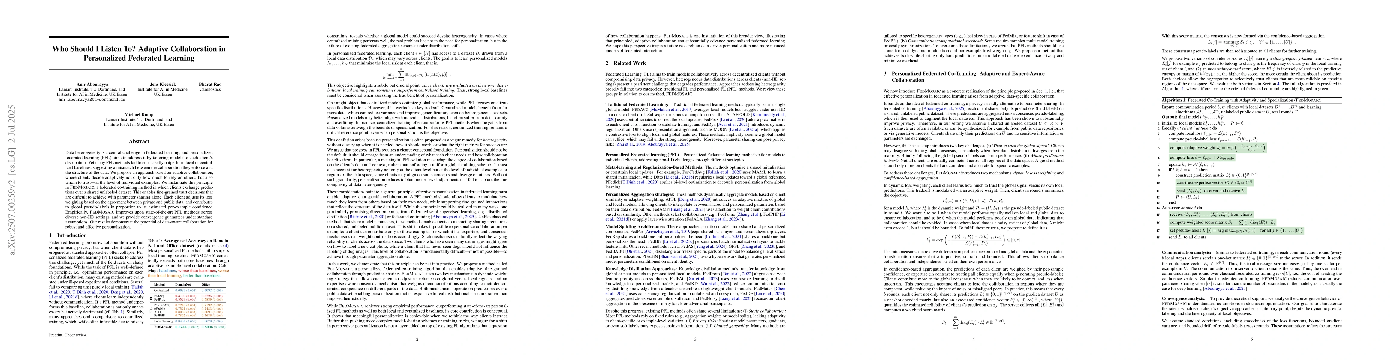
Data heterogeneity is a central challenge in federated learning, and personalized federated learning (PFL) aims to address it by tailoring models to each client's distribution. Yet many PFL methods fa...

Reliable end-to-end clinical report generation has been a longstanding goal of medical ML research. The end goal for this process is to alleviate radiologists' workloads and provide second opinions to...
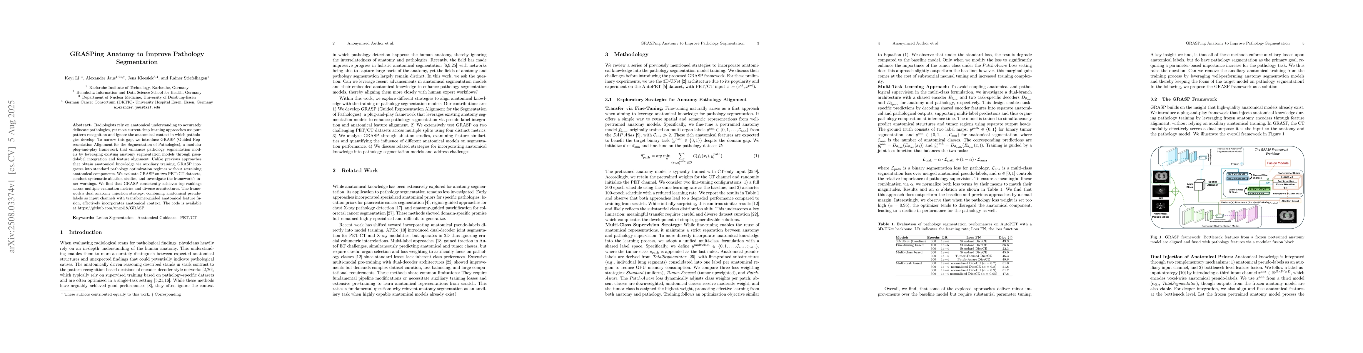
Radiologists rely on anatomical understanding to accurately delineate pathologies, yet most current deep learning approaches use pure pattern recognition and ignore the anatomical context in which pat...
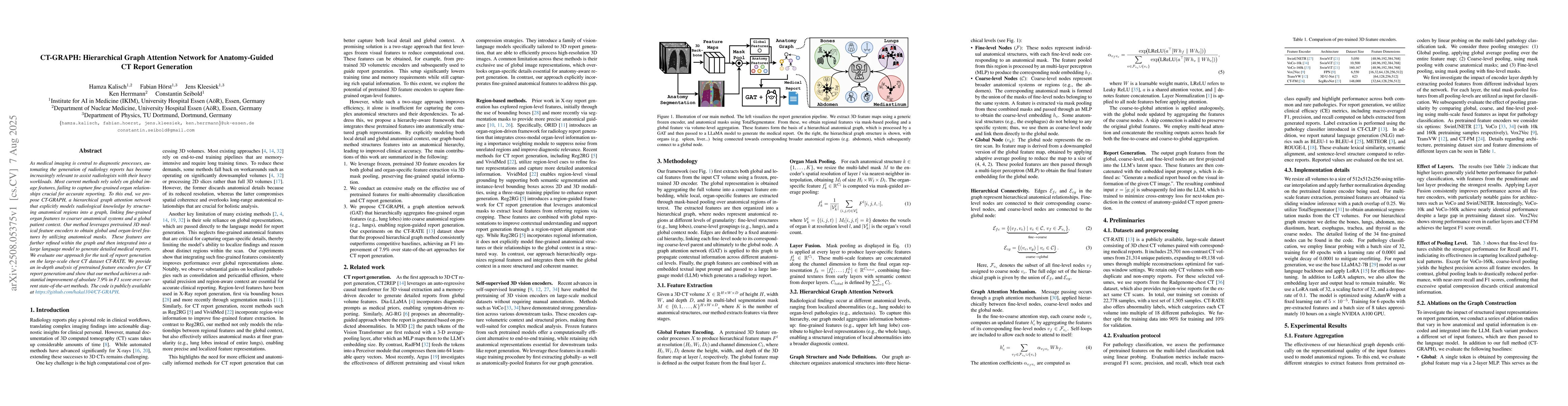
As medical imaging is central to diagnostic processes, automating the generation of radiology reports has become increasingly relevant to assist radiologists with their heavy workloads. Most current m...
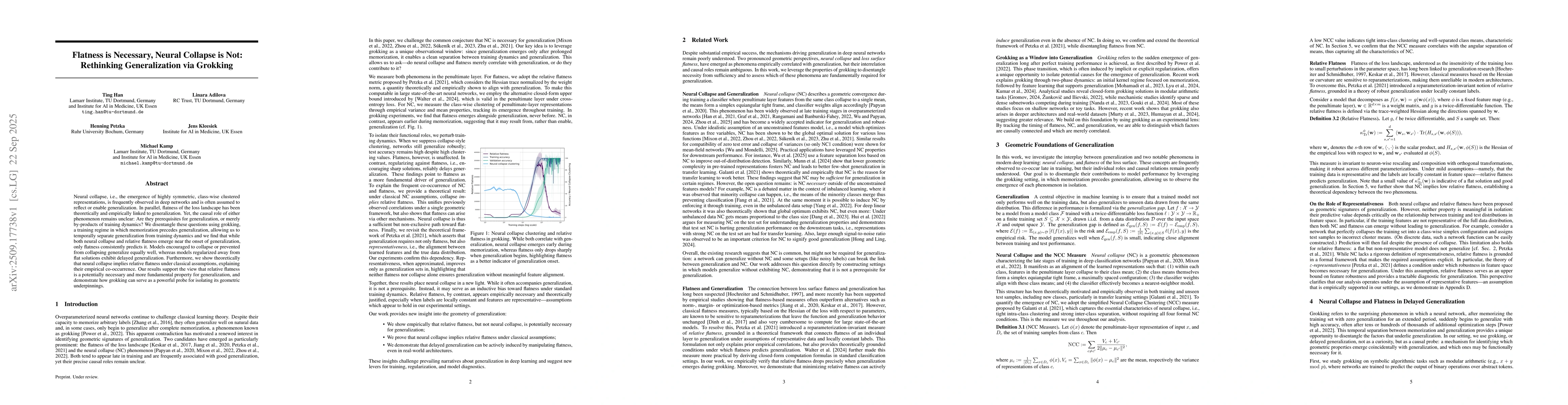
Neural collapse, i.e., the emergence of highly symmetric, class-wise clustered representations, is frequently observed in deep networks and is often assumed to reflect or enable generalization. In par...
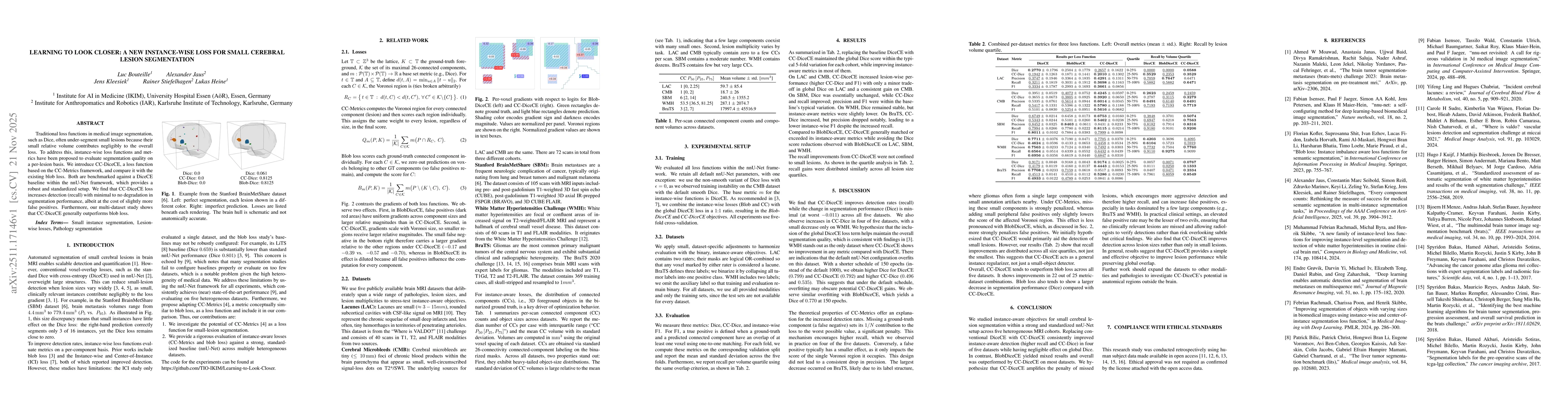
Traditional loss functions in medical image segmentation, such as Dice, often under-segment small lesions because their small relative volume contributes negligibly to the overall loss. To address thi...
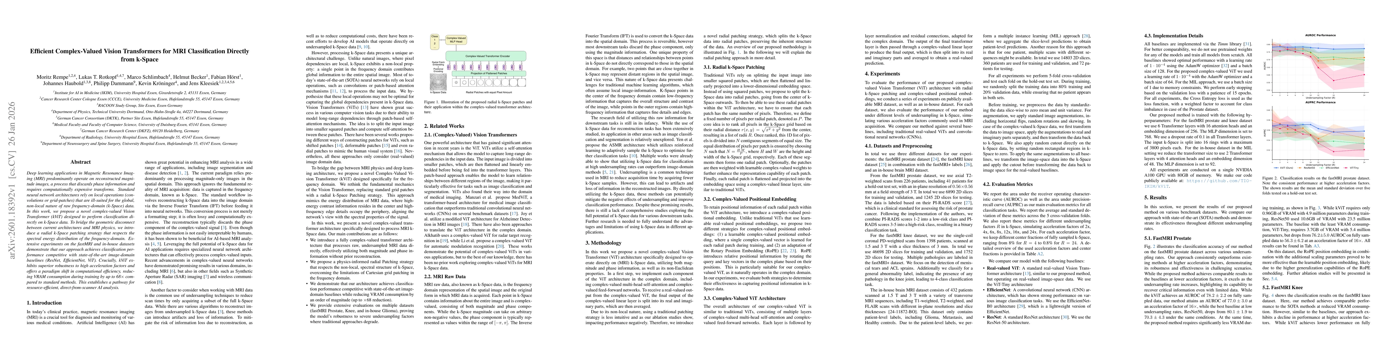
Deep learning applications in Magnetic Resonance Imaging (MRI) predominantly operate on reconstructed magnitude images, a process that discards phase information and requires computationally expensive...
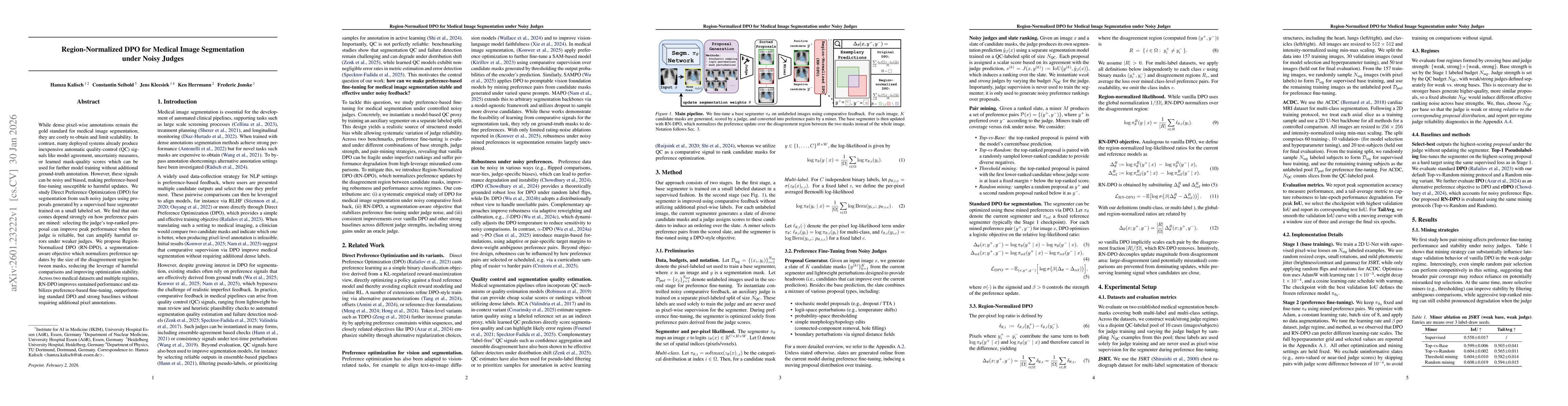
While dense pixel-wise annotations remain the gold standard for medical image segmentation, they are costly to obtain and limit scalability. In contrast, many deployed systems already produce inexpens...
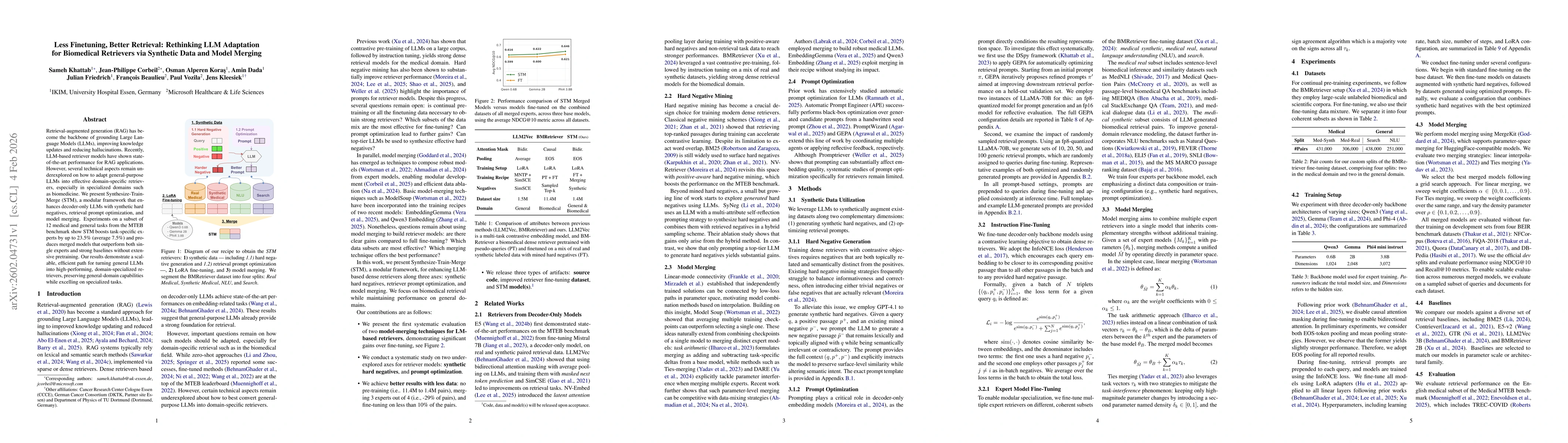
Retrieval-augmented generation (RAG) has become the backbone of grounding Large Language Models (LLMs), improving knowledge updates and reducing hallucinations. Recently, LLM-based retriever models ha...
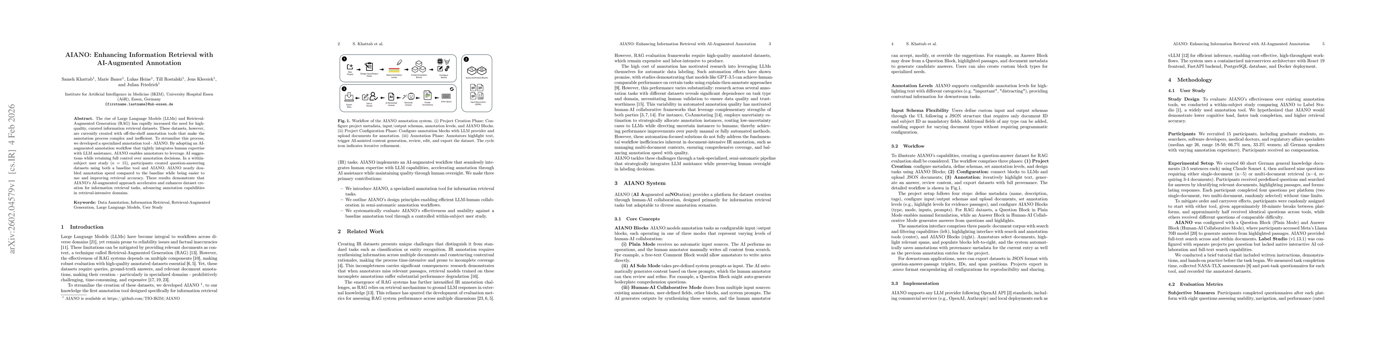
The rise of Large Language Models (LLMs) and Retrieval-Augmented Generation (RAG) has rapidly increased the need for high-quality, curated information retrieval datasets. These datasets, however, are ...

Objective. Standard Magnetic Resonance Imaging (MRI) reconstruction pipelines discard phase information captured during acquisition, despite evidence that it encodes tissue properties relevant to tumo...
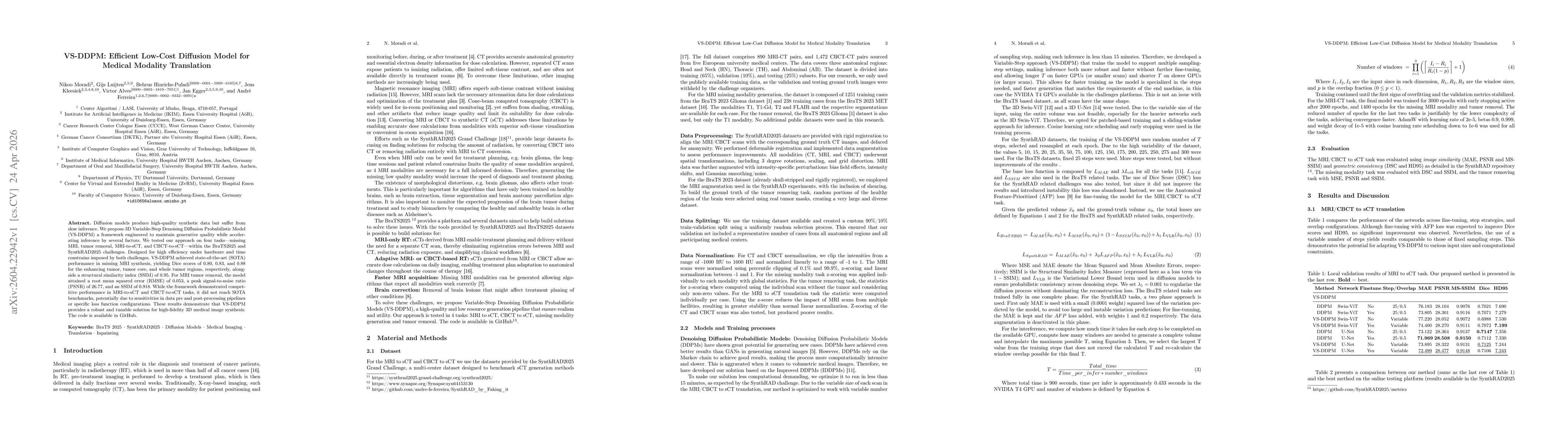
Diffusion models produce high-quality synthetic data but suffer from slow inference. We propose 3D Variable-Step Denoising Diffusion Probabilistic Model (VS-DDPM) a framework engineered to maintain ge...
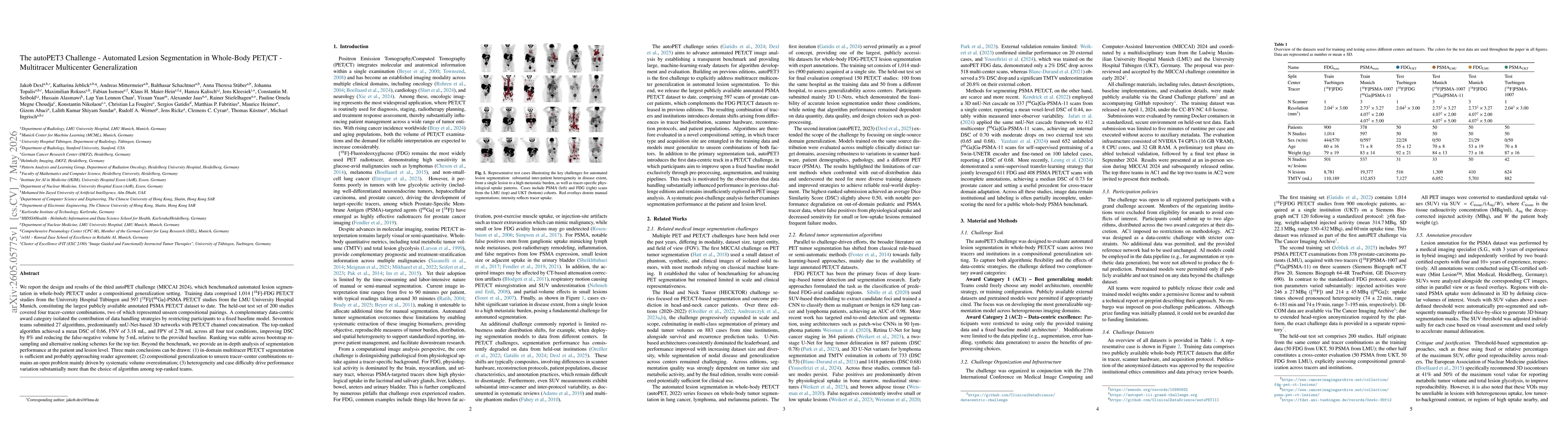
We report the design and results of the third autoPET challenge (MICCAI 2024), which benchmarked automated lesion segmentation in whole-body PET/CT under a compositional generalization setting. Traini...
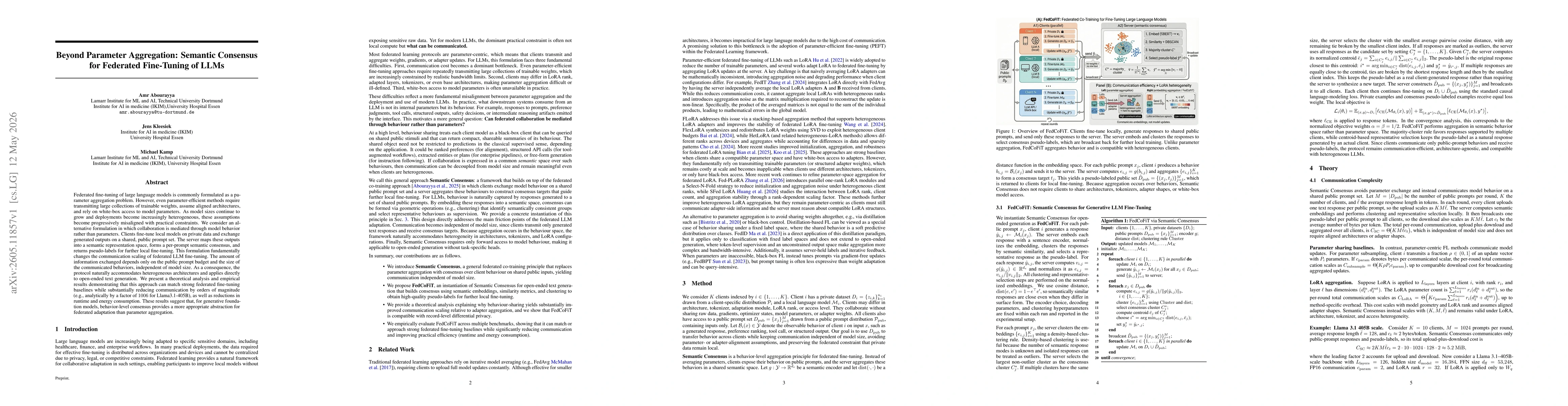
Federated fine-tuning of large language models is commonly formulated as a parameter aggregation problem. However, even parameter-efficient methods require transmitting large collections of trainable ...
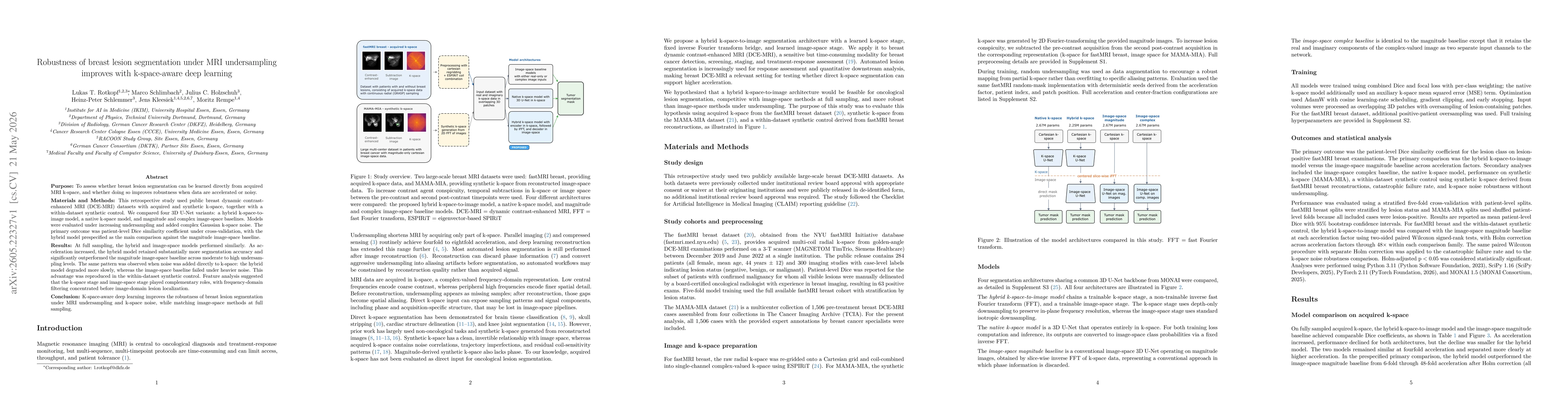
Purpose: To assess whether breast lesion segmentation can be learned directly from acquired MRI k-space, and whether doing so improves robustness when data are accelerated or noisy. Materials and Me...
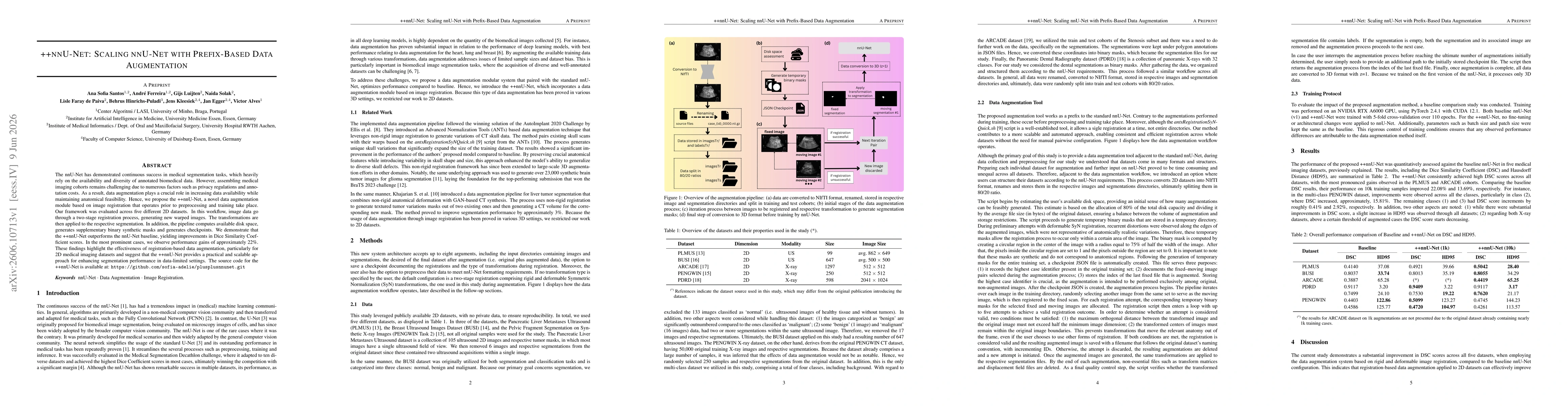
The nnU-Net has demonstrated continuous success in medical segmentation tasks, which heavily rely on the availability and diversity of annotated biomedical data. However, assembling medical imaging co...
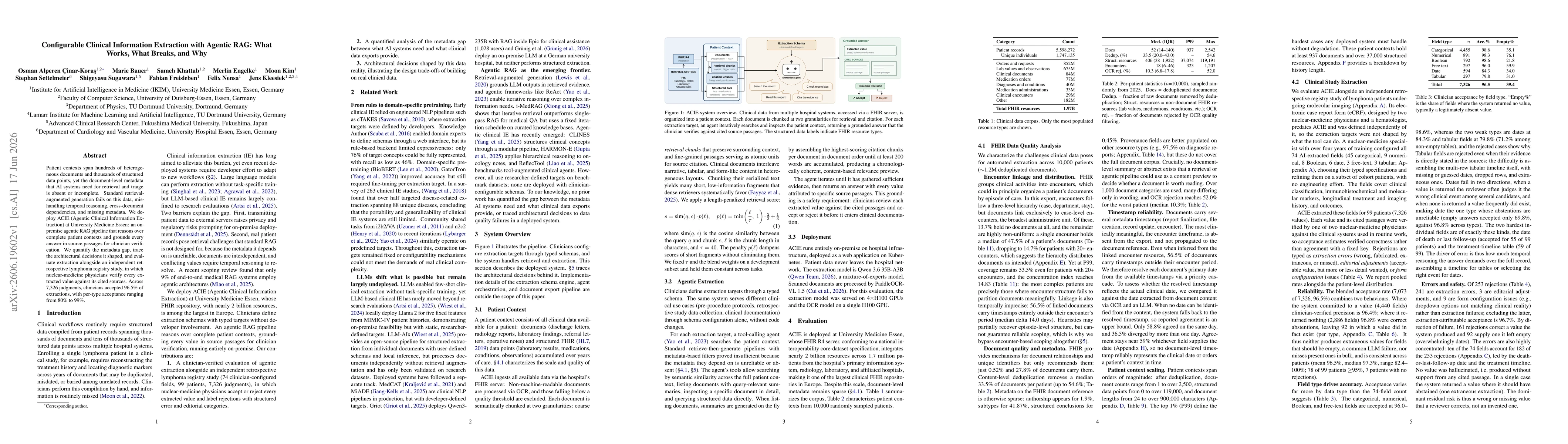
Patient contexts span hundreds of heterogeneous documents and thousands of structured data points, yet the document-level metadata that AI systems need for retrieval and triage is absent or incomplete...

Patient contexts span hundreds of heterogeneous documents and thousands of structured data points, yet the document-level metadata that AI systems need for retrieval and triage is absent or incomplete...